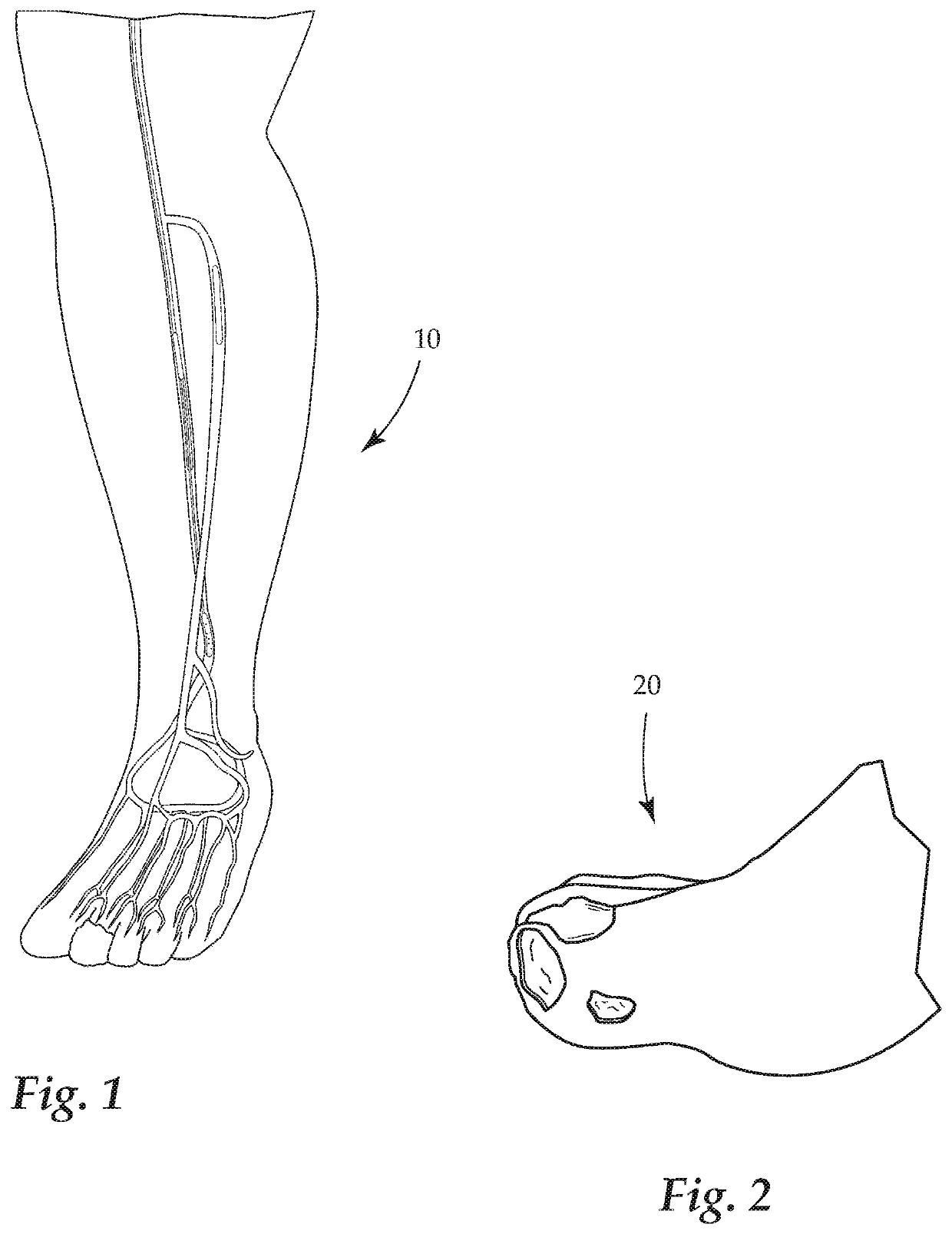
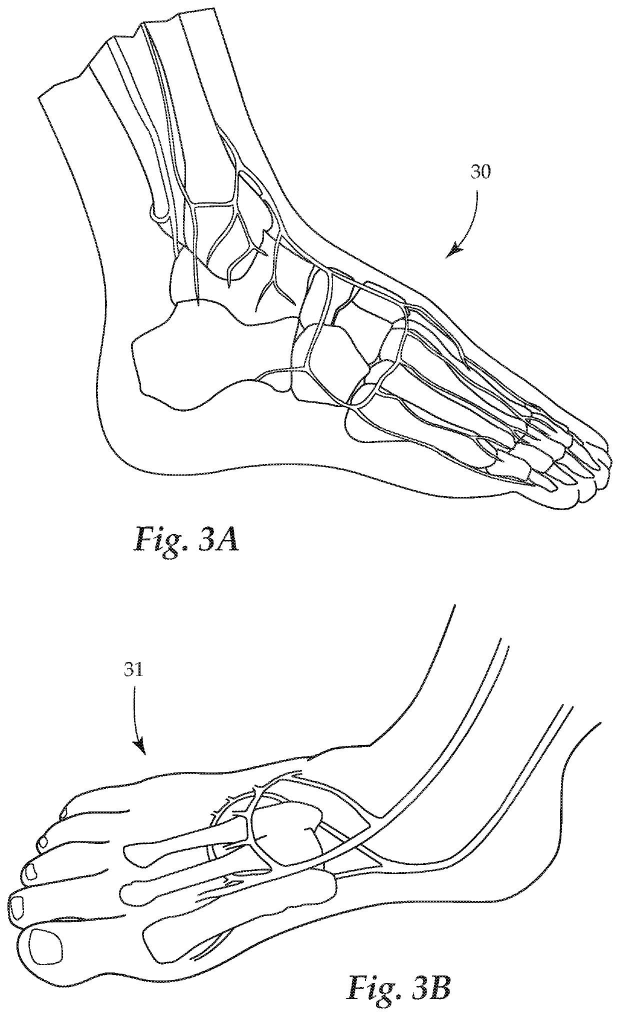
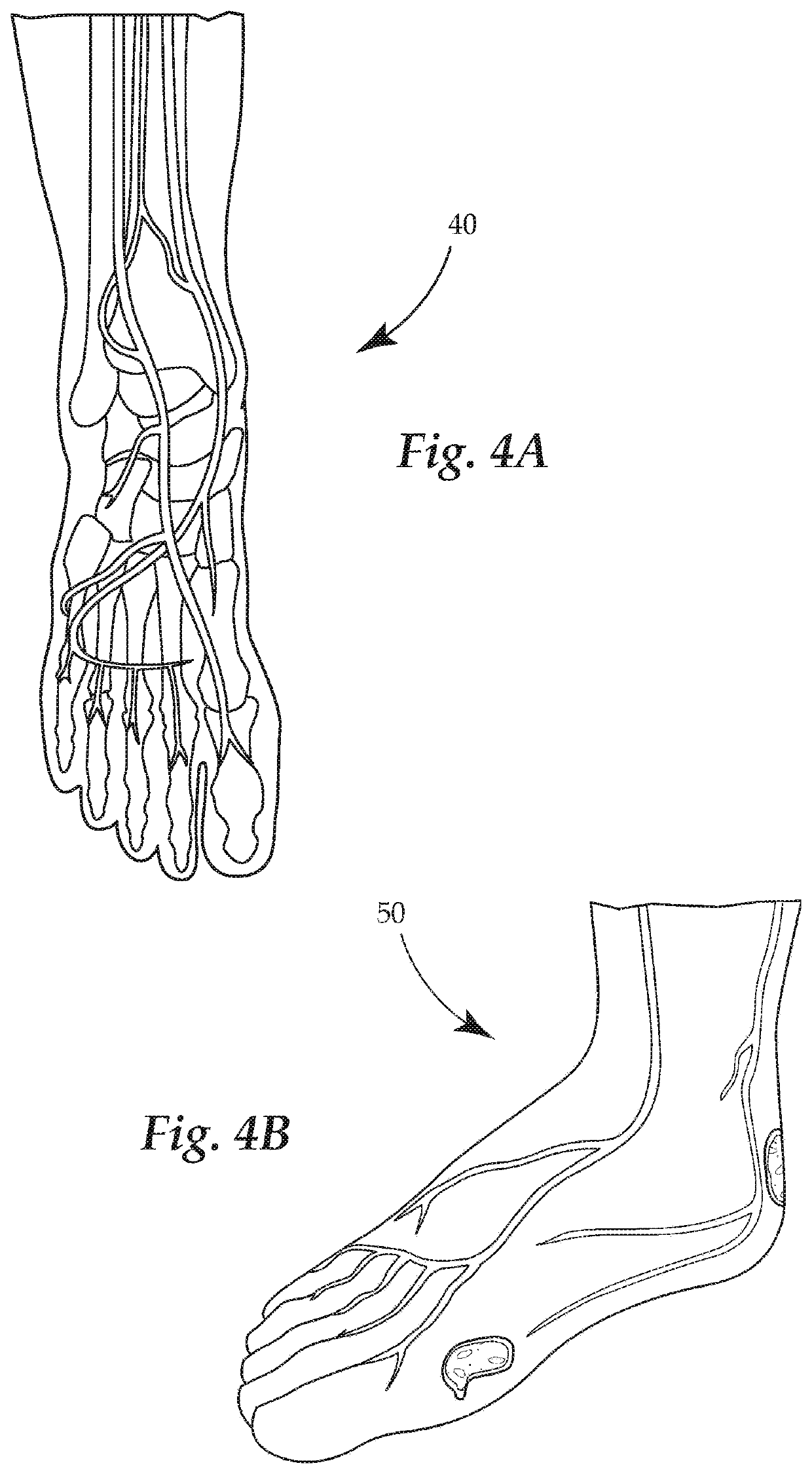
Currently there are no favorable stents on the market to meet needs for below the
ankle due to
ankle strap crush effect and the ankle area is susceptible to
high stress, torsion, extension, and
body weight.
If
blood flow to your
toe, foot, or leg is completely blocked, the tissue begins to die.
This buildup of plaque, also known as atherosclerosis, narrows or blocks
blood flow, reducing circulation of blood to the legs, feet or hands and markedly reduces blood flow to the extremities (hands, feet and legs) and has progressed to the point of
severe pain and even
skin ulcers or sores.
This condition will not improve on its own.
Concurrently, when dealing with patients in the throes of critical
limb ischemia and exhibiting those issue delineated above, the treating
medical team must decide which patient is a candidate for limb salvage, and this has always been an open-ended question due to the limitations of the treatment methods, as well as the restrictive nature of access to the pertinent arteries and veins within the lower extremities.
PAD is a major contributor to morbidity, reduced
quality of life (QOL), and mortality in an increasing elderly demographic in the Western world.
The chief challenge in the management of
peripheral arterial disease would be retooling of the health
system to focus on identifying patients with PAD and taking the enormous opportunity and responsibility to refine and aggressively manage the atherosclerotic risk factors in these patients.
As a general rule, in patients with combined inflow and outflow
disease, inflow problems are corrected first, since improvement of the inflow may diminish the symptoms of
claudication and reduce the likelihood of distal graft
thrombosis from low flow.
However, the presence of severe
vascular calcification, particularly in the infrainguinal vasculature, presents a significant procedural challenge to current endovascular strategies.
However, early elastic
recoil, frequent dissections, and poor primary and secondary patency rates for long lesions, with 40-50% of cases requiring bail-out stenting, limit
balloon angioplasty of “severely” calcified lesions, despite the high procedural success rates.
Furthermore, the presence of rigid calcified plaques may result in incomplete
stent expansion and significant residual
stenosis.
Distal
embolization remains a concern with these devices, given that these devices require retrieval of removed plaque, and the use of
distal protection devices may be needed, particularly in cases of heavily calcified lesions.
Despite different choices of
atherectomy devices and advanced technologies, there have been no comparative
efficacy or safety studies evaluating the four FDA-approved
atherectomy devices.
Atherectomy devices can reduce the burden of soft atheromatous or calcific plaque, change the vessel compliance, reduce vessel wall trauma, leading to a decrease in the need for bail-out stenting.11 On the other hand, atherectomy devices carry significantly higher
capital equipment-related costs, particularly when used in conjunction with
distal protection filters, and lead to an increase in procedure duration and
exposure to
radiation.
The challenge for the vascular specialist is to determine whether the nature and severity of presenting symptoms correlate with the degree of chronic arterial insufficiency present or whether alternative etiologies, such as neuropathy,
inflammation, infection, lymphatic or venous
disease, and repetitive trauma, are more likely responsible.
In practice, conduit availability is almost always a critical, rate-
limiting factor because good quality, autogenous
vein conduit is preferred in almost every circumstance.
Although the proximal superficial femoral and deep femoral arteries can be exposed via this incision, such a curvilinear or oblique incision limits further distal arterial
exposure.
An increasing incidence of femoral incisional complications, including
wound edge necrosis and separation, lymphatic leaks, femoral neuropraxia, and venous injuries are associated with incorrectly placed inguinal incisions for femoral
exposure.
Caution is necessary in this area, as a prominent
femoral vein tributary crosses anteriorly over the CFA in this area and is prone to injury if not identified, ligated, and divided early in the
dissection.
Inadvertent injury to this “
vein of pain” produces retraction and troublesome bleeding.
Transection may result in medial
thigh discomfort.
However, in certain patient's not suitable for
surgery, and with improving technology,
angioplasty is being used more frequently.
Other
key issues broach whether
drug-delivery balloons and the current
stent designs are not the correct systems for the SFA and whether changes to stent technologies—woven stents, alternatively designed stents or very conformable stents—define the future treatment directives.
An isolated
occlusion or
stenosis of the SFA often results in decreased
perfusion of the leg, resulting in demand related, reversible, ischemic pain localized to the calf.
Because the popliteal
artery is deep, it may be difficult to feel the popliteal pulse.
A popliteal
aneurysm (abnormal dilation of all or part of the popliteal
artery) usually causes
edema and pain in the popliteal fossa.
Because the artery is closely applied to the popliteal surface of the
femur and the
joint capsule, fractures of the
distal femur or dislocations of the knee may rupture the artery, resulting in hemorrhage.
Furthermore, because of their proximity and confinement within the fossa, an injury of the artery and
vein may result in an
arteriovenous fistula (communication between an artery and a vein).
Failure to recognize these occurrences and to act promptly may result in the loss of the leg and foot.
This repetitive trauma may result in stenotic artery degeneration, complete artery
occlusion or even formation of an
aneurysm.
These clots can
pose serious and even fatal risks.
Weighing the risks and complications of not treating, the obstructions within the vein go untreated, some serious complications can occur.
These may include a
pulmonary embolism occurs when a blood clot travels up to a vein within a
lung from another part of the body, causing a blockage.
Another risks and complications of not treating could be postphlebitic syndrome which occurs as a complication of damage to the vein caused by a blood clot.
The damage results in inhibited blood flow in the affected areas of the vein.
Although the indications for CB-PTA in the SFA includes significant residual stenosis or in-
stent restenosis, there are currently no published randomized controlled trials (RCT) comparing PTA vs.
cutting balloon angioplasty (CB-PTA) for any specific condition.
Angioplasty disrupts the atherosclerotic plaque by displacing it radially and results in stretching of the
adventitia thereby increasing the
lumen diameter in the treated vessel.
The presence of a stent may impede endovascular re-intervention if a re-stenosis results in occlusion of the stented arterial segment.
Problems that may happen include
severe pain in the hip,
thigh, calf, or foot, and trouble when walking.
Having these problems may decrease a person's ability to do his daily activities and affect his
quality of life.


