Use of osteopontin for the treatment and/or prevention of neurologic diseases

a neurologic disease and osteopontin technology, applied in the field of neurologic diseases and disorders, can solve the problems of loss of neurologic function, paralysis and death, and high vulnerability of oligodendrocytes to glutamate application, and achieve the effects of promoting myelination and nerve regeneration, promoting glial cell proliferation and differentiation, and promoting neuroprotection

Inactive Publication Date: 2007-05-15
MERCK SERONO SA
View PDF4 Cites 8 Cited by
  • Summary
  • Abstract
  • Description
  • Claims
  • Application Information

AI Technical Summary

Benefits of technology

[0123]The invention is based on the finding that the protein osteopontin promotes glial cell proliferation and differentiation, thus promoting myelination and regeneration of nerves. In accordance with the present invention, it has further been found that osteopontin has a beneficial effect in animal models of multiple sclerosis and peripheral neuropathies.

Problems solved by technology

While of various etiologies, such as infectious pathogens or autoimmune attacks, demyelinating diseases all cause loss of neurologic function and may lead to paralysis and death.
Moreover oligodendrocytes display high vulnerability to glutamate application (McDonald et al.
Even a brief interruption to the blood flow can cause decreases in brain function (neurologic deficit).
If the blood flow is decreased for longer than a few seconds, brain cells in the area are destroyed (infarcted) causing permanent damage to that area of the brain or even death.
A stroke may be caused by reduced blood flow (ischemia) that results in deficient blood supply and death of tissues in that area (infarction).
Damage to the brain is often more severe than with a stroke caused by cerebral thrombosis.
Damage to small unmyelinated and myelinated fibers results primarily in loss of temperature and pain sensation; damage to large myelinated fibers results in motor or proprioceptive defects.
A sensory deficit in the palmar aspect of the first three fingers may follow; the muscles that control thumb abduction and opposition may become weak and atrophied.
In severe cases, there are objective signs of sensory loss, typically with stocking-and-glove distribution.
Painless ulcers on the digits or Charcot's joints may develop when sensory loss is profound.
Sensory or proprioceptive deficits may lead to gait abnormalities.
Motor involvement results in distal muscle weakness and atrophy.
Wasting and symmetric weakness of the distal extremities is usually insidious but can progress rapidly, sometimes accompanied by sensory loss, paresthesias, and pain.
Aching, cramping, coldness, burning, and numbness in the calves and feet may be worsened by touch.
Multiple vitamins may be given when etiology is obscure, but they have no proven benefit.
Less common sensorimotor neuropathies begin at birth and result in greater disability.
The main problem is pedal mutilation due to pain insensitivity, with frequent infections and osteomyelitis.
Genetic analysis is available, but no specific treatment.
It is the most common cause of intellectual decline with aging.
Insufficient dopamine disturbs the balance between dopamine and other transmitters, such as acetylcholine.
Without dopamine, the nerve cells cannot properly transmit messages, and this results in the loss of muscle function.
In addition to the loss of muscle control, some people with Parkinson's disease become severely depressed.
The CAG repeat length in Huntington's disease families does exhibit some instability that is particularly marked when children inherit the huntingtin gene from affected fathers.
In HD, it is not known how this widely, expressed gene, results in selective neuronal death.
In particular, the question of how these widely expressed genes cause selective neuronal death remains unanswered.
The nerves controlling these muscles shrink and disappear, which results in loss of muscle tissue due to the lack of nervous stimulation.
There may be a loss of nervous stimulation to semi-voluntary muscles, such as the muscles that control breathing and swallowing.
It mostly happens during car or bicycle accidents, but may also occur as the result of near drowning, heart attack, stroke and infections.
In this case, the skull hits a stationary object and the brain, which is inside the skull, turns and twists on its axis (the brain stem), causing localised or widespread damage.
Also, the brain, a soft mass surrounded by fluid that allows it to “float,” may rebound against the skull resulting in further damage.
Due to the twisting and rebounding, the traumatically brain injured patient usually receives damage or bruising to many parts of the brain.
There may be small hemorrhagic lesions or diffuse damage to axons, which can only be detected microscopically.
Secondary brain damage occurs as a result of complications developing after the moment of injury.
This type of head injury is more likely to damage a specific area of the brain.
Although medical care administered may be minimal, persons with brain injury without coma may experience symptoms and impairments similar to those suffered by the survivor of a coma injury.
Perforating injuries can cause extensive disruption and hemorrhage.
Unless the biochemical defect can be corrected or compensated for, permanent, often widespread, neurologic deficits result.
Mutations in the gene for the lysosomal enzyme galactocerebrosidase (GALC) result in low enzymatic activity and decreased ability to degrade galactolipids found almost exclusively in myelin.
However, remyelination occurs in many instances, and repair, regeneration, and complete, recovery of neural function can be rapid.
Mental deterioration, spasticity, and blindness may occur.
Adrenoleukodystrophy is invariably fatal.
In both diseases, axonal damage can add to a primarily demyelinating lesion and cause permanent neurologic deficits.
There is currently no clear curative treatment for the severest forms of MS, which are generally fatal.
This causes widespread demyelination and, subsequent loss of conduction efficiency among the axons of the CNS.
However, the myelinating or remyelinating oligodendrocyte is a cell under extreme metabolic stress, which under pressure of even minor additional insults can be irreversibly damaged (Scolding and Lassmann, 1996).
This decreases the probability of spontaneous repair in an active MS lesion, where inflammation and other detriments pose obstacles to remyelination.
In addition, it appears that the endogenous oligodendrocyte precursor populations adjacent to MS lesions are depleted during the chronic phases of the disease, due to inhibition of these precursors' ability to proliferate and differentiate (Wolswijk, 1998).
It would also be important to identify molecules that inhibit myelin synthesis, since these could lower the effectiveness of repair strategies such as oligodendroglial cell transplantation in MS.
However, in most cases they act by affecting the induction or transcription of certain genes, thus affecting the immune system.

Method used


Image

  • Use of osteopontin for the treatment and/or prevention of neurologic diseases
  • Use of osteopontin for the treatment and/or prevention of neurologic diseases
  • Use of osteopontin for the treatment and/or prevention of neurologic diseases

Examples


example 1

Osteopontin is Differentially Expressed in in Vivo and in Vitro Models of Demyelinating Diseases

[0271]Methods

[0272]In Vitro Model Systems

[0273]The Oli-neu cell line has been established via immortalization of oligodendroglial precursors with a replication-defective retrovirus encoding the t-neu oncogene, a constitutively active tyrosine kinase: this cell line was shown to be induced to differentiate in the presence of 1 mM dibutyryl-cAMP in the culture medium (Jung et al., 1995). This provided the possibility of studying oligodendrocytes as an isolated cell type.

[0274]The morphology and antigenic characteristics of cells of the mouse oligodendrocyte cell line Oli-neu, derived from A2B5 mouse oligodendrocyte precursors, in the untreated condition and after 6 days of pro-differentiating treatment with 1–5 mM dibutyryl-cAMP differs substantially. Whereas untreated Oli-neu cells have a round shape and are mostly bipolar like oligodendrocyte precursor cells, cAMP treated cells generate m...

example 2

Confirmation of Differential Gene Expression of Osteopontin by Real-Time Quantitative Reverse Transcriptase (RT)-PCR Assay (TaqMan®)

[0330]Methods

[0331]cDNA Template Generation

[0332]The cDNA templates for TaqMan® analysis were generated from total RNA samples via reverse-transcription (RT) using the TaqMan® reverse transcription reagents (P / N N808–0234). All RT reactions were performed in a 100-μl volume containing: 10 μl TaqMan RT buffer, 22 μl 25 mM MgCl2 solution (5.5 mM), 20 μl deoxyNTPs mixture (500 μM of each dNTP), 5 μl random hexamers (2.5 μM), 2 μl RNase inhibitor (0.4 U / μl), 2.5 μl. MultiScribe™ Reverse Transcriptase (1.25 U / μl) and 38.5 μl RNA sample (1 μg total) in RNase-free H2O. Reactions were performed on an Eppendorf MasterCycler at 25° C. for 10 min (incubation step), 48° C. for 30 min (reverse transcription), and 95° C. for 5 min (inactivation step). All synthesized cDNAs were stored at −20° C. in 20 μl volumes.

[0333]Primer Design and Verification

[0334]SYBR Green Re...

example 3

Confirmation of Differential Osteopontin Expression by Northern Blot

[0364]Methods

[0365]Blot Preparation

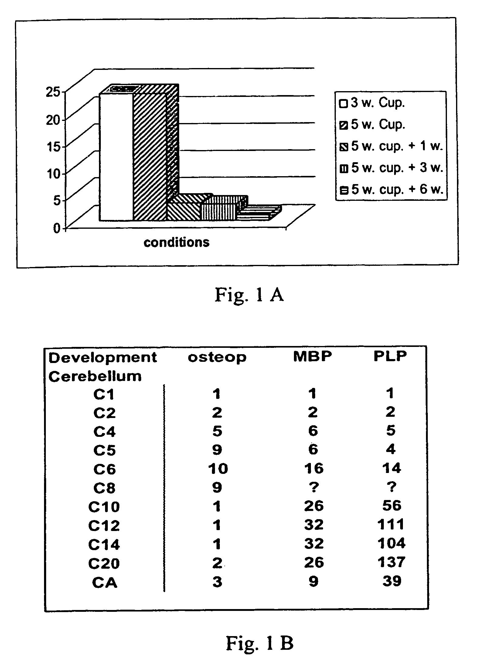
[0366]For specific genes, tissue specificity of expression was assayed using mouse Multi-Tissue Northern blots (Clontech Labs, 1020 East Meadow Circle, Palo Alto, Calif.). These contained 2 μg of poly(A)+ RNA per lane from different tissues of the adult mouse. Separate blots were prepared for analysis of differential gene expression in both in vitro and in vivo situations. RNA isolated from the brains of cuprizone treated mice at 3 weeks, 5 weeks and the 1, 3 and 6-week time points during the recovery process (up to 6 weeks) was used on one set of blots. Whole brain RNA from different postnatal day stages was used on a second set. Finally, a time-course series of RNAs was prepared from Oli-neu cells grown in culture and treated for different lengths of time with dibutyryl-cAMP. This RNA was used to prepare a third set of blots. New blots were used with each gene-specific probe to e...


PUM

PropertyMeasurementUnit
Tmaaaaaaaaaa
concentrationaaaaaaaaaa
volumeaaaaaaaaaa

Abstract

The invention relates to the use of osteopontin, or of an agonist of osteopontin activity, for treatment or prevention of a neurologic diseases.

Description

[0001]This application is a national stage application of PCT / EP02 / 05081, filed May 8, 2002, which claims foreign priority to European application 01111296.8, filed May 17, 2001.FIELD OF THE INVENTION[0002]The present invention is generally in the field of neurologic diseases and disorders. It relates to neuroprotection, nerve myelination and generation or regeneration of myelin producing cells. In particular, it relates to demyelinating and neurodegenerative diseases, neuropathies, traumatic nerve injury, stoke and neurologic diseases caused by congenital metabolic disorders. More specifically, the present invention relates to the use of osteopontin, or of an agonist of osteopontin activity, for the manufacture of a medicament for treatment and / or prevention of a neurologic disease.BACKGROUND OF THE INVENTION[0003]Nerve myelination is an essential process in the formation and function of the central nervous system (CNS) and peripheral nervous system (PNS) compartments. The myelin s...

Claims


Application Information

Patent Timeline
Patent Type & Authority Patents(United States)
IPC IPC(8): C07K14/00A61K31/7052C12N15/09A61K35/00A61K38/00A61K38/19A61K38/21A61K45/00A61K48/00A61P3/00A61P9/10A61P21/04A61P25/00A61P25/02A61P25/14A61P25/16A61P25/28C07K14/52
CPCA61K38/21C07K14/52A61K35/00A61K48/00C07K2319/30C07K2319/00A61K2300/00A61P19/10A61P21/04A61P25/00A61P25/02A61P25/14A61P25/16A61P25/18A61P25/28A61P3/00A61P3/10A61P43/00A61P9/10
Inventor BOSCHERT, URSULAFEGER, GEORGSELVARAJU, RAGHURAMBERNASCONI, LILIAPAPOIAN, RUBEN
Owner MERCK SERONO SA
Why Eureka
Try Eureka