Phage ligand sensor devices and uses thereof

a phage ligand and sensor technology, applied in the field of phage ligand sensor devices and their use, can solve the problems of limited longevity, low sensitivity, and previously reported biosensors, and achieve the effects of low yield, simple, routine steps, and efficient and convenient production

Inactive Publication Date: 2007-09-11
AUBURN UNIV
View PDF11 Cites 39 Cited by
  • Summary
  • Abstract
  • Description
  • Claims
  • Application Information

AI Technical Summary

Benefits of technology

[0031]Another advantage of the present invention is the ease with which phage can be produced for use in a PLSD device. Filamentous phage are efficiently and conveniently produced using bacterial cell cultures. The yield of wild-type phage particles from bacterial cultures regularly reaches 300 mg/liter, although engineered phage particles tend to have lower yields, e.g., 20 mg/liter for engineered or landscape phage. The phage particles are secreted from the cell nearly free of intracellular components, and further purification is easily accomplished by simple, routine steps that are applicable to any phage.
[0032]Thus, the invention provides compositions and assays for the rapid discovery of ligands specific to various peptides and finds use in the detection of a wide range of biological, organic, and other materials. A ligand is any compound, particle, or organism that binds at some measurable level to at least one foreign peptide displayed on landscape phage of the PLSD, thereby producing a detectable signal by a PLSD of the invention. Thus, ligand binding is detected and can be quantitated using a PLSD of the invention. Ligands as well as peptides of interest may be isolated or derived from any organism or species, including but not limited to mammals, reptiles, amphibians, plants, bacteria, viruses, amoeba, rickettsia, etc.
[0033]The PLSDs of the invention find use in detecting ligands such as, for example, enzymes, bacteria, viruses and other biological or organic agents and/or compounds as well as synthetic or artificial agents and/or compounds. Any ligand that is capable of binding to a foreign peptide displayed on a landscape phage may be detected and evaluated using the compositions and methods of the invention. More than one ligand may bind to a particular foreign peptide.
[0034]Thus, a ligand that binds to a particular foreign peptide may be but is not limited to microorganisms, including bacteria, viruses, fungi, and protozoa as well as organic and inorganic chemical compounds. Thus, ligands may include pathogens or harmful agents which are viruses, bacteria, fungus, prions, rickettsia, amoeba, and natural and synthetic toxins. Ligands may also include biochemical compounds, such as, for example, proteins, peptides, and nucleic acids. The term “virus” as used herein encompasses any virus, for example, smallpox virus, yellow fever virus, cholera virus, and hemorrhagic fever viruses such as Ebola virus, Marburg virus, and Lassa fever virus. The term “bacteria” as used herein encompasses bacterial spores and includes any species of bacteria, such as, for example, those bacteria known to cause bubonic plague (e.g., Yersinia pestis), pneumonic plague, and anthrax (e.g., Bacillus anthracis). Harmful agents and toxins include but are not limited to organic toxins such as ricin, botulism toxin (e....

Problems solved by technology

Biosensors previously reported in the literature are somewhat limited because the reported devices have low sensitivity, limited longevity, and/or long response times. Decker et al.
These long response times are not compatible with rapid screening and make large-scale screening unwieldy.
In addition, reported biosensors generally suffer from disadvantages such as l...

Method used


Image

  • Phage ligand sensor devices and uses thereof
  • Phage ligand sensor devices and uses thereof
  • Phage ligand sensor devices and uses thereof

Examples


example 1

PLSDs for Detection of β-Galactosidase

[0059]β-galactosidase from Escherichia coli is a tetrameric protein of identical 1,023-residue polypeptides, with molecular dimensions of 17.5-13.5-9 nm. Phage that bind to this protein and to small haptens were isolated from phage display libraries as described in Petrenko and Smith (2000) Prot. Eng. 13: 589-592. Binding of phage to the antigen was confirmed by enzyme linked immunosorbent assay (ELISA) in which the phage were immobilized on the plastic surface of the ELISA wells and reacted with their antigens in solution (see Petrenko and Smith, id.). The phage was biotinylated using commercially available reagents and standard procedures (see, e.g., 2002 catalog from Pierce Biotechnology, Inc., Rockford, Ill.).

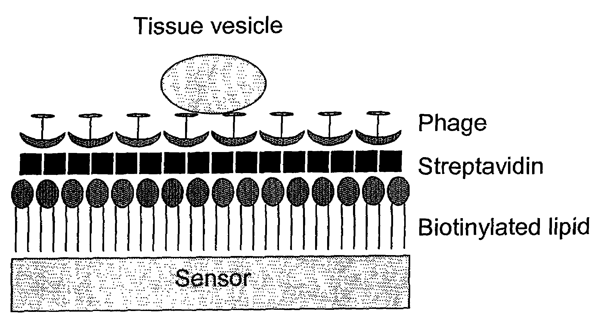
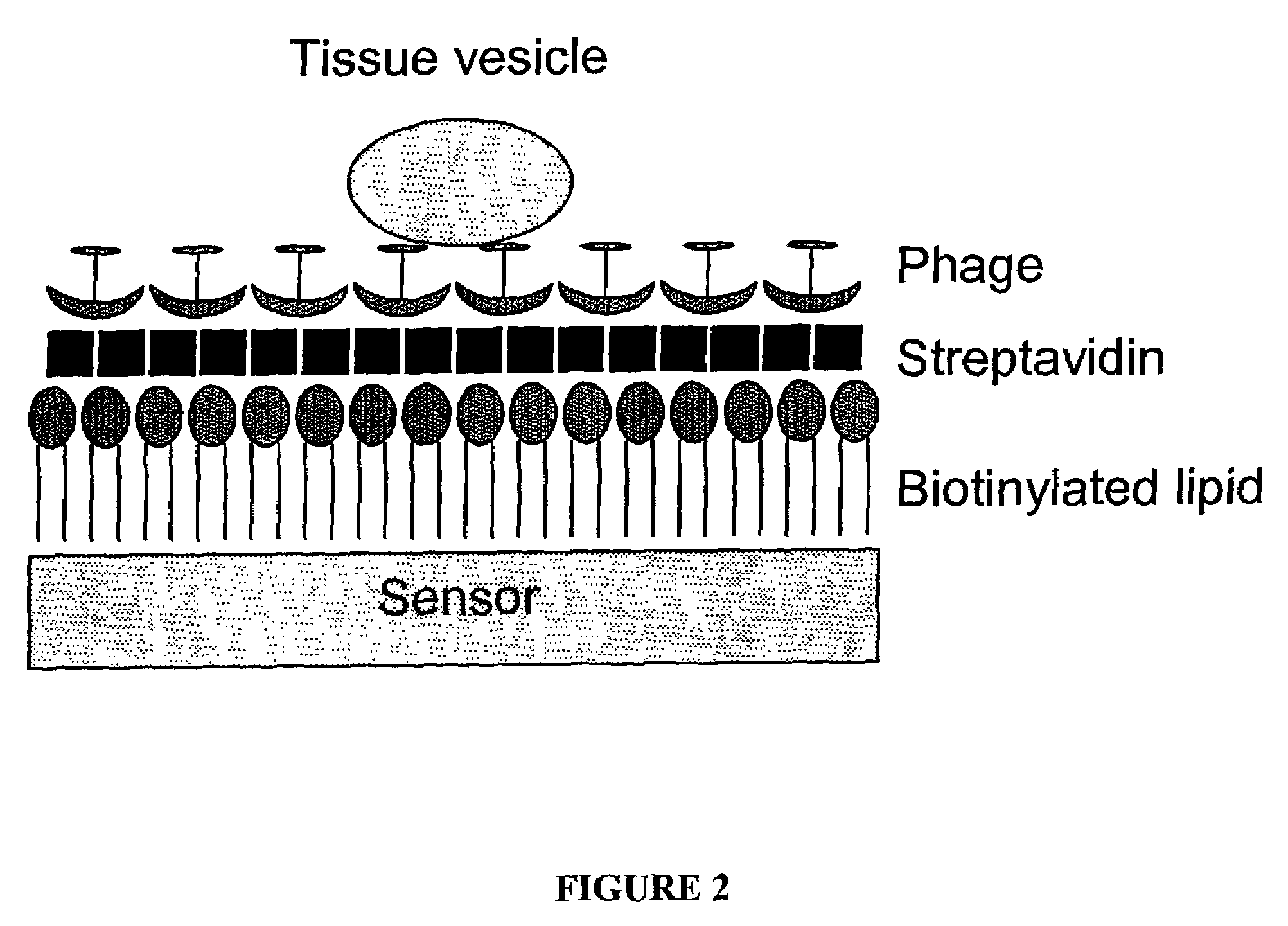
[0060]To prepare the sensor component of the device, AT-cut planar quartz crystals with a 5 MHz nominal oscillating frequency were purchased from Maxtek, Inc. Circular gold electrodes were deposited on both sides of the crystal sensor f...

example 2

Construction of Engineered Phage Libraries

[0070]A large (109-clone) phage library was constructed by splicing a degenerate coding sequence into the beginning of the pVIII coat-protein gene, replacing wild-type codons 2-4 (Petrenko et al. (1996) Protein Engineering 9: 797-801). The degenerate coding sequence used is underlined below in Table 1:

[0071]

TABLE 1Sequence Used to Generate Phage LibraryDNAGCAGNKNNKNNKNNKNNKNNKNNKNNGGATCCCGCAAAAGCGGCCTTTGACTCC(SEQ ID NO:1)pVIII amino acid sequence A  X  X  X  X  X  X  X  X  D  P  A  K  A  A  F  D  S(SEQ ID NO:2) 1                          5  6  7  8  9  10 11 12 13

[0072]In the DNA sequence, each N represents an equal frequency of all four nucleotides (A, G, C and T), while K represents an equal frequency of G and T. As a result of this modification, every pVIII subunit in an engineered phage was five amino acids longer than the wild-type pVIII subunit and displayed a “random” sequence of eight amino acids, or an octamer (X8 above). In any sin...

example 3

Selection of Phage that Bind Particular Lilands

[0075]Model ligands selected for creating PLSDs were: streptavidin (from the bacterium Streptomyces avidinii), avidin (from chicken egg white) and β-galactosidase (from Escherichia coli). Each protein was absorbed to the surface of a 35-mm polystyrene Petri dish and exposed to the phage library. Unbound phage were washed away, and bound phage was eluted with acid buffer and amplified by infecting fresh bacterial host cells. After three rounds of selection, individual phage clones were propagated and partially sequenced to determine the amino acid sequence of the displayed foreign peptide. The selected phage were further characterized by enzyme linked immuno sorbent assay (ELISA) in which the phage were immobilized on the plastic surface of ELISA wells and reacted with the ligands in solution phase. Neutravidin and streptavidin were labeled with alkaline phosphatase, while β-galactosidase was unlabeled. The data (graphed in FIG. 6) demon...


PUM


Abstract

Methods and compositions for identifying and characterizing one or more ligands of a peptide are provided. In particular, the invention provides a phage ligand sensor device (PLSD) comprising a sensor coupled to a binding element of interest. Binding elements of interest comprise phage displaying at least one foreign peptide. The PLSD and assays find particular use in identifying and characterizing ligand-peptide interactions.

Description

CROSS-REFERENCE TO RELATED APPLICATIONS[0001]This Application claims the benefit of U.S. Provisional Application No. 60 / 340,017, filed Nov. 7, 2001, and U.S. Provisional Application No. 60 / 415,037, filed Oct. 1, 2002, which disclosures are herein incorporated.FEDERALLY SPONSORED RESEARCH OR DEVELOPMENT[0002]The research underlying this invention was supposed in part with funds from the National Institutes of Health, Grant No. NIH-1-R21-A105564501, and the Department of Army, Grant No. DAAD 19-01-1-0454. The United States Government may have an interest in the subject matter or this invention.FIELD OF THE INVENTION[0003]The invention relates to sensors for the detection of ligands of interest and uses thereof.BACKGROUND OF THE INVENTION[0004]Many applications require detection and identification of ligands, or molecules having particular binding properties. The binding properties of a particular ligand may be detected and characterized by the use of appropriate sensors. “Biosensors” ...

Claims


Application Information

Patent Timeline
IPC IPC(8): G01N33/543C12M1/34C12Q1/70G01N33/483G01N33/53G01N33/569
CPCG01N33/54373G01N33/569
Inventor PENTRENKO, VALERY A.VODYANOY, VITALY J.SAMOYLOV, ALEXANDRE M.SOROKULOVA, IRYNANANDURI, VISWAPRAKASHCHIN, BRYAN A.BARBAREE, JAMES M.NEELY, W. CHARLES
Owner AUBURN UNIV
Why Eureka
Try Eureka