Novel bone tissue engineering scaffold and preparation method thereof

A bone tissue engineering and tissue technology, applied in the field of biomedical tissue engineering, can solve the problems of expensive preservation, cumbersome operation process, difficult preservation, etc., achieve good biomechanical properties, reduce immune rejection, and maintain the effect of roughly morphological structure

Pending Publication Date: 2020-08-07
XIANGYA HOSPITAL CENT SOUTH UNIV
View PDF9 Cites 7 Cited by
  • Summary
  • Abstract
  • Description
  • Claims
  • Application Information

AI Technical Summary

Problems solved by technology

However, with the deepening of research, some theoretical and technical problems have been encountered: 1) traditional tissue engineering technology needs to rely on the collection, separation and expansion of seed cells, and the extraction process is time-consuming and difficult to preserve; The introduction of derived seed cells has the risk of tumorigenesis, and the seed cells are loaded on the scaffold in vitro, the operation process of constructing tissue engineering grafts is cumbersome, and the storage cost is expensive; 3) The biological induction activity of the existing scaffold materials is not good, and the bone tissue regeneration effect not ideal

Method used


Image

  • Novel bone tissue engineering scaffold and preparation method thereof
  • Novel bone tissue engineering scaffold and preparation method thereof
  • Novel bone tissue engineering scaffold and preparation method thereof

Examples


Embodiment 1

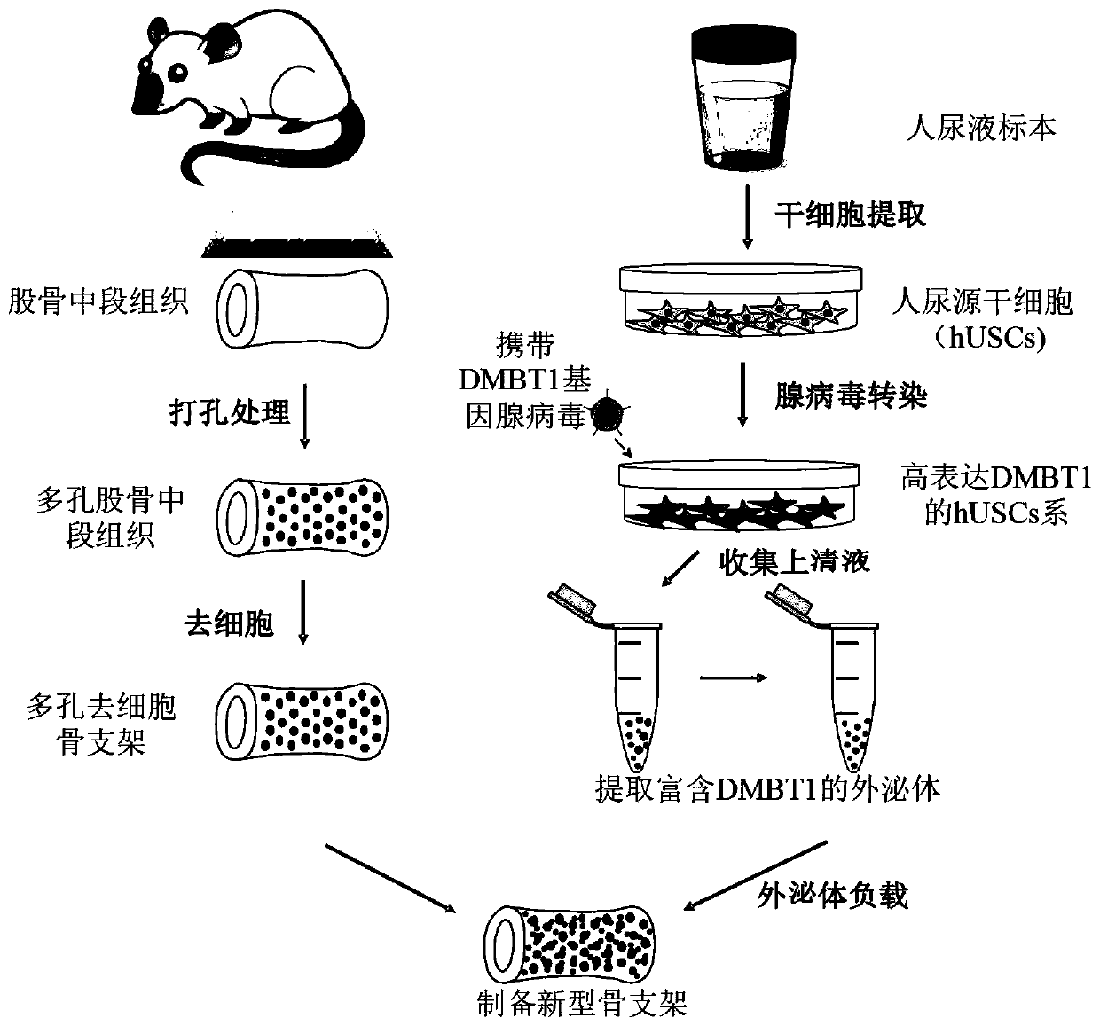
[0103] Example 1 Preparation and evaluation of decellularized porous femur

[0104] 1. Porous femur tissue acquisition

[0105] After euthanasia of adult (12-week-old) SD rats, the middle section of the right femur was collected, trimmed to a length of 10 mm, washed with 1% double-antibody PBS solution to remove bone marrow and blood, and punched with a device (for the device, refer to patent application CN201520084830. 7) Drill holes in the segment of bone tissue, and decalcify with 10% EDTA buffer solution for 15 days.

[0106] Wherein the aperture of the hole of the femur tissue obtained after punching is 100 μm on average, the hole depth is about 1mm, the spacing of the holes is 0.5mm on average, and the porosity of the bone material is 50%.

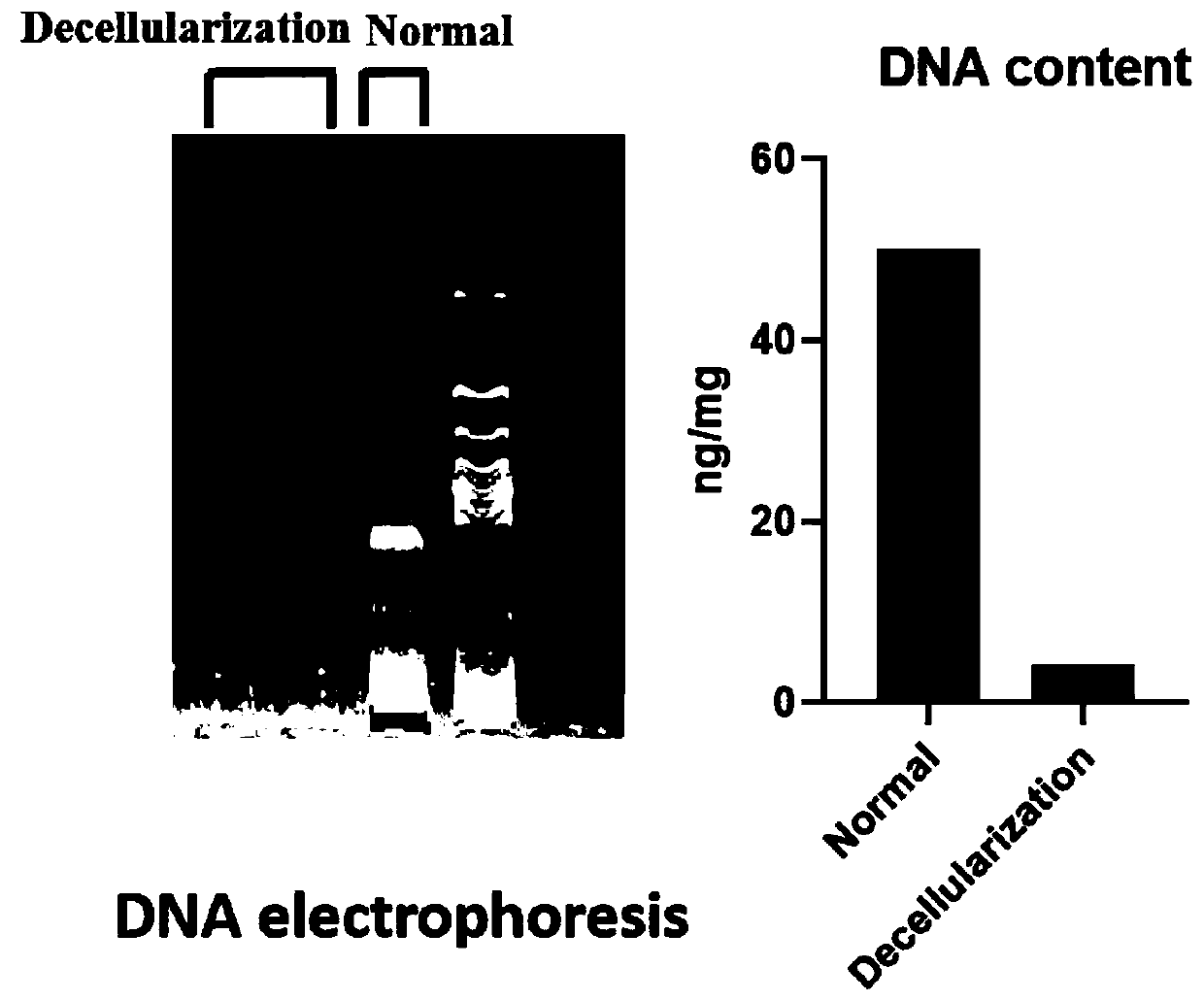
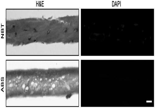
[0107] 2. Decellularization of porous femoral tissue

[0108] The porous femoral tissue was placed in a PBS solution containing 0.1% (w / v) SDS+0.1% (v / v) Triton X-100, shaken slowly at 4°C for 24 hours, and rinsed with 1% (w / v) dou...

Embodiment 2

[0116] The preparation of embodiment 2 novel support

[0117] 1. Isolation and culture of human urinary stem cells (hUS Cs)

[0118] Collect 200 mL of sterile fresh middle urine from 3 healthy adults, add 5 mL of penicillin and streptomycin to mix, divide into 4 tubes of 50 mL centrifuge tubes, centrifuge at 400 rpm for 10 min, discard the supernatant, add 20 mL of PBS solution, blow and mix, Continue to centrifuge at 400 rpm for 10 min, discard the supernatant, resuspend the hUSCs medium and inoculate them on 0.1% gelatin-coated 6-well plates, and culture them statically in a 37°C, 5% CO2 incubator. hUSCs medium components: DMEM / F12, 2% fetal bovine serum, 10ng / ml epidermal growth factor (EGF), 2ng / ml platelet-derived growth factor (PDGF), 1ng / ml transforming growth factor (TGF), 2ng / ml ml recombinant human fibroblast growth factor (hFGF), 0.5 mmol / L hydrocortisone, 24 mg / ml insulin, 20 mg / mL transferrin, 549 ng / mL epinephrine, 125 ng / mL triiodothyronine. After the primary ...

Embodiment 3

[0131] Example 3 In vivo evaluation of decellularized bone scaffolds

[0132] 1. Establishment of SD rat femoral shaft defect model establishment and surgical treatment

[0133] Adult male SD rats were taken, and after satisfactory intraperitoneal anesthesia with 0.3% pentobarbital sodium, the hair on the right lower limb was removed, disinfected with povidone iodine, and sterile towels and hole towels were spread. A minimally invasive longitudinal incision was made on the lateral side of the femur to expose the femoral shaft, and the bone tissue with a length of 0.8 cm was removed with a small animal pendulum saw. Then, select the lateral patellar incision of the right knee joint to incise the skin, separate it layer by layer, expose the femoral condyle, use an electric drill to drill a 2.0mm Kirschner wire retrogradely into the femoral medullary cavity through the intercondylar fossa of the femur, and penetrate the skin of the greater trochanter of the femur proximally. The...


PUM

PropertyMeasurementUnit
pore sizeaaaaaaaaaa
pore sizeaaaaaaaaaa
pore sizeaaaaaaaaaa

Abstract

The invention relates to the technical field of biomedical tissue engineering, in particular to a novel bone tissue engineering scaffold and a preparation method thereof. The bone scaffold comprises abone material and an exosome-loaded fibrin gel compound, the bone material is provided with holes, and the gel compound is distributed in the holes. The invention researches a bone material which ishighly similar to a natural bone matrix and has osteogenesis and vascularization activities. The porous decellularized tissue engineering scaffold is closer to a normal bone, has good biomechanical properties, is suitable for bone defect repair of a load bearing area, and maximally retains inherent components of the scaffold. According to the method, cell components with most antigens in tissues can be effectively removed, the immunological rejection reaction of grafts is reduced, the approximate morphological structure of the tissues can be maintained, and most tissue matrix components and bioactive factors are retained.

Description

technical field [0001] The invention relates to the technical field of biomedical tissue engineering, in particular to a novel bone tissue engineering scaffold and a preparation method thereof. Background technique [0002] The repair and functional reconstruction of bone defects caused by diseases, trauma, infection, osteonecrosis, congenital deformities, bone tumors, etc., especially critical bone defects, has always been one of the clinical problems in orthopedics. Traditional biological treatments for bone defects include autologous bone grafting and allogeneic bone grafting. [0003] The number of patients with large segmental bone defects due to various diseases such as trauma, tumors and congenital heredity in my country is as high as more than 3 million every year. At present, the clinical treatment methods for bone defects mainly include bone transport, Masquelet technique, vascularized autologous bone graft, autologous bone graft, and allogeneic bone graft, etc. ...

Claims


Application Information

Patent Timeline
Patent Type & Authority Applications(China)
IPC IPC(8): A61L27/38A61L27/20A61L27/22A61L27/36A61L27/56
CPCA61L27/3608A61L27/38A61L27/225A61L27/20A61L27/56A61L2430/02C08L89/00C08L5/08C08L5/04
Inventor 史强赵瑞波朱勇陈灿许琰闫少杭
Owner XIANGYA HOSPITAL CENT SOUTH UNIV
Why Eureka
Try Eureka