Radiographic assessment of tissue after exposure to a compound

a tissue and radiographic technology, applied in the field of radiographic assessment of tissue after exposure to a compound, can solve the problems of inability to perform serial measurements on an individual animal during and after repeated administration of a test compound, and the approach to characterization of pharmacological activity has several significant limitations

Inactive Publication Date: 2002-04-04
VERITAS PHARM INC
View PDF0 Cites 36 Cited by
  • Summary
  • Abstract
  • Description
  • Claims
  • Application Information

AI Technical Summary

Problems solved by technology

This approach to characterization of pharmacological activity has several significant limitations.
First, anatomical and histological studies may not precisely reflect significant changes in the activity of relevant biochemical pathways in normal and abnormal tissue after administration of test compounds.
Second, because animals must be killed for anatomical and histological studies, serial measurements cannot be performed on an individual animal during and after repeated administration of a test compound.
Finally, processing of tissue for histological examination is labor-intensive, time consuming, and expensive, and often represents a serious rate-limiting step in applications such as high-throughput drug discovery.
However, certain practical limitations of these modalities have reduced their economic feasibility and restricted their widespread use.
These limitations include very high procedure costs, limited availability, need for dedicated imaging devices, and relatively low spatial resolution.
The spatial limitations of radiopharmaceutical-based imaging modalities are particularly problematic for lead validation studies in small animals.
In these experimental models, the problem of low spatial resolution is more serious because of the small size of the subject relative to the fixed spatial resolution of the imaging modality.
Alternative, relatively time-consuming and labor-intensive non-imaging-based methods have therefore been devised for metabolic measurements in small animals [Green LA et al.
Disadvantages of this method include the limited sensitivity and reproducibility of manual measurements, the difficulty of accurately determining tumor volume from two-dimensional surface measurements, the confounding effects of necrotic and scar tissue on tumor dimensions, and the inability to use tumor models that are typically localized deep within internal organs.
In a typical clinical setting, a course of chemotherapy may be administered for many weeks and often costs tens of thousands of dollars.
Currently, the outcome of cancer chemotherapy varies widely in different tumor types and is unpredictable for any individual patient.
In addition, the administration of chemotherapy is frequently accompanied by significant side effects to the patient including severe systemic toxicity.
While PET imaging has demonstrated very high accuracy in the measurement of glucose metabolism in normal and malignant tissue, it suffers from disadvantages that diminish its utility for routine monitoring of chemotherapy.
PET imaging devices are generally located only in large medical centers, and are therefore not available for widespread use.
PET scans are very expensive, costing approximately $2500 per scan.
In addition, the spatial resolution of PET is relatively low, and there is a consequent decrease in accuracy in imaging of tumors and metastases that are smaller than approximately 1 cm.
Similarly, SPECT imaging procedures are relatively expensive, and require the use of radiopharmaceuticals and a gamma camera.
The spatial resolution of SPECT is also low, and the imaging technique is therefore inaccurate in imaging small tissue structures.
The time required to evaluate the efficacy of a particular therapeutic regimen may be significantly reduced, which may lower the risk of metastasis in the interim.

Method used


Image

  • Radiographic assessment of tissue after exposure to a compound
  • Radiographic assessment of tissue after exposure to a compound
  • Radiographic assessment of tissue after exposure to a compound

Examples


example 1

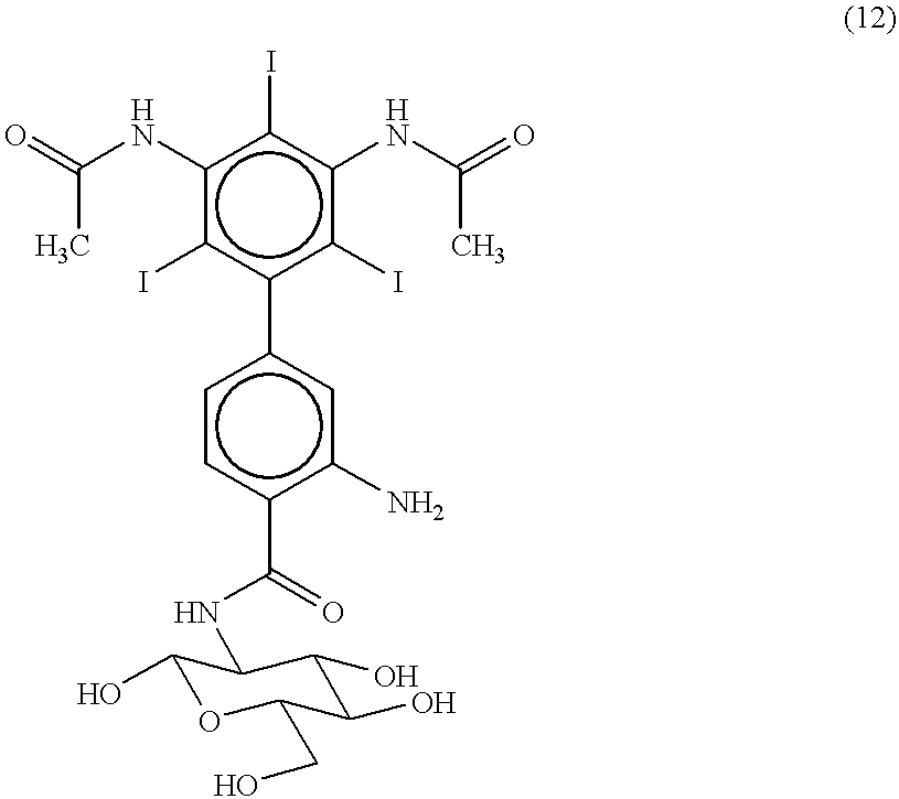
[0123] 2-Amino-4-[3',5'-bis(N-acetamido)-2',4',6'-triiodophenyl]-benzoyl-D--glucosamine (12) 3

[0124] The nitro groups of 2-chloro-1,3,5-trinitrobenzene (1) are reduced by reaction with tin and hydrochloric acid to yield 2-chloro-1,3,5-triaminobenzene (2).

[0125] The amino groups of 2-chloro-1,3,5-triaminobenzene (2) are acetylated by reaction with acetic anhydride to yield 2-chloro-1,3,5-triacetamidobenzene (3).

[0126] 2-chloro-1,3,5-triacetamidobenzene (3) is nitrated by reaction with a mixture of concentrated sulfuric and nitric acids to yield 2-chloro-1,3,5-triacetamido-3,5-dinitrobenzene (4).

[0127] 2-chloro-1,3,5-triacetamido-3,5-dinitrobenzene (4) is coupled with 4-iodotoluene by treatment with copper bronze at 200.degree. C. to yield 2',4',6'-triacetamido-3',5'-bis(nitro)-4-methylbiphenyl (5).

[0128] 2',4',6'-triacetamido-3',5'-bis(nitro)-4-methylbiphenyl (5) is treated with 6N hydrochloric acid to yield 2',4',6'-triamino-3',5'-bis(ni-tro)-4-methylbiphenyl (6).

[0129] The amino gr...

example 2

[0136] 2,6-Diamino4-[3',5'-bis(N-methylacetamido)-2',4',6'-triiodophenyl]--benzoyl-D-glucosamine (16) 6

[0137] 2',4',6'-triiodo-3',5'-bis(amino)-4-methylbiphenyl (8) is reacted with a mixture of nitric acid and sulfuric acid. Nitration under more vigorous conditions than those of Example 1 result in a product with a higher yield of nitro groups on both the 3- and 5-positions of the lower biphenyl ring. The product is treated with sodium cyanoborohydride and formaldehyde. The resulting product is treated with acetic anhydride to yield 2',4',6'-triiodo-3',5'-bis(N-methylacetamido)-3,5-dinitro-4-methylb-iphenyl (13).

[0138] The 4-methyl group of 2',4',6'-triiodo-3',5'-bis(N-methylacetamido)--3,5-dinitro-4-methylbiphenyl (13) is oxidized by reaction with potassium permanganate to yield 2',4',6'-triiodo-3',5'-bis(N-methylacetamido)-3,5-d-initrobiphenyl-4-carboxylic acid (14).

[0139] 2',4',6'-triiodo-3',5'-bis(N-methylacetamido)-3,5-dinitrobiphenyl-4--carboxylic acid (14) is coupled with 1,3...

example 3

[0142] 2-Amino-4-[3',5'-bis(2,3-dihydroxypropylmethylcarbamoyl)-2',4',6'-t-riiodophenyl]-benzoyl-D-glucosamine (20) 8

[0143] 2',4',6'-triiodo-3',5'-bis(amino)-4-methylbiphenyl (8) is reacted with a mixture of nitric acid and sulfuric acid. The product is treated with copper cyanide and sodium nitrite to yield 2',4',6'-triiodo-3',5'-bi-s(cyano)-3-nitro-4methylbiphenyl (17).

[0144] 2',4',6'-triiodo-3',5'-bis(cyano)-3-nitro-4-methylbiphenyl (17) is treated with sodium hydroxide. The product is treated with hydrochloric acid and methanol. The 4-methyl group of the product is oxidized by potassium permanganate to yield 2',4',6'-triiodo-3',5'-bis(carboxymethyl)--3-nitrobiphenyl-4-carboxylic acid (18).

[0145] 2',4',6'-triiodo-3',5'-bis(carboxymethyl)-3-nitrobiphenyl-4-carboxy-lic acid (18) is coupled with 1,3,4,6-tetra-O-acetyl-D-glucosamine in the presence of 1-[3-(dimethylamino)propyl]-3-ethylcarbodiimide hydrochloride and N-hydrosuccinamide to yield 2-nitro-4-[3',5'-bis(carboxy)-2',4',6'-t...


PUM


Abstract

Radiographic system and method for noninvasively assessing the response of tissue to a compound, such as a therapeutic compound. In one embodiment, a non-radioactive, radio-opaque imaging agent accumulates in tissue in proportion to the tissue concentration of a predefined cellular target. The imaging agent is administered to a live organism, and after an accumulation interval, radiographic images are acquired. The tissue being examined is transilluminated by X-ray beams with preselected different mean energy spectra, and a separate radiographic image is acquired during transillumination by each beam. An image processing system performs a weighted combination of the acquired images to produce a first image. The image processing procedure isolates the radiographic density contributed solely by differential tissue accumulation of the imaging agent. A compound is administered to the organism, and after a selected interval, a second radiographic image of the tissue is acquired. Radiographic density contributed by accumulated imaging agent in corresponding areas of tissue in the first and second images are compared. Differences in radiographic density between the images reflect changes in the concentration of the cellular target that have occurred after administration of the compound. The system and method may be used to assess therapeutic efficacy of compounds in the drug discovery process, in clinical trials, and in the evaluation of clinical treatment. In other embodiments, pharmacological and toxicological effects of a wide variety of compounds on tissue may be noninvasively assessed.

Description

[0001] This application is a continuation-in-part of co-pending U.S. patent application Ser. No. 09 / 149,734 which was filed Sep. 8, 1998, and this application hereby claims the benefit of this prior filing date. This application is also a continuation-in-part of co-pending U.S. patent application Ser. No. 60 / 190,330 which was filed Mar. 16, 2000; this application claims the benefit of the provisional's filing date under 35 U.S.C. .sctn.119(e).[0002] This invention relates to a radiographic system and method for assessing the response of tissue in vivo to compounds, including therapeutic compounds.DESCRIPTION OF THE RELATED ART[0003] The ability to accurately assess the effects of compounds in vivo is essential in a wide range of pharmacological and related studies. Assessment of tissue response to compounds is particularly important in each stage of drug discovery, development, and clinical application. Noninvasive studies of tissue response may be particularly useful in the early v...

Claims


Application Information

Patent Timeline
Patent Type & Authority Applications(United States)
IPC IPC(8): A61B6/00A61B6/03
CPCA61B6/4035A61B6/4241A61B6/508A61B6/4092A61B6/481
Inventor SALB, JESSE
Owner VERITAS PHARM INC
Why Eureka
Try Eureka