Scaffold for tissue engineering, artificial blood vessel, cuff, and biological implant covering member

a biological implant and tissue engineering technology, applied in the field of tissue engineering scaffolds, can solve the problems of inability to adjust the distribution of engraftments, inability to obtain uniform cell engraftments on the whole surface, and inability to meet the requirements of scaffolds comprising appropriate three-dimensional network structures, etc., to achieve the effect of reducing the foreign-body reaction

Inactive Publication Date: 2005-05-19
JAPAN AS REPRESENTED BY PRESIDENT OF NAT CARDIOVASCULAR +2
View PDF2 Cites 67 Cited by
  • Summary
  • Abstract
  • Description
  • Claims
  • Application Information

AI Technical Summary

Benefits of technology

[0033] The biological implant covering member may be used for purposes other than clinical purposes and may be used to cover the outer sur

Problems solved by technology

In conventional artificial blood vessels, tubes made of polyester resin mesh or PTFE resin mesh have been in practical use from a long time ago, and works challenging for smaller caliber or for better patency rate has been proceeding.
Collagen gels for embedding culture do not have a porous structure such as three-dimensional network structure, and there remains a problem that it is impossible to obtain uniform cell engraftment on the whole surface or impossible to adjust the distribution of engraftment are not achieved.
Although methods employing salt or bubbles are known as method of preparing a porous material having a three-dimensional network structure, any has difficulty in strictly and discretionary adjusting the pore diameter and pore density, so a scaffold comprising appropriate three-dimensional network structure is still not fulfilled.
Cell engraftment structure achieved by collagen gel embedding culture can not used for applications to be subjected to mechanical load such as artificial blood vessel while it is available for evaluating cell function because collagen gel as the scaffold thereof does not have physical strength.
Though artificial blood vessels as alternative materials for autologous blood vessels are used in clinical application broadly, smaller diameter artificial blood vessels have poor patency rate.
In addition, since wall does not have a hole through which the cell would enter, even if the pannus extends from the inosculated part, it would not be bonded to the wall and would float and many cases of resulting occlusion of blood vessels have been reported.
There is fabric velour impregnated with collagen and objected for robust bonding.
Troubles that would be caused due to these stresses causing a lowering of adhesion between the cuff and the subcutaneous tissue include, as typical trouble, infection trouble such as tunnel infection.
In cases of ventricular assist device therapy, such infection trouble experiences are being very frequent

Method used


Image

  • Scaffold for tissue engineering, artificial blood vessel, cuff, and biological implant covering member
  • Scaffold for tissue engineering, artificial blood vessel, cuff, and biological implant covering member
  • Scaffold for tissue engineering, artificial blood vessel, cuff, and biological implant covering member

Examples


example 1

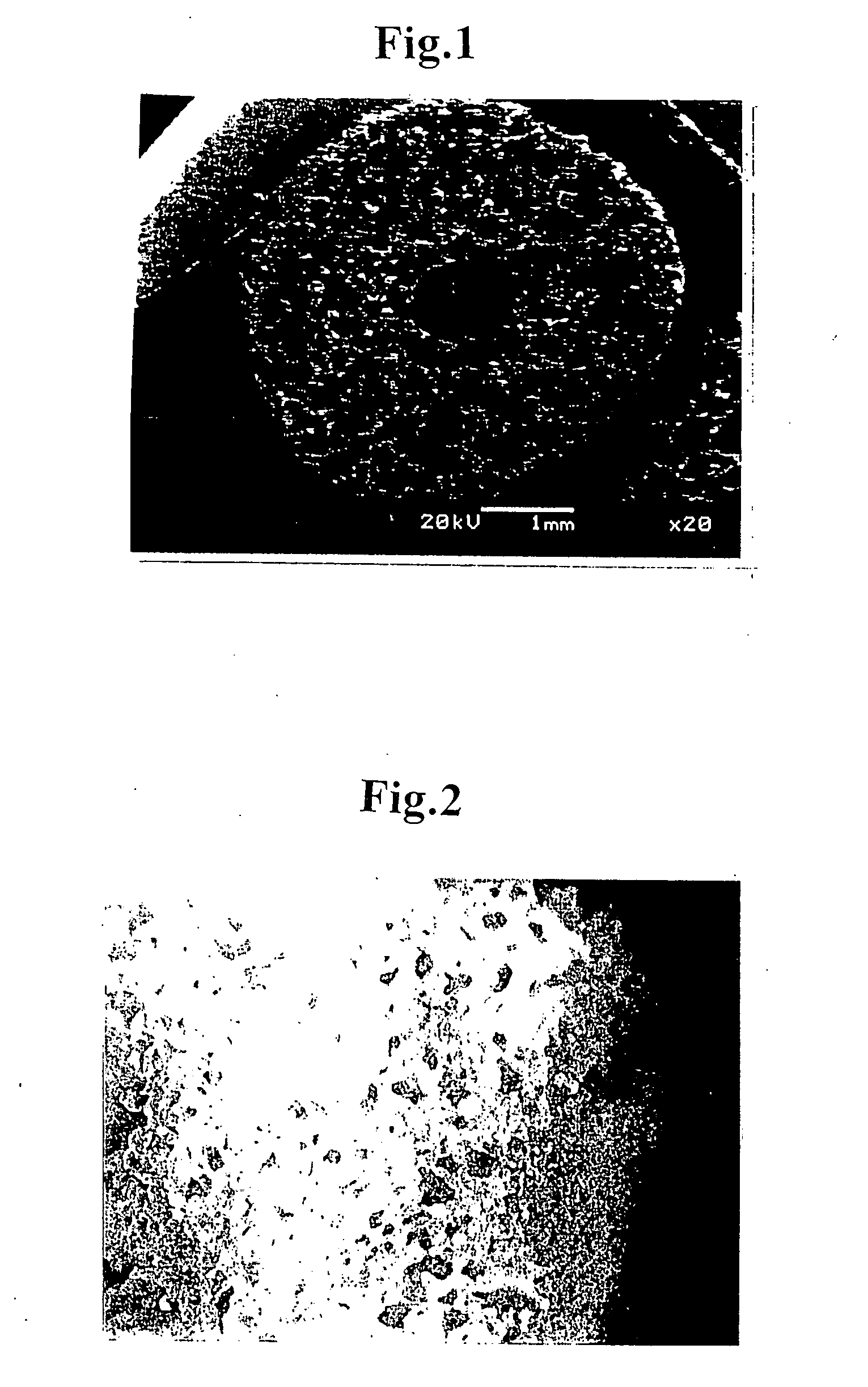
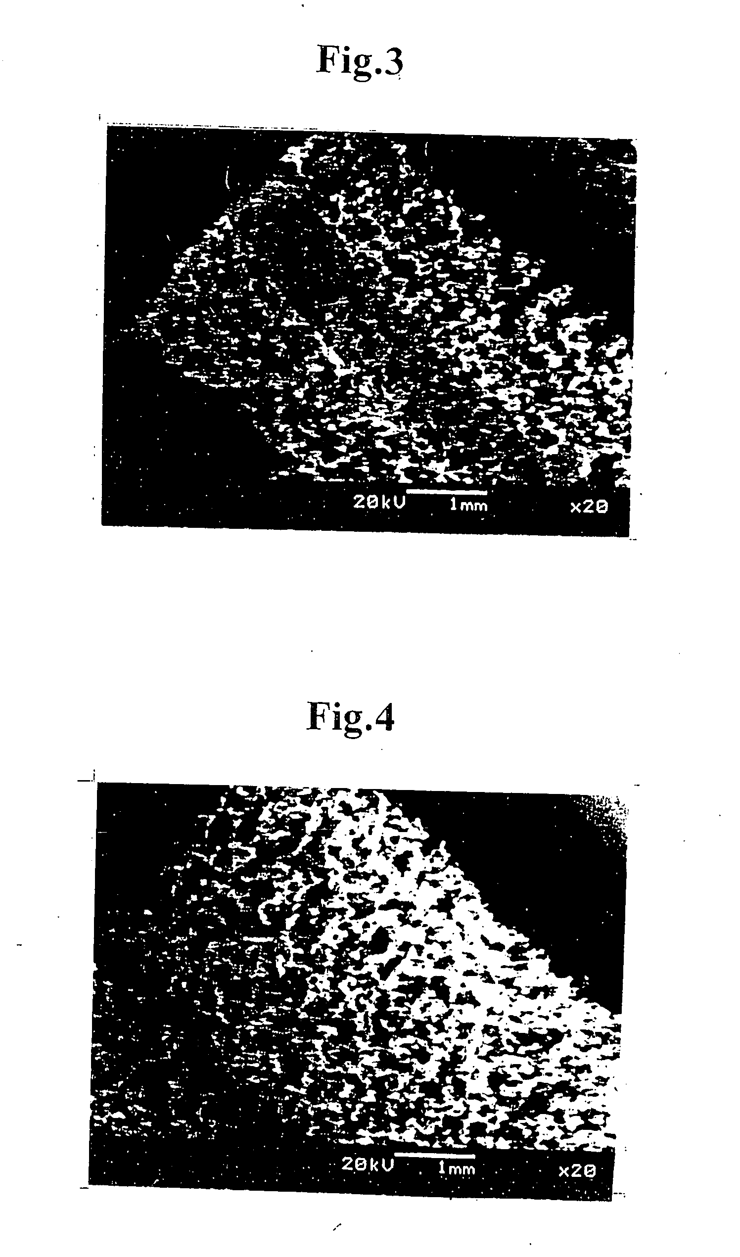
[0071] A thermoplastic polyurethane resin (MIRACTRAN E980PNAT available from Nippon Miractran Co., Ltd.) was dissolved into N-methyl-2-pyrrolidinone (reagent for peptide synthesis, NMP available from Kanto Kagaku) by using a dissolver (about 2000 rpm) at room temperature to obtain 5.0% solution (weight / weight). 1.0 kg of this NMP solution was measured and entered into a planetary mixer (PLM-2 type, capacity 2.0 liters, available from Inoue Mfg., Inc.) and was mixed with methylcellulose (reagent, 25 cp grade, available from Kanto Kagaku) of an amount corresponding to the amount of polyurethane resin at a temperature of 40° C. for 20 minutes. With the agitation being continued, the defoaming was conducted by reducing the pressure to 20 mmHg (2.7 kPa) for 10 minutes, thereby obtaining polymer dope.

[0072] A tube forming jig was prepared which comprised cylindrical paper tube of 3.5 mmφ in inner diameter, 4.6 mmφ in outer diameter, and 60 mm in length made of a chemical experimental pap...

example 2

[0079] DMEM (culture component) solution (containing FCS (cow embryo blood serum) 10%) of smooth muscle cells from cow's blood vessel (cell density: 6×10⁶ cells / mL) and collagen type I solution (0.3% acid solution available from Koken Co., Ltd.) are mixed in equivalent quantities while being cooled on ice, thereby preparing suspension solution of smooth muscle cells (cell density: 3×10⁶ cells / mL).

[0080] The scaffold of tubular porous three-dimensional network structure (inner diameter: 1.2 mmφ, outer diameter: 3.2 mmφ, length: 2 cm) prepared in Example 1 was clamped at its one end and the suspension solution of smooth muscle cells (1 mL) was injected at the other end into the scaffold until leaking out through a side wall of the tubular structure. All of the injection operation was conducted on ice. By repeating the injection operation several times, the collagen solution containing smooth muscle cells well penetrated all over the tubular structure including the inside thereof. Aft...

example 3

[0082] The scaffold of tubular porous three-dimensional network structure (inner diameter: 1.2 mmφ, outer diameter: 3.2 mmφ, length: 2 cm) prepared in Example 1 was clamped at its one end and the collagen type I solution (0.15 wt. %) was injected at the other end into the scaffold until the collagen solution penetrated all over the scaffold including the inside thereof. After that, the clamping was cancelled, a mandrel of 1.2 mmφ made of SUS440 was inserted into the tubular body of the scaffold at the center thereof, and the tubular structure of the scaffold was held inside an incubator at a temperature of 37° C. to make the collagen solution to gel, thereby obtaining a tubular body of which network structure was filled with collagen gel.

[0083] A piece of about 3 cm was exfoliated from aorta abdominalis of a rat and was clamped at its both ends to block the blood stream. After that, a middle portion of the aorta was cut. The tubular body was inserted between the cut ends of the aor...


PUM


Abstract

The invention provides a porous scaffold for tissue engineering which allows easy cell engraftment and cell culture and thus enables stable organization and an artificial blood vessel which exhibits high patency rate even if the inner diameter is small. The scaffold for tissue engineering is made of thermoplastic resin which forms a porous three-dimensional network structure having communication property, wherein the porous three-dimensional network structure has an average pore diameter of from 100 to 650 μm and an apparent density of from 0.01 to 0.5 g/cm3. The artificial blood vessel is composed of this scaffold.
The invention provides a cuff which allows easy infiltration of cells from living subcutaneous tissues, easy engraftment of cells, and neovascularization of capillary vessels so as to obtain robust bonding with subcutaneous tissues and, as a result, ensures separation of a wounded portion from the outside, thereby blocking exacerbation factors such as bacterial infection on healing and inhibiting progression of downgrowth. That is, the invention provides a cuff with none or little infection trouble such as tunnel infection. The cuff comprises a porous three-dimensional network structure which is made of thermoplastic resin or thermosetting resin and has communication property, wherein the porous three-dimensional network structure has an average pore diameter of from 100 to 1000 μm and apparent density of from 0.01 to 0.5 g/cm3.
The invention provides a biological implant covering member which allows easy infiltration of cells from living subcutaneous tissues, easy engraftment of cells, and organization, thereby obtaining robust bonding with native tissues and therefore protecting a living body from adverse effect which may occur due to the insertion of a biological implantation member into the living body. The biological implant covering member comprises a porous three-dimensional network structure which is made of thermoplastic resin or thermosetting resin and has communication property, wherein the porous three-dimensional network structure has an average pore diameter of from 100 to 1000 μm and apparent density of from 0.01 to 0.5 g/cm3.

Description

CROSS REFERENCE TO RELATED APPLICATION [0001] This is a continuation application of PCT / JP03 / 03594 filed on Mar. 25, 2003.TECHNICAL FIELD [0002] The present invention relates to a scaffold material for tissue engineering, an artificial blood vessel, a cuff and a biological implant covering member. BACKGROUND ART [0003] The present invention relates, in the first place, to a porous scaffold for tissue engineering which allows easy cell engraftment and cell culture and thus enables stable organization, and to an artificial blood vessel using this scaffold. The scaffold and the artificial blood vessel of the present invention are effectively used not only for basic studies on biotechnologies, but also for biomedical materials used as artificial bone structure substrates for substitute medicine by an artificial internal organ or for regenerative medicine by tissue engineering, and especially, for an artificial blood vessel which can exhibit high patency rate even if the inner diameter i...

Claims


Application Information

Patent Timeline
IPC IPC(8): A61L27/38A61L27/50A61L27/56
CPCA61L27/38A61L27/56A61L27/507
Inventor NAKAYAMA, YASUHIDETATSUMI, EISUKENEMOTO, YASUSHI
Owner JAPAN AS REPRESENTED BY PRESIDENT OF NAT CARDIOVASCULAR
Why Eureka
Try Eureka