Interactive wound cover

a wound cover and active technology, applied in the field of interactive wound covers, can solve the problems of inferior scars, inability to repair damaged areas, cosmetically and functionally, and inability to manufacture wound covers, etc., and achieve the effect of accelerating skin tissue regeneration and rapid production of wound covers

Inactive Publication Date: 2007-11-08
RELIANCE LIFE SCI PVT
View PDF6 Cites 10 Cited by
  • Summary
  • Abstract
  • Description
  • Claims
  • Application Information

AI Technical Summary

Benefits of technology

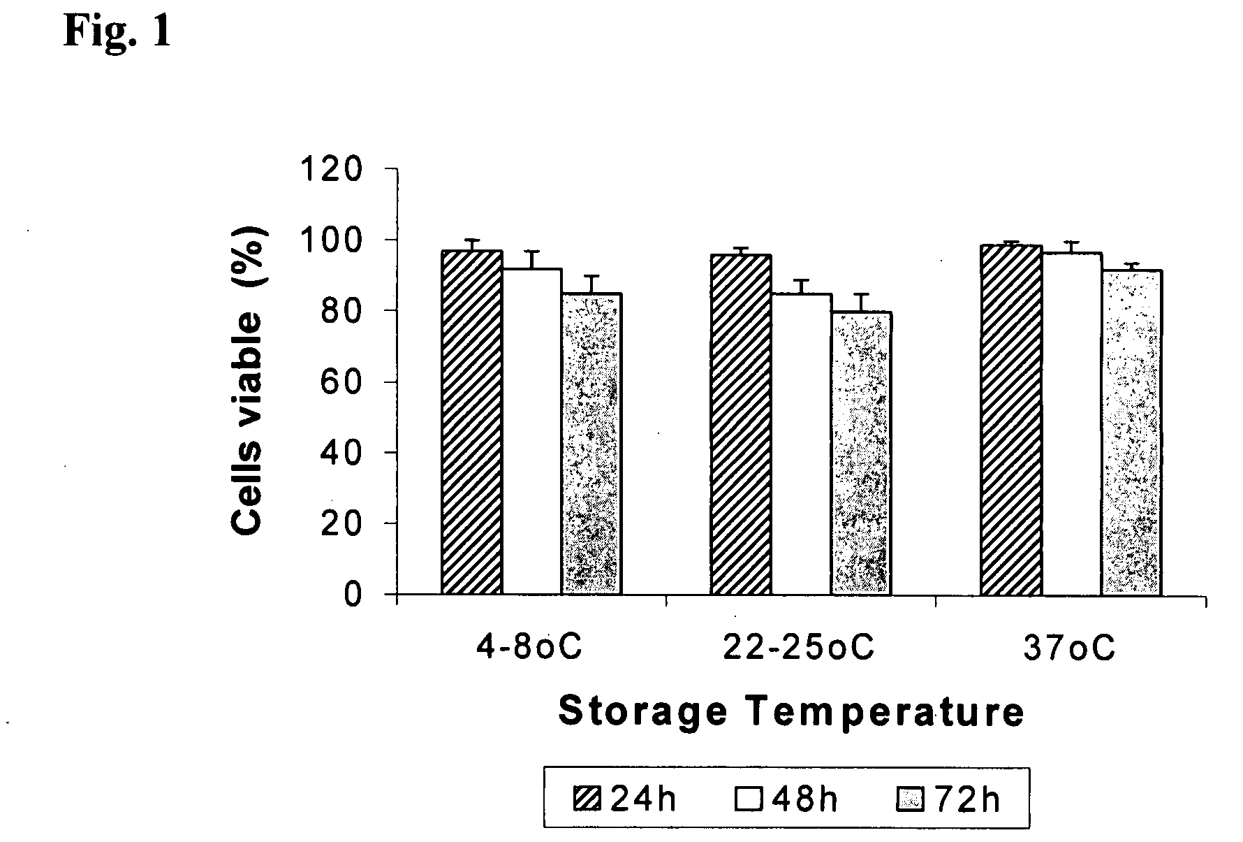
[0044] In related embodiments at least 90% of said cells are actively proliferating keratinocytes. The epithelial on the biocompatible polymer are, in some embodiments, preconfluent, including less than 20%, less than 50%, less than 75%, less than 80%, less than 90%, less than 95%, and less than 99% confluent. The present compositions possess a number of advantages over other compositions in the art, including that it (a) adheres quickly to the wound; (b) mimics the physiology and some of the mechanics of the normal skin; (c) is highly effective in accelerating tissue regeneration and wound repair; and (d) can be made available in less time with a commercially viable process. The present wound treatment compositions are also effective and safe. An additional advantage of the present invention is that, in some embodiments, the keratinocytes retain substantial viability on the wound cover for at least 72 hours after application to a wound. Substantial viability is 75-90% viability.
[0045] In related embodiments, the present invention provides for methods and processes of preparing a wound cover. In one embodiment, a process for the preparation of a wound cover comprises the steps of: (a) isolating epidermal cells from the donor skin; (b) preparing a suspension of proliferating basal cells (c) expanding the cells (d) cryopreserving the cells and thawing when needed; (e) seeding cells onto PLA sheets in a transport device comprising transport media. In one embodiment, keratinocytes are seeded onto PLA sheets in at a concentration of 0.10×10⁶ to 0.5×10⁶ cells / cm.2 In related embodiments, the wound cover is transferred to a transport device made of polycarbonate. The transport media may comprises carbon dioxide enriched media. In embodiments where the donor skin is autologous, for the construction of an autologous skin graft, cryopreservation may be used. When the donor skin is allogenic, cryopreservation is more common, so that a bank of cryopreserved cells can be available for rapid production of wound covers, as needed. In further embodiments, the invention comprises a kit for the preparation of wound covers. Such a kit includes, in one embodiment, (a) sterile sheets of biocompatible polymers; (b) a device for growth and subsequent transport of the wound preparation. The kit may also include appropriate media, instructions, and may even include stocks of frozen keratinocytes which have been pre-screened to be suitable for forming the wound preparation in that they are viable and free from pathogens and other contaminants.
[0046] The presently inventive composition find use in a variety of therapeutic modalities, such as a tissue substitute. It addresses the problem of severe of chronic wounds by accelerating skin tissue regeneration and wound healing by stimulating the recipient's own wound bed-derived skin cells. This includes wounds such as a post-operative wound, a burn, a diabetic ulcer, a bed sore, and a traumatic injury. The present invention is suitable for use in a variety of animals, including domesticated and agricultural animals, in veterinary practice, or on human patients.

Problems solved by technology

One of the persistent problems in the treatment of large wounds, like burns, is the availability of skin cover to repair damaged areas.
Scar formation under such conditions can often result in a cosmetically and functionally inferior scar.
However, more tissue distortion and an extensive, cosmetically unsatisfactory, scar often causes an impairment of function.
These were developed due to limitations associated with the use of autografts, including the creation of a donor site, which is at risk of developing pain, scarring, infection and / or slow healing, for example.
While autologous tissue transfers can be highly effective in securing wound healing, such procedures (e.g., grafts and flaps) are invasive, painful, and expensive, and are not within the purview of many wound care practitioners.
Autologous skin grafts, though successful, have limitations due to the limited donor sites as well as creation of fresh wounds at the donor site.
Limitations of this treatment is related to sterility, difficulty in handling, risk of viral transmission and immune rejection by the host.
The grafting of such labor-intensive sheets has faced mixed fortunes, however, with reports of lower than anticipated efficiencies.
Problems with the lack of “take” and long-term durability, time required for film preparation and high cost to produce such grafts, as well as difficulty in handling have led to the development of alternate delivery systems to transfer keratinocytes to the wound bed.
However, the limitations of this graft include a 3-week delay for graft cultivation, the lack of a dermal component, and high cost.
For a number of technical reasons, this technology is not commercially viable.
The disadvantages of allografts are that they do not survive permanently on the wound bed, and there is a possibility of disease transmission, although, as with blood transfusions, this risk can be minimized with extensive screening.
Yet the disposable silicone sheet can allow the accumulation of exudate, increasing the risk of infection.
It also does not provide a real epidermal component; and the silicone sheet must be surgically removed and ultimately replaced with an autograft or allograft.
This is not successful enough to make Dermagraft™ a routinely acceptable skin replacement.
The additional disadvantage of this skin substitute is the need to wait 3 to 4 weeks to produce the cultured grafts.
The disadvantages include the possibility of disease transmission and poor drainage from the wound, increasing the accumulation of exudate and resultant risk of infection.
There is a scarcity of clinical data supporting the tolerability and effectiveness of these types of dermal matrices.
However, disadvantages associated with this method include that it causes morbidity at the donor site and that there are limited donor sites in extensive burns.

Method used


Image

  • Interactive wound cover
  • Interactive wound cover
  • Interactive wound cover

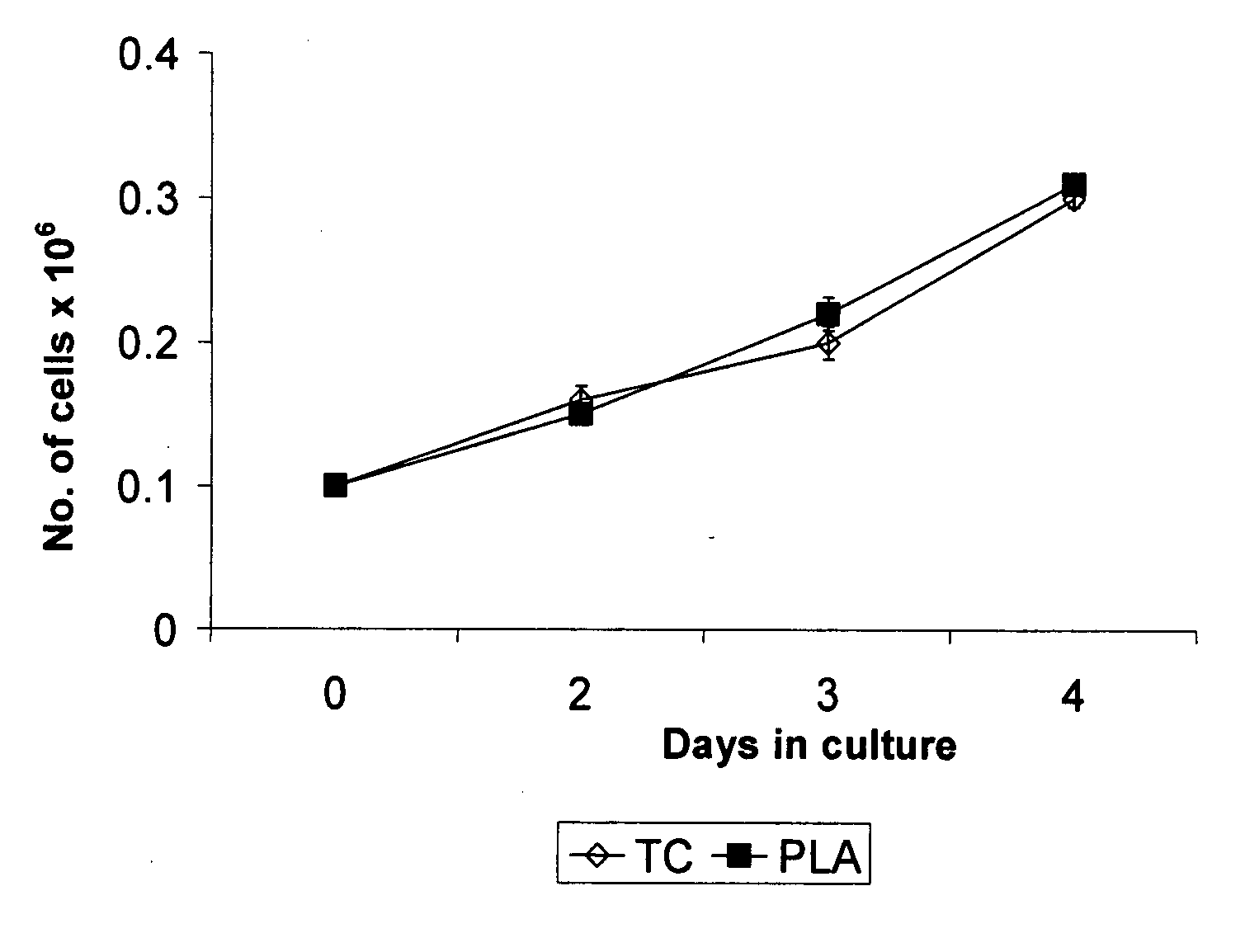
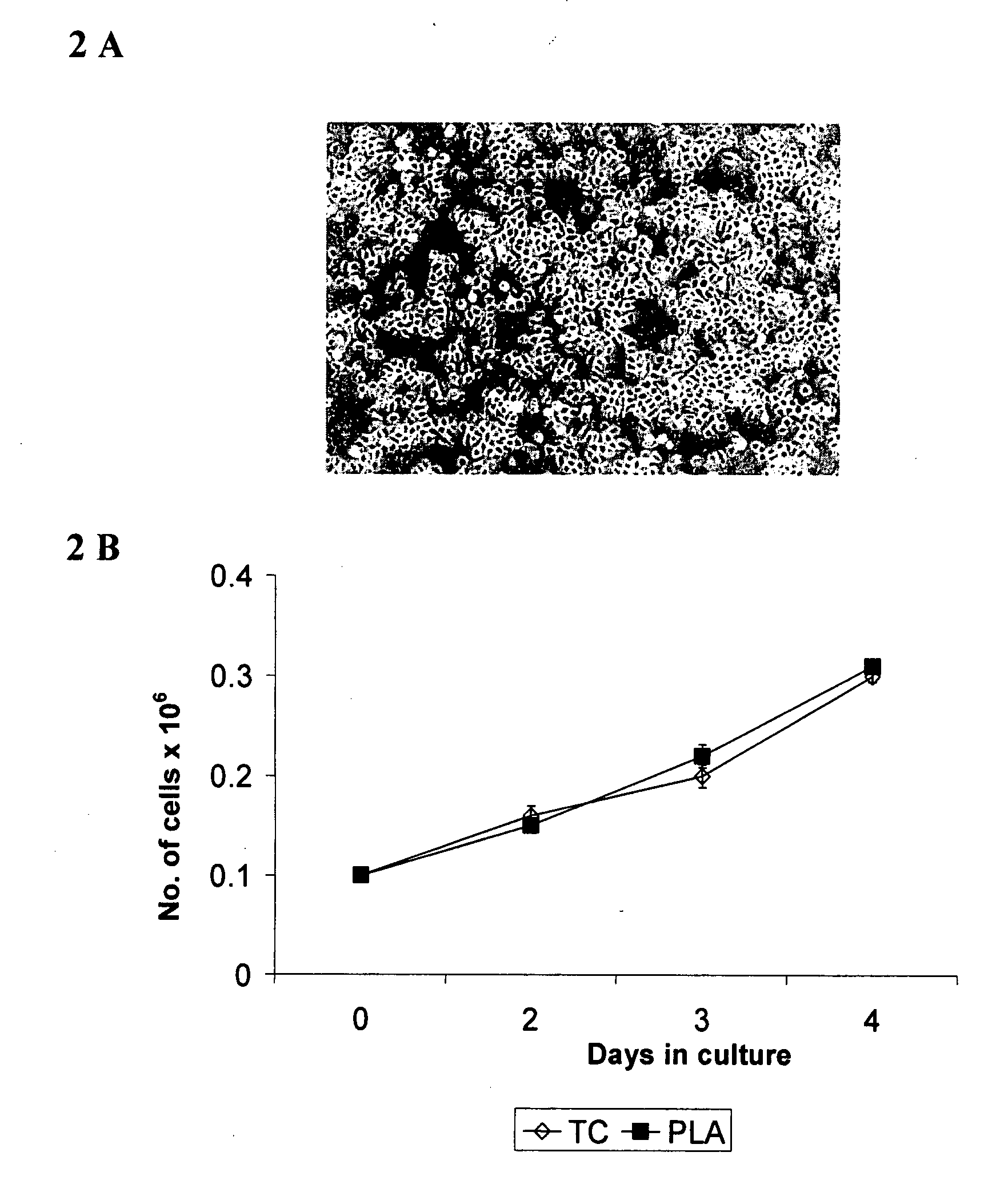
Examples


example 1

Preparation of the Wound Cover

A) Preparation of the PLA Sheets

[0094] The polymer was specially made by mixing equal amounts of L and DL dilactide, and polymerized to give PLA with a molecular weight of about 50,000-150,000 Da. A 20% (w / v) solution of the polymer was prepared in acetone and clarified to remove any suspended particles by centrifugation. The polymer film was then prepared by solvent casting method by pouring the solution over a solid support such as glass or steel plates. The sheets were allowed to dry by evaporation of solvent at ambient temperature; rinsed in sterile water and dried at ambient temperature. PLA film was sterilized with Ethylene Oxide (ETO).

[0095] Physical properties of ETO sterilized films were characterized using standard methods (IS / ASTM). Barrier property of the films against microbes (bacteria, yeast and fungi) was assessed by the ability of the films to prevent contamination of the underlying nutrient mix.

[0096] Physical characteristics of t...

example 2

Product Characterization and Testing

A) Microbial Testing:

[0102] 1) Bioburden: The microbial contamination in the sample was determined in terms of numbers of colonies appearing on plates of solid media. The test involved the addition of sterile molten Casein Digest Agar to 1 ml of test sample in a petri dish. After solidification of the medium the plate was incubated at 30-35° C. in the inverted position for 48 hours. After this period the plates were visually inspected. No bacterial growth was observed.

[0103] 2) Sterility testing: Sterility testing was performed to detect the presence of aerobic and anerobic microbes by inoculating the test samples in two different sterile nutritive medias namely Fluid Thioglycolate Medium (FTM) and Soybean Casein Digest Medium (SCDM). The result showed absence of growth during a period of 14 days for microorganisms such as bacteria, yeast and mold and for pathogens like E. coli, S. aureus, P. aeruginosa and Salmonella indicating sterility of t...

example 3

In Vivo Testing

A) Migration of Keratinocytes from PLA

[0119] To enhance wound healing, keratinocytes should be delivered to the wound bed. Once the wound cover is applied to the wound bed, cells must migrate from the PLA membrane to the wound bed. To confirm this event, an in vivo experiment was performed on guinea pigs. Briefly, partial thickness punch wound biopsies were created on the posterior portion of the animals. Cells in a wound cover of the present invention were labeled with a red fluorescent dye (CyDiI, Molecular Probes, USA) and then applied on the wound bed. At the end of 3 days, the wound was excised and processed for histological analysis. As shown in FIG. 6, fluorescent-labeled cells were visualized in the wound bed, indicating migration of keratinocytes from the PLA sheet to the wound bed.

B) Preclinical Evaluation:

[0120] The toxicity testing was done with a PBS solution with the PBS sheet for 72 h at 37° C. (hereinafter “test substance”). All toxicity studies ...


PUM

PropertyMeasurementUnit
molecular weightaaaaaaaaaa
melting pointaaaaaaaaaa
temperatureaaaaaaaaaa

Abstract

The present invention relates to an interactive wound cover wherein a cultured monolayer of keratinocytes is delivered using a biopolymer. The present invention describes the composition, method of preparation and its properties relating to safety, and efficacy. The wound cover of the present invention is useful in the treatment of wounds and in skin tissue regeneration.

Description

CROSS-REFERENCES TO RELATED APPLICATIONS [0001] This application claims benefit of the filing date under 35 U.S.C. § 119(e) to provisional Indian Application No. 205 / MUM / 2006, filed on Feb. 14, 2006, which is entirely incorporated by reference herein. FIELD OF THE INVENTION [0002] The present invention relates to an interactive wound cover, its composition and method of preparation. The present invention particularly relates to the delivery of a cultured monolayer of keratinocytes on polymer for skin regeneration. BACKGROUND OF THE INVENTION The Structure of the Skin [0003] Skin is a bilayer organ comprising an outer, thinner, epidermis and an inner, thicker, dermis, both of which have specific properties. [0004] The epidermis is composed mainly of epithelial cells. Keratinocytes are the outermost cells of the epidermis and produce the protein keratin. Keratinocytes are produced from a layer of basal epidermal cells that are anchored to the basement membrane by adhesion molecules, ...

Claims


Application Information

Patent Timeline
Patent Type & Authority Applications(United States)
IPC IPC(8): A61K45/00
CPCA61L15/26A61L15/40A61L27/18A61L27/3813A61L27/60C08L67/04
Inventor GHOSH, DEEPAKUCHROO, PUSHPASHENOY, SUDHEERRANGASWAMY, VIDHYAVENKATA RAMA KRISHNA, SONTI
Owner RELIANCE LIFE SCI PVT
Why Eureka
Try Eureka