System for the adaptation of cell-based assays for analysis on automated immuno-assay platforms

a cell-based assay and automated immunoassay technology, applied in the direction of animals/human proteins, receptors of cytokines/lymphoines/interferons, animals/human proteins, etc., can solve the failure of patients to respond to therapy, allergic reactions, severe autoimmune reactions, etc., and achieve the effect of being easily measured

Inactive Publication Date: 2021-09-23
SVAR LIFE SCI AB
View PDF0 Cites 1 Cited by
  • Summary
  • Abstract
  • Description
  • Claims
  • Application Information

AI Technical Summary

Benefits of technology

[0036]In a further embodiment of the invention in order to facilitate the detection and quantification of the ADCC activity of therapeutic antibodies the ADCC effector reporter-gene cell line and drug-specific target cells are mixed together with suitable dilution(s) of the drug to be analyzed. The effector cells (E) and target cells (T) may be either cultivated continuously in the laboratory or frozen separately and thawed immediately prior to use and then mixed at a suitable target cell ratio (E:T ratio) together with a suitable dilution(s) of the drug to be analyzed and incubated for an appropriate time prior to lysing the cells and addition of the FL specific antibody (monoclonal or polyclonal) pair and addition of a sample of the cell lysate supernatant to the assay platform for quantification of ADCC activity. Alternatively, the ADCC effector reporter-gene cell line and drug-specific target cells can be pre-mixed at the optimal effector (E): target cell (T) ratio prior to freezing the cells (“Combo”) either alone or together with a pair of anti-luciferase antibodies at the appropriate concentrations. Thus, upon thawing of the “Combo” vial of ADCC effector and drug-specific target cells frozen at the optimal E:T ratio are mixed directly with an appropriate dilution(s) of the drug for which ADCC activity is to be quantified and incubated for an appropriate time prior to lysing the cells and addition of the FL specific antibody (monoclonal or polyclonal) pair (in the case that the antibodies were not included in the vial of cells prior to freezing) and addition of a sample of the cell lysate supernatant to the assay platform for quantification of ADCC activity. The ADCP and / or CDC activity of therapeutic antibodies may be quantified in a similar manner using a target cell containing a protease cleavable reporter-gene product such as a luciferase that responds to the release of a protease and that once secreted can be quantified allowing the assessment of ADCP or CDC activity. Alternatively, the protease may induce a conformational change in the reporter-gene protein product that results in the appearance of a hidden epitope that can be detected by the antibody pair used in the platform detection system. This invention is also applicable to the quantification of the activity of cells used in adoptive therapy such as CAR-T cells using an automated assay platform. CAR-T activity would be quantified using the same type of target cell as that described above for an ADCP or CDC assay.

Problems solved by technology

In addition to adversely affecting pharmacokinetics, pharmacodynamics, bioavailability, and efficacy, ADAs can also cause immune complex disease, allergic reactions, and in some cases severe autoimmune reactions.
ADAs can result in the failure of the patient to respond to therapy and may even prove to be life threatening in the case of NAbs that cross-react with essential non-redundant endogenous proteins such as EPO or thrombopoietin (2,3).
ADAs can also persist for long periods after cessation of treatment, thereby limiting subsequent treatment with the same drug (5).
Although immuno-assays such as bridging ELISA based screening or confirmatory assays can be readily adapted to run on automated assay platforms such as Meso Scale Discovery electro-chemiluminescence (MSD-ECL), Luminex, SMC, Alpco, AlphaLISA or Gyros, or label-free detection systems such as SPR systems including Biacore, cell-based assays are less amenable to adaptation to such platform technologies.
Assessment of antibody mediated immune effector functions require the use of cell-based assays that are difficult to adapt to analysis on an automated immuno-assay platforms.

Method used


Image

  • System for the adaptation of cell-based assays for analysis on automated immuno-assay platforms
  • System for the adaptation of cell-based assays for analysis on automated immuno-assay platforms
  • System for the adaptation of cell-based assays for analysis on automated immuno-assay platforms

Examples


example 1

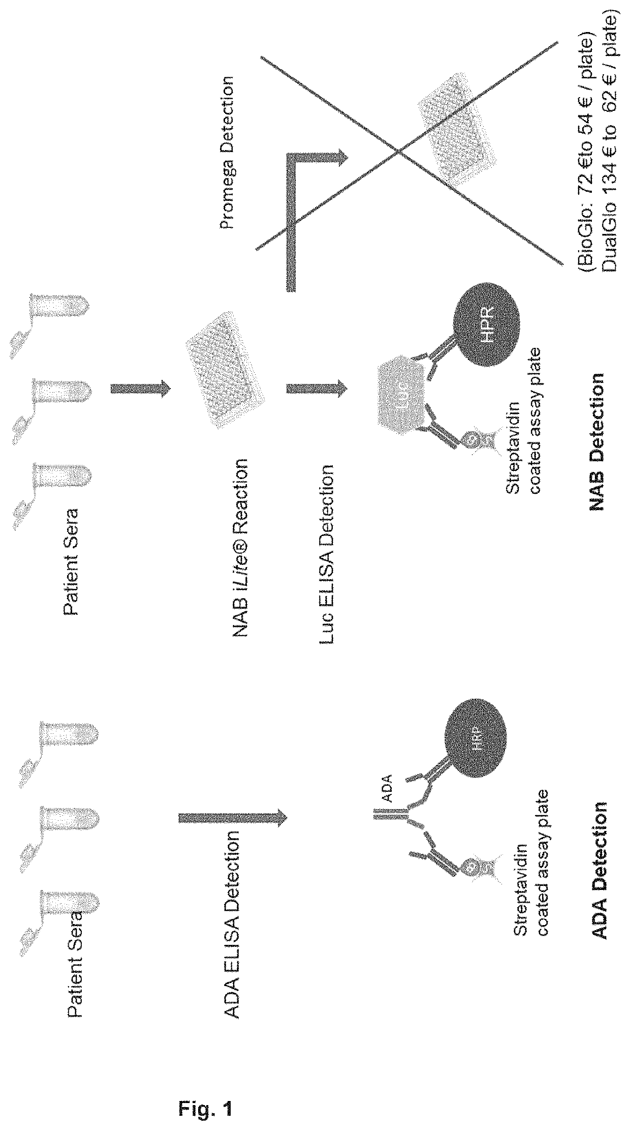
[0133]The bridging ELISA in which a drug (small molecule, peptide, or biopharmaceutical) is detected by the formation of a bridge between the drug, and one molecule of an antibody (monoclonal or polyclonal) directed against one epitope of the drug attached either directly to a solid surface, usually a 96, or 384-well micro-titer plate, or an antibody (monoclonal or polyclonal) directed against one epitope of the drug labelled with biotin that it is turn bound to a streptavidin coated surface, usually a 96, or 384-well micro-titer plate or streptavidin coated bead, and another molecule of an antibody (monoclonal or polyclonal) directed against a second epitope of the drug labelled with horse radish peroxidase (HRP) or another suitable marker is used widely for the quantification of the level a drug (FIG. 1). Similarly, the presence of anti-drug antibodies in a sample can be detected by the formation of a bridge between two molecules of the drug and one molecule of an antibody (monocl...

example 2

[0136]The Gyros platform technology is based on the use of centrifugal control of capillary action using a CD engineered to incorporate nanoliter microfluidics and a detection system based on laser activated fluorescence. Immunogenicity assays using the Gyros platform are based on a bridging ELISA in which anti-drug antibodies are detected by the formation of a bridge between two molecules of the drug labelled with biotin and another molecule of the drug labelled with a florescent marker such as Alexa-647 (FIG. 6). The presence of anti-drug antibodies in a sample will form a bridge allowing the Alexa labelled drug molecule to be bound to the biotin labelled drug molecule that it is turn bound to a streptavidin coated bead. The fluorescence emitted by the Alexa labelled bound drug is then quantified following activation with a laser. Alternatively, the potency of a therapeutic antibody may be quantified using an anti-drug antibody pair, specific for the therapeutic antibody, labelled...

example 3

[0138]The MSD platform technology is based on the use of electrochemiluminescence and a detection system based on a Sulfo-Tag labelled antibody that detects an analyte bound to a capture antibody that is in turn bound to a carbon or gold coated 96-well plate with an embedded electrode. Immunogenicity assays using the MSD platform are based on a bridging ELISA in which anti-drug antibodies are detected by the formation of a bridge between two molecules of the drug consisting one molecule of which is labelled with biotin bound to a streptavidin coated gold plate and another molecule of the drug (a monoclonal or polyclonal therapeutic antibody) with a Sulfo-Tag (FIG. 11). The presence of anti-drug antibodies in a sample will form a bridge allowing the Sulfo-Tag labelled drug molecule to be bound to the biotin labelled drug molecule that it is turn bound to a streptavidin coated plate. The light signal emitted by the Sulfo-Tag labelled bound drug is then quantified. Alternatively, the p...


PUM

PropertyMeasurementUnit
concentrationsaaaaaaaaaa
concentrationaaaaaaaaaa

Abstract

Cell based assays are required for the detection of biological activity and hence are required for use as potency assays, detection of neutralizing antibodies and detection and quantification of the effector cell function of therapeutic antibodies or the quantification of the potency or neutralizing antibody response to virus vectors such as AAV vectors used in gene therapy. Cell-based assay are also required for the quantification of antibody mediated effector functions including complement-dependent cytotoxicity (CDC), antibody-dependent cellular phagocytosis (ADCP), and antibody-dependent cell-mediated cytotoxicity (ADCC). Cell based assays are difficult to adapt for use on automated immuno-assay platforms. Reporter-genes that encode readily visible proteins that can be quantified using fluorescence or luminescence such as luciferases (firefly, Renilla, Gaussia, Nano Luciferase etc), fluorescent proteins such as green fluorescent protein (GFP) or dsRED or an enzyme such as chloramphenicol acetyltransferase (CAT) or a protease and respond to signal transduction, that is directly related to the mechanism of action of a drug, can be used to quantify the potency of a drug following binding of a drug to a soluble target or cell surface target (receptor or other cell-surface molecule), anti-drug neutralizing antibodies, and therapeutic antibody induced effector cell function. The principal of the invention is that reporter-gene product or by-product produced either during the course of the cell-based assay or onconclusion of the cell based assay is quantified either in the cell medium or cell supernatant, for a secreted protein, or following lysis of the cells with a suitable passive lysis buffer. The reporter-gene product such as firefly luciferase is then detected in the cell medium or cell supernatant or cell lysate using an antibody pair (monoclonal or polyclonal) specific for the gene product such as a firefly luciferase labelled with the dual detection system specific for an ELISA or a particular automated assay platform such as Meso Scale Discovery electro-chemiluminescence (MSD-ECL), Luminex, SMC, Alpco, AlphaLISA, Gyros or label free detection using SPR such as the Biacore platform. The expression of the reporter-gene product such as firefly luciferase can be normalized with respect to the expression of a second reporter gene product such as Renilla luciferase or Nano Luciferase under the control of a constitutive promoter.

Description

CROSS-REFERENCE TO RELATED APPLICATIONS[0001]This application is a national phase application under 35 U.S.C. § 371 of PCT International Application No. PCT / EP2018 / 058890, filed Apr. 6, 2018, which claims priority to European Application No. 17165502.0, filed Apr. 7, 2017, which are hereby incorporated by reference in its entirety.INCORPORATION-BY-REFERENCE OF MATERIAL SUBMITTED ELECTRONICALLY[0002]Incorporated by reference in its entirety herein is a computer-readable nucleotide / amino acid sequence listing submitted concurrently herewith and identified as follows: One 5,432 Byte ASCII (Text) file named “38525-251_ST25.TXT,” created on Mar. 10, 2021.FIELD OF THE INVENTION[0003]The present invention relates to a novel system and its use in methods for determining the potency, neutralizing antibody response, and effector cell function of a therapeutic antibody using an automated immuno-assay platform. The system according to the invention may be used in a kit or a kit of parts that ma...

Claims


Application Information

Patent Timeline
Patent Type & Authority Applications(United States)
IPC IPC(8): G01N33/50C07K14/735C12N15/86C07K14/47
CPCG01N33/5008C07K14/70535C12N2750/14143C07K14/4705C12N15/86C07K14/715C07K2317/732C07K2317/76C12Q1/6897C07K16/283C12Q1/66G01N33/5088
Inventor TOVEY, MICHAELLALLEMAND, CHRISTOPHE
Owner SVAR LIFE SCI AB
Why Eureka
Try Eureka