Tissue engineered biografts for repair of damaged myocardium

a biograft and myocardium technology, applied in the field of tissue engineered cardiac biografts, can solve the problems of limited success in restoring heart function after myocardial injury, inability to regenerate myocardium, and limited cardiac transplantation strategies, so as to improve cardiac function, prolong the survival of cells, and the effect of high degree of neovascularization

Inactive Publication Date: 2007-05-08
UNIV OF THE NEGEV BEN GURION +1
View PDF5 Cites 23 Cited by
  • Summary
  • Abstract
  • Description
  • Claims
  • Application Information

AI Technical Summary

Benefits of technology

[0022]The tissue-engineered biografts of the present invention present a number of novel features, some of which will be illustrated in the following description and Examples. One such relevant feature is the formation of a contracting cardiac-like tissue, characterized by cell—cell interactions and extracellular matrix components within the tissue-engineered biograft prior to transplantation. Another important feature is the extensive neovascularization that is observed following implantation of the bioengineered cardiac tissue into the infarcted myocardium. It appears that the high degree of neovascularization contributes to the prolonged survival of the cells in the grafts. A further structural characteristic of the tissue-engineered biograft technique is the integration of the tissue with the surrounding myocardial tissue following transplantation. This integration is seen both in terms of the formation of intercellular junctions and tissue ingrowth from the host into the biograft. Furthermore, when the cells used are fetal cardiomyocytes, it is possible to observe differentiation of said fetal cells into mature cardiomyocytes, together with their organization into bundles of myofibers. As a result of all these integration processes, re-modeling of the damaged myocardium can take place. Such re-modeling, in turn, causes an improvement in cardiac function, as witnessed by the attenuation of ventricular dilation and the cessation of further deterioration in contractility and critical pressure / flow parameters that would otherwise occur as part of the normal response to the myocardial lesion.
[0023]Many of the advantageous features of the tissue-engineered biograft of the present invention are attributable to the use of polysaccharides such as alginate matrices as scaffolds for the seeded cells. The scaffolds are hydrophilic in nature, which permits their rapid wetting by aqueous media and efficient cell seeding. In addition, as a consequence of the large pore diameter (between 100 and 150 μm), the physical obstruction of liquid flow by the tortuous pore pathway is minimal, allowing relatively free movement of cells into the scaffold pores, with their subsequent uniform distribution throughout the scaffold volume. These matrices display a highly-porous structure, characterized by a high degree of pore interconnectivity. The connectivity of the pores in the alginate scaffolds allows the re-organization of the dispersed cardiac cells into multicellular aggregates, with 3-D cell—cell interactions. Furthermore, ECM components that are secreted by the cells contribute to the compaction of the cell aggregates into a tissue-like form. The contractility of the multicellular aggregates indicates that the newly-organized tissue maintains the characteristics of a cardiac tissue.

Problems solved by technology

Despite recent advances in the treatment of acute myocardial infarction (MI), attempts to repair extensive myocardial damage and to treat heart failure are often met with limited success.
One of the reasons for the lack of success is that the myocardium is unable to regenerate because cardiomyocytes do not have the capacity for replication after injury, and furthermore because there are apparently no muscle stem cells in the myocardium.
Existing strategies for restoring heart function after myocardial injury are practically limited to cardiac transplantation.
However, this approach does not permit the formation of tissue-engineered cardiac biografts having the desired shape, size and consistency.
When transplanted into rodent post-infarction myocardial tissue, however, no improvement in cardiac function could be observed.
In both cases, the biograft did not provide tissue characteristics such as cell—cell interactions and the formation of extracellular matrix.
In addition, the methods of U.S. Pat. No. 6,099,832 do not provide the ability to determine the composition and consistency of the biograft.
However, cells were not implanted within a scaffolding-type matrix prior to transplantation, and therefore there was no creation of tissue-engineered cardiac biograft prior to transplantation.

Method used


Image

  • Tissue engineered biografts for repair of damaged myocardium
  • Tissue engineered biografts for repair of damaged myocardium
  • Tissue engineered biografts for repair of damaged myocardium

Examples


example 1

Tissue Engineering of a Cardiac Tissue from Fetal or Neonatal Cardiac Cells within 3-D Alginate Scaffolds

Preparation of 3-D Alginate Scaffolds:

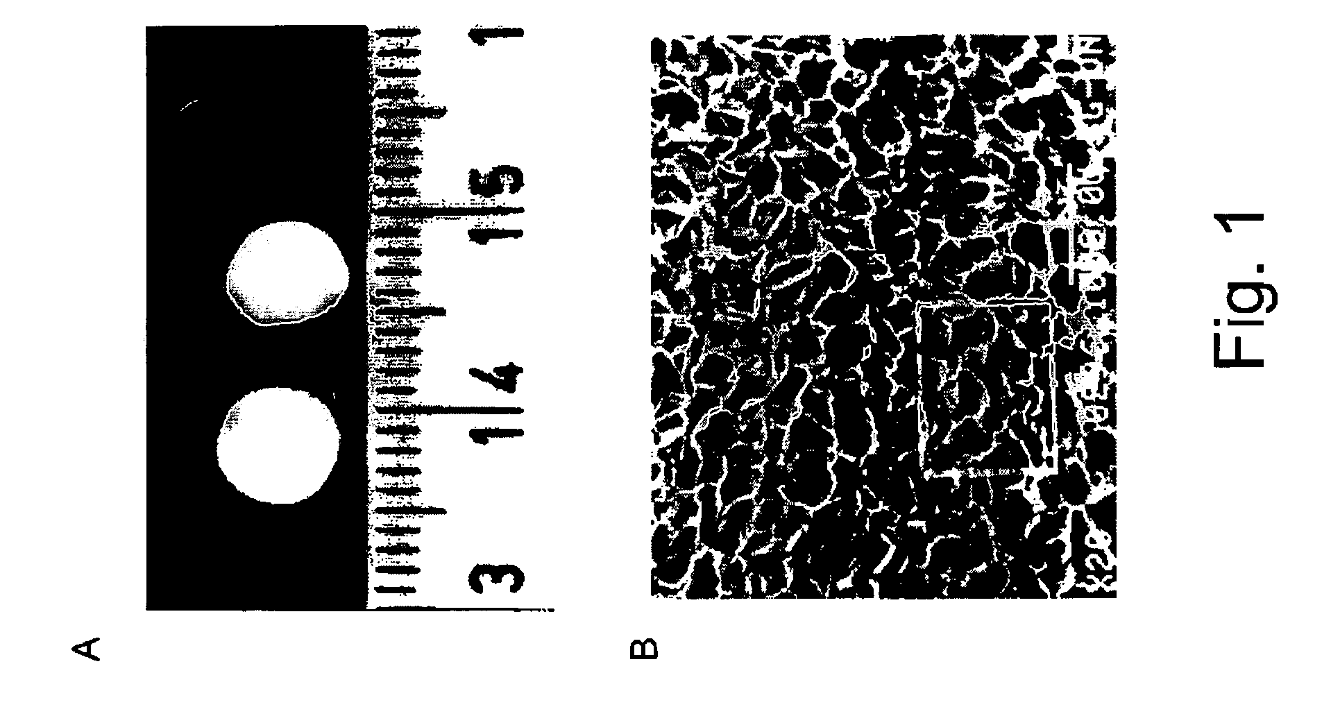
[0047]The 3-D scaffolds were prepared as previously described (Shapiro and Cohen., Biomaterials, 18 583–90, 1997) from a pharmaceutical-grade alginate, Protanal LF 5 / 60 (Pronova Biopolymers, Drammen, Norway), which has a G contents (65–75%) and solution viscosity (1% w / v, 25° C.) of 50 cP. Scaffold preparation consists of (i) Preparation of sodium alginate stock solutions, at concentrations of 1–3% (w / v). (ii) Cross-linking of the alginate by adding, dropwise, the bivalent cross-linker, e.g., calcium gluconate. (iii) Freezing the cross-linked alginate and (iv) lyophilization to produce a sponge-like scaffold. The sponges were sterilized using ethylene oxide gas apparatus. The residual ethylene oxide was removed by aeration of the samples with warm air flow. The sponges were stored in laminated bags, at room temperature, until use. A photograp...

example 2

Tissue Engineering of Capillary-Like Tubes from Endothelial Cells Seeded within 3-D Alginate Scaffolds, In Vitro

Culturing of Endothelial Cells within the 3-D Alginate Scaffolds:

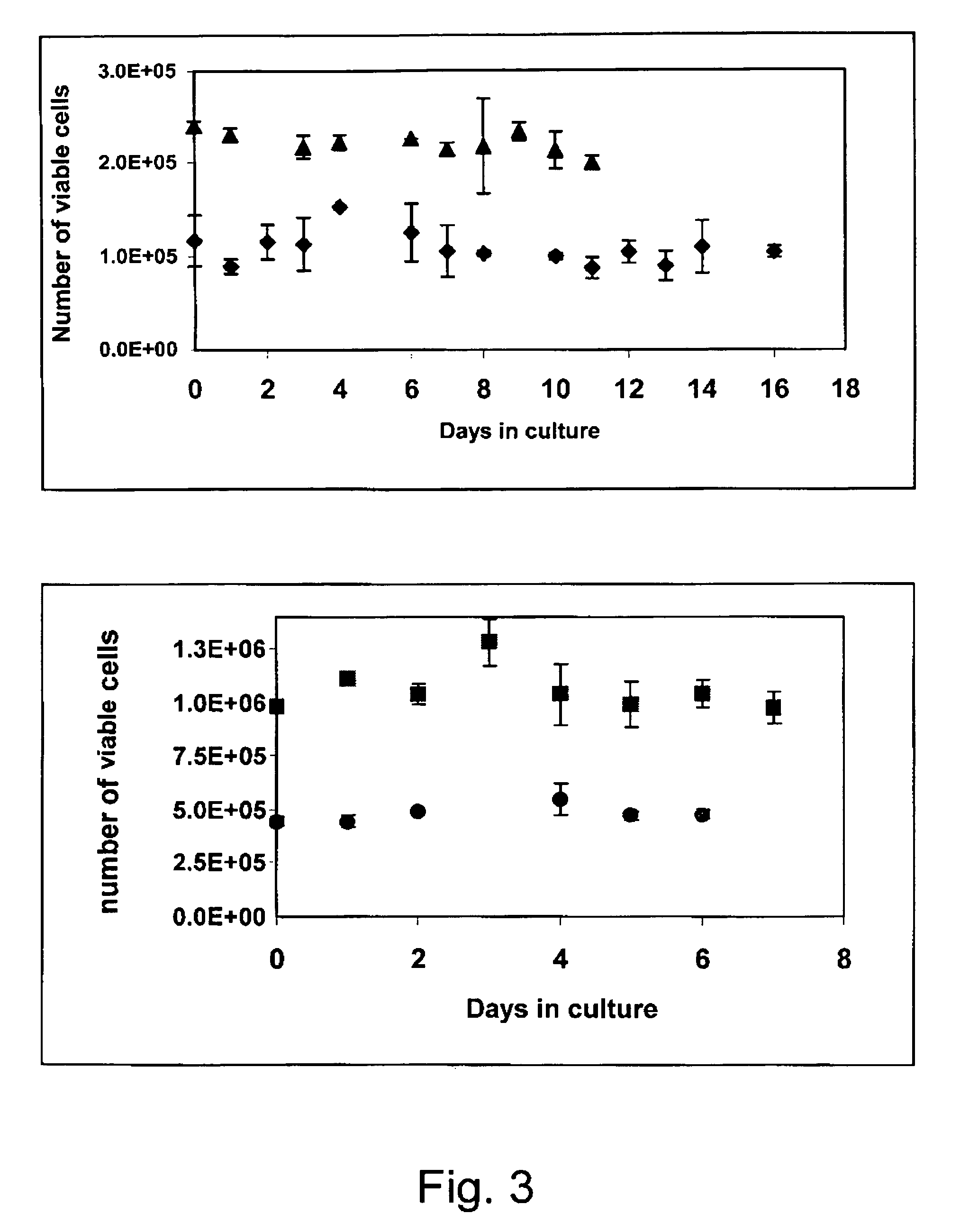
[0053]For the isolation of Aortic Endothelial Cells, the aorta was aseptically collected, stripped of adventitia and sliced into rings. The rings were cultured on a tissue flask in an incubator, at 37° C., without medium. After an hour incubation, DMEM supplemented with 10% FBS, 0.02 μg / ml bFGF, 100 U / ml nystatin, 5 μg / ml insulin, 5 μg / ml transferrin, 5 ng / ml sodium selenite and penicillin-streptomycin, was added. The endothelial cells grew as colonies from the aortic rings, and then were expanded as pure population of endothelial cells. Purity of the cells was analyzed by FACS using anti-von Willebrant Factor antibodies. The endothelial cells were seeded onto the alginate scaffolds at an initial cell density of 1 million per scaffold. The growth medium was supplemented with rHuVEGF165 VEGF (50 ng / ml) (produc...

example 3

Preparation of Composite Alginate Scaffolds Containing Microspheres with Controlled Release Growth Factors

[0055]The microspheres containing the growth factors are prepared from poly(D,L-Lactide-(o-glycolide) (PLGA) (RG502H, Boehringer Ingelheim, Germany) by the solvent evaporation method, based on a double emulsion (Cohen et al., Pharmaceutical Research, 8, 713–720, 1991). The polymer was dissolved in a volatile organic solvent, methylene chloride. An aqueous solution containing the growth factors was added to the polymer solution, and the mixture was homogenized to create an inner emulsion. This emulsion was further emulsified in a second aqueous phase that contained a surface active agent such as poly(vinyl alcohol). The resulting double emulsion was stirred until all organic solvent was evaporated, leaving solid microspheres. To formulate the composite scaffold, the microspheres were suspended in the alginate solution, and scaffold preparation proceeds as described in Example 1.

[...


PUM

PropertyMeasurementUnit
pore diameteraaaaaaaaaa
pHaaaaaaaaaa

Abstract

The invention is directed to tissue-engineered biografts, methods for preparing the biografts of the invention, and methods for repairing a damaged myocardium in a mammal. The methods of the invention can include providing a three-dimensional porous polysaccharide matrix; introducing mammalian cells into said matrix; growing said cells in said matrix in vitro, until a tissue-engineered biograft is formed; and transplanting the tissue-engineered biograft onto myocardial tissue or myocardial scar tissue of said mammal. The tissue-engineered biograft of the invention can form a contracting tissue. The methods of the invention can optionally include removing scar tissue or dead tissue from the site of implantation prior to transplanting the biograft.

Description

FIELD OF THE INVENTION[0001]The present invention relates to the use of tissue-engineered cardiac biografts for transplantation into damaged myocardium.BACKGROUND OF THE INVENTION[0002]Despite recent advances in the treatment of acute myocardial infarction (MI), attempts to repair extensive myocardial damage and to treat heart failure are often met with limited success. One of the reasons for the lack of success is that the myocardium is unable to regenerate because cardiomyocytes do not have the capacity for replication after injury, and furthermore because there are apparently no muscle stem cells in the myocardium.[0003]Existing strategies for restoring heart function after myocardial injury are practically limited to cardiac transplantation. Since the supply of donor hearts is limited, tissue engineering appears to be a promising approach for the formation of new functional tissue to replace lost or failing tissue. Earlier studies indicated the possibility of transplanting isola...

Claims


Application Information

Patent Timeline
Patent Type & Authority Patents(United States)
IPC IPC(8): C12N5/08A61K9/14C12N5/071C12N5/077
CPCA61L27/20A61L27/3804A61L27/3873A61L27/3886A61L27/56C12N5/0657C12N5/0691C08L5/04C12N2501/165C12N2533/74
Inventor COHEN, SMADARDAR, AYELETETZION, SHARONPERETS, ANATSHAPRUT, SIGALITLEOR, JONATHAN
Owner UNIV OF THE NEGEV BEN GURION
Why Eureka
Try Eureka