
Light activated fluorescent probe having protein label positioning function as well as preparation method and application thereof

A fluorescent probe and light-activated technology, applied in the field of fluorescent probes, can solve the problems of no spontaneous transmembrane ability, low degree of specific labeling, inability to use live cell imaging, etc., achieve specific positioning, and the preparation method is simple and easy Line, high yield effect

Inactive Publication Date: 2015-07-01
EAST CHINA UNIV OF SCI & TECH
View PDF0 Cites 28 Cited by
  • Summary
  • Abstract
  • Description
  • Claims
  • Application Information

AI Technical Summary

Problems solved by technology

However, there are still many deficiencies in this probe system worthy of our attention: 1) Cy3-Cy5 probe pair is labeled on the antibody by immunofluorescence labeling method, and the antibody does not have transmembrane ability, so it can only be applied to fixed cells Or the surface of the cell membrane, which cannot be used for live cell imaging; 2) The photoconversion mechanism of Cy3-Cy5 probes is not clear, and the requirements for the surrounding environment are very harsh, because the photoconversion needs to be carried out under reducing conditions, which is Need to add mercapto-containing reducing agent and oxygen scavenger; 3) The operation is relatively difficult, because the distance between Cy3 and Cy5 must meet 1-3nm to complete the pairing
In addition, compared with fluorescent proteins, although small molecule fluorescent dyes have many advantages, they also have some disadvantages: 1) They are chemically synthesized and added to cells by exogenous sources, and their specific labeling degree is relatively low, often requiring a lot of washing and fluorescence The background is high; 2) Many small molecule fluorescent probes (such as sulfonated Cy dyes) do not have the ability to spontaneously transmembrane, which limits their application to fixed cells or cell membrane surfaces

Method used


Image

  • Light activated fluorescent probe having protein label positioning function as well as preparation method and application thereof
  • Light activated fluorescent probe having protein label positioning function as well as preparation method and application thereof
  • Light activated fluorescent probe having protein label positioning function as well as preparation method and application thereof

Examples


Embodiment 1

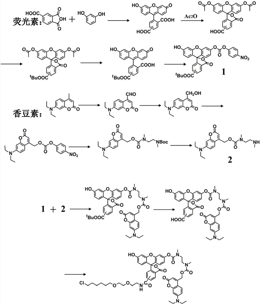
[0030] A method for synthesizing a light-activated fluorescent probe with protein label positioning, the specific molecule is composed of the following structural formula I:

[0031]

[0032] Concrete synthetic steps are as follows:

[0033] (1) Add 35g resorcinol and 240ml methanesulfonic acid into a 500ml round bottom flask, add 30g trimellitic anhydride under nitrogen protection, mix well and stir at 145°C for 24h, pour into ice water after cooling, filter, Washed with water, dried, and recrystallized from ethanol to obtain 30g, yield 60%. The resulting product 5(6)-carboxyfluorescein, after drying, take 5g of 5(6)-carboxyfluorescein (12.8mmol) in a 50mL round bottom flask, and then add 24mL of Ac 2 O (0.26mol), heated to reflux for 3h, cooled to room temperature after complete reaction, then poured the reaction liquid into about 200mL of ice water, extracted with ethyl acetate (150mL×3), washed the organic phase with water (150mL×3), anhydrous Na 2 SO 4 After drying,...

Embodiment 2

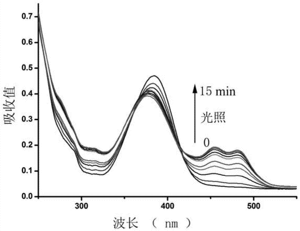
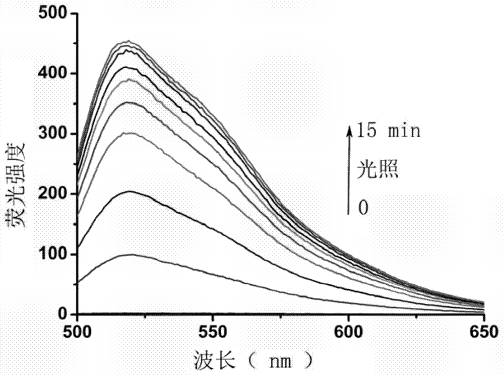
[0044]UV absorption and fluorescence properties of a photoactivatable fluorescent probe with protein tag localization in methanol. Fluorescent probe of the present invention in methanol (concentration is 10 -5 mol / L) UV-Vis absorption spectrum- figure 2 (0 minute curve) and fluorescence emission spectrum- image 3 (0 minute curve).

[0045] figure 2 It is the ultraviolet-visible light absorption spectrum of the photoactivation process of the probe of the present invention. at 10mw / cm 2 Under the irradiation of 365nm light with light intensity, after about 10 minutes, the absorption peak of fluorescein at about 490nm reaches the peak value, which shows that the 4-position ester bond of coumarin is broken, and N,N'-dimethylethylenediamine quickly Self-elimination, releasing fluorescein.

[0046] image 3 It is the fluorescence emission spectrum of the photoactivation process of the probe of the present invention. It can be seen from the figure that the background fluor...

Embodiment 3

[0048] Light-activatable fluorescent probes with protein tags localized for imaging detection in living cells. After the target protein and Halo-tag (dehalogenase) are fused and expressed by gene fusion technology, the Halo-tag ligand (this ligand is on the probe of the present invention) can be directly and specifically labeled on the fusion protein under physiological conditions on the Halo-tag.

[0049] Figure 4 In order to use light-activated fluorescent probes with protein label localization for imaging detection in living cells, the cell line used is 293 cells that have expressed the NLS-Halo label in the nucleus in advance, 10uM of the probe of the present invention is incubated with the cells for 30min, and then Wash 3 times and perform light-activated fluorescence test with microscope light source. Quantitative analysis of the cell area and background area of ​​the imaging data found that the probe can effectively enter the cell and specifically localize in the nuc...


PUM


Abstract

The invention discloses a light activated fluorescent probe having a protein label positioning function, and further discloses a preparation method and application thereof. The light activated fluorescent probe having the protein label positioning function has excellent photophysical properties, such as high-efficiency light control property and high fluorescence quantum yield, and characteristics, such as simple small molecule dye synthesis; furthermore, in combination with a chemical label technology, the light activated fluorescent probe is capable of positioning target molecular specificity in living cells precisely; real-time super-resolution fluorescence imaging in the living cells can be realized; and therefore, the light activated fluorescent probe has important application value in the field of super-resolution, especially super-resolution imaging in the living cells.

Description

technical field [0001] The invention relates to the technical field of fluorescent probes, in particular to a light-activated fluorescent probe with protein label positioning and its preparation method and application. Background technique [0002] In the field of biological research, light microscopy is a widely used and indispensable imaging tool. Light microscopy, especially fluorescence microscopy, has unique advantages over other microscopes: non-invasive (allows observation of living cells, tissues, and organisms) and high precision (precisely specific observation of targeted molecules through fluorescent labeling) ). We know that conventional optical microscopes such as confocal fluorescence microscopes have never been able to break through the limit resolution of 200nm due to the diffraction effect of light waves, that is, limited by the diffraction limit. In order to study the structural characteristics of intracellular molecules at the nanoscale, improving the re...

Claims


Application Information

Patent Timeline
IPC IPC(8): C09K11/06C07D493/10A61K49/00
Inventor 林秋宁宋江江包春燕朱麟勇
Owner EAST CHINA UNIV OF SCI & TECH
Learn More