Drug releasing coatings for medical devices

a technology of medical devices and coatings, applied in the field of coating medical devices, can solve the problems of affecting the effect of treatment, so as to avoid the dependence on lipolysis and other disadvantages, facilitate rapid drug elution, and improve the effect of drug infiltration into the tissues

Active Publication Date: 2008-05-22
LUTONIX INC
View PDF94 Cites 368 Cited by
  • Summary
  • Abstract
  • Description
  • Claims
  • Application Information

AI Technical Summary

Benefits of technology

[0019]The present inventor has found that coating the exterior surface of a medical device, and particularly of a balloon catheter or a stent, for example, with a layer comprising a therapeutic agent and an additive that has both a hydrophilic part and a hydrophobic part is useful in solving the problems associated with the coatings discussed above. Surprisingly, the present inventor has found that the at least one additive according to embodiments of the present invention, which comprises a hydrophilic part and a hydrophobic part, in combination with a therapeutic agent, forms an effective drug delivery coating on a medical device without the use of oils and lipids, thereby avoiding the lipolysis dependence and other disadvantages of conventional oil-based coating formulations. Moreover, the additives according to embodiments of the present invention facilitate rapid drug elution and superior permeation of drug into tissues at a disease site. Thus, coatings according to embodiments of the present invention provide an enhanced rate and / or extent of absorption of the hydrophobic therapeutic agent in diseased tissues of the vasculature or other body lumen.

Problems solved by technology

While these medical devices initially appear successful, the benefits are often compromised by the occurrence of complications, such as late thrombosis, or recurrence of disease, such as stenosis (restenosis), after such treatment.
Over time, de-endotheliaziation and injury to smooth muscle cells results in thrombus deposition, leukocyte and macrophage infiltration, smooth muscle cell proliferation / migration, fibrosis and extracellular matrix deposition.
The widespread use of coronary stents has altered the vascular response to injury by causing a more intense and prolonged inflammatory state, due to chronic irritation from the implanted foreign body, and in the case of drug eluting stents (DES), from insufficient biocompatibility of the polymer coating.
Moreover, local drug delivery may allow for the use of certain effective therapeutic agents, which have previously been considered too toxic or non-specific to use systemically.
Thus, a considerable disadvantage of a DES, or any other implanted medical device designed for sustained release of a drug, is that the drug is incapable of being rapidly released into the vessel.
Additionally, while drug-eluting stents were initially shown to be an effective technique for reducing and preventing restenosis, recently their efficacy and safety have been questioned.
A life-threatening complication of the technology, late thrombosis, has emerged as a major concern.
Drug eluting stents cause substantial impairment of arterial healing, characterized by a lack of complete re-endothelialization and a persistence of fibrin when compared to bare metal stents (BMS), which is understood to be the underlying the cause of late DES thrombosis.
Concerns have also been raised that the polymeric matrix on the stent in which the anti-proliferative drug is embedded might exacerbate inflammation and thrombosis, since the polymers used are not sufficiently biocompatible.
Thus, combining a therapeutic agent with a polymer in a medical device coating may have significant disadvantages.
Another important limitation of the DES is that the water insoluble drugs are not evenly distributed in the polymeric matrix of the coating.
The non-uniform distribution of drug causes non-uniform drug release to the tissue of the vessel walls.
This may cause tissue damage and thrombosis in areas exposed to excess drug and hyperplasia and restenosis areas that are undertreated.
Yet another important limitation of the DES is that only a limited amount of an active agent can be loaded into the relatively small surface area of the stent.
Any component of the coating that inhibits rapid release, such as a lipid or polymer or an encapsulating particle, is necessarily disadvantageous to the intended use of the balloon catheter, which is inflated for a very brief period of time and then removed from the body.
However, a polymeric hydrogel coating can not effectively deliver water insoluble drugs (such as paclitaxel and rapamycin), because they can not mix with the hydrogel coating.
These agents are associated with the risk of bradycardia, ventricular arrthymia, hypotension, heart block, sinus arrest, sinus tachycardia, and fibrillation.
Iodine contrast agents may also induce renal failure, and as a result there are significant efforts to remove these contrast agents from the vascular system after diagnostic procedures.
They are therefore not optimally effective at carrying hydrophobic drugs such as paclitaxel into cells, and the percent of paclitaxel reported to be taken up by vascular tissue after deployment of these devices is only 5-20%.
All of these drug delivery formulations have significant disadvantages.
Unlike hydrophilic contrast agents, oils and lipids mix well with water-insoluble drugs such as paclitaxel or rapamycin, but the particle sizes of oils used for solubilizing the therapeutic agents are relatively unstable, ranging in a broad particle size distribution from several hundred nanometers to several microns in diameter.
Loading capacity of conventional micelles is low.
Another disadvantage of oil-based liposome formulations is the dependence of drug absorption on the rate and extent of lipolysis.
Lipolysis of oil-based triglycerides is difficult and dependent upon many factors, and triglycerides must be digested and drug released in order to be absorbed by diseased tissue.
The amount of hydrophobic drug delivered to tissues by these agents will be low, because liposomes and micelles cannot efficiently release hydrophobic drug, which they carry away before it can be absorbed by tissues.
Oils and lipids are therefore not effective at rapidly and efficiently facilitating tissue uptake of drug during a very brief device deployment time, and no report has shown these types of coatings to be effective.
Drug that is encapsulated in polymeric particles may take even longer to diffuse from the coating and will have further difficulty permeating target tissues rapidly.
Thus, these polymeric microspheres are useful for sustained release of drug over a long period of time but cannot rapidly release drug and facilitate tissue uptake.
Combining drugs and medical devices is a complicated area of technology.
It involves the usual formulation challenges, such as those of oral or injectable pharmaceuticals, together with the added challenge of maintaining drug adherence to the medical device until it reaches the target site and subsequently delivering the drug to the target tissues with the desired release and absorption kinetics.
The coating thickness must also be kept to a minimum, since a thick coating would increase the medical device's profile and lead to poor trackability and deliverability.
Thus, formulations that are effective with pills or injectables might not work at all with coatings of medical device.
If the drug releases from the device too easily, it may be lost during device delivery before it can be deployed at the target site.

Method used


Image

  • Drug releasing coatings for medical devices
  • Drug releasing coatings for medical devices

Examples


example 1

Preparation of Coating Solutions

[0227]Solution 1—50-150 mg (0.06-0.18 mmole) paclitaxel, 2-6 ml acetone (or ethanol), 25-100 mg ascorbyl palmitate, 25-100 mg L-ascorbic acid and 0.5 ml ethanol are mixed.[0228]Solution 2—50-150 mg (0.05-0.16 mmole) rapamycin, 2-6 ml acetone (or ethanol), 50-200 mg polyglyceryl-10 oleate and 0.5 ml ethanol are mixed.[0229]Solution 3—50-150 mg (0.06-0.18 mmole) paclitaxel, 2-6 ml acetone (or ethanol), 50-200 mg octoxynol-9 and 0.5 ml ethanol are mixed.[0230]Solution 4—50-150 mg (0.05-0.16 mmole) rapamycin, 2-6 ml acetone (or ethanol), 50-200 mg p-isononylphenoxypolyglycidol and 0.5 ml ethanol are mixed.[0231]Solution 5—50-150 mg (0.06-0.18 mmole) paclitaxel, 2-6 ml acetone (or ethanol), 50-200 mg Tyloxapol and 0.5 ml ethanol are mixed.[0232]Solution 6—50-150 mg (0.05-0.16 mmole) rapamycin in 2-6 ml acetone (or ethanol), 50-150 mg L-ascorbic acid in 1 ml water or ethanol, both, then are mixed.[0233]Solution 7—50-150 mg (0.06-0.18 mmole) paclitaxel, 2-6 ...

example 2

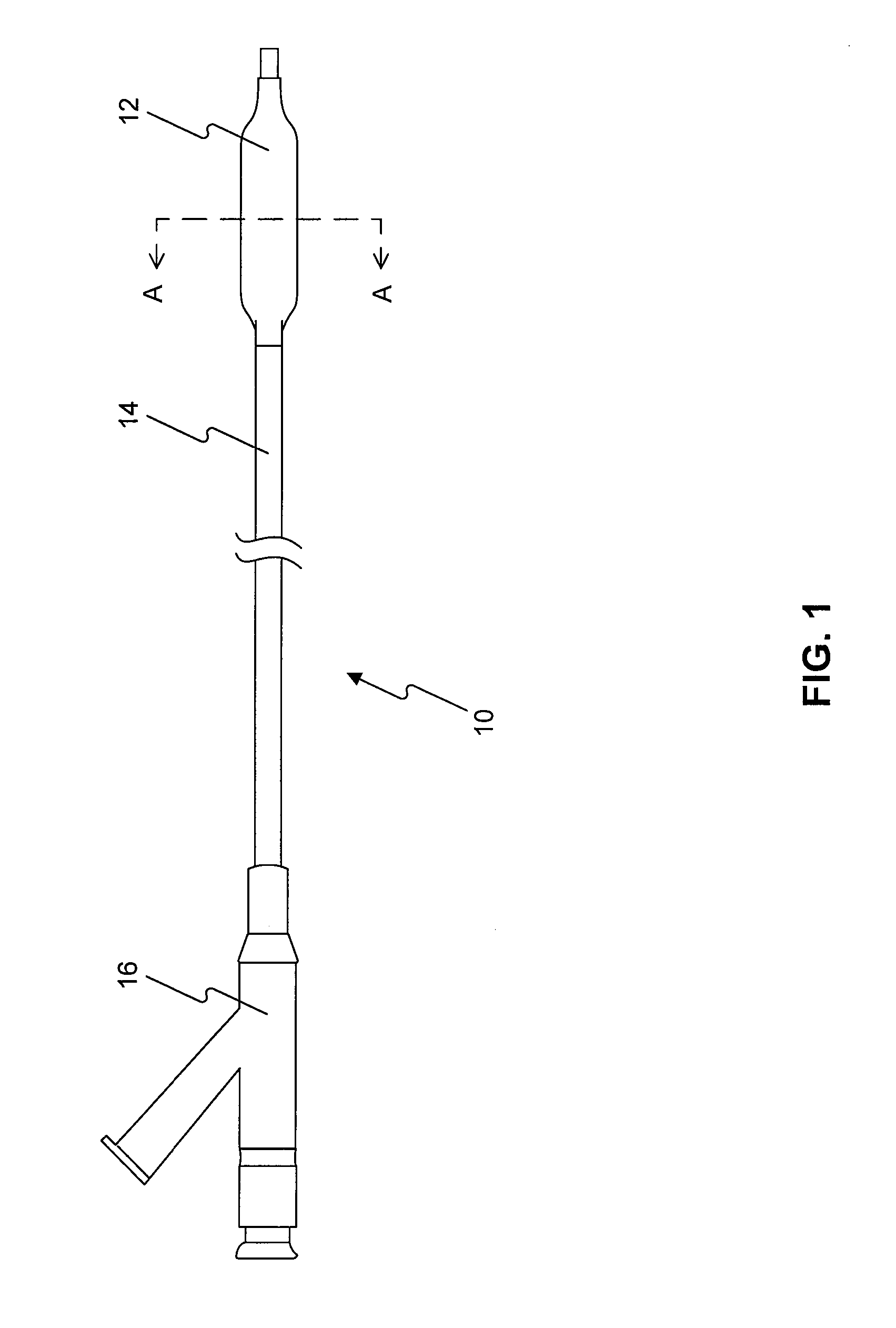
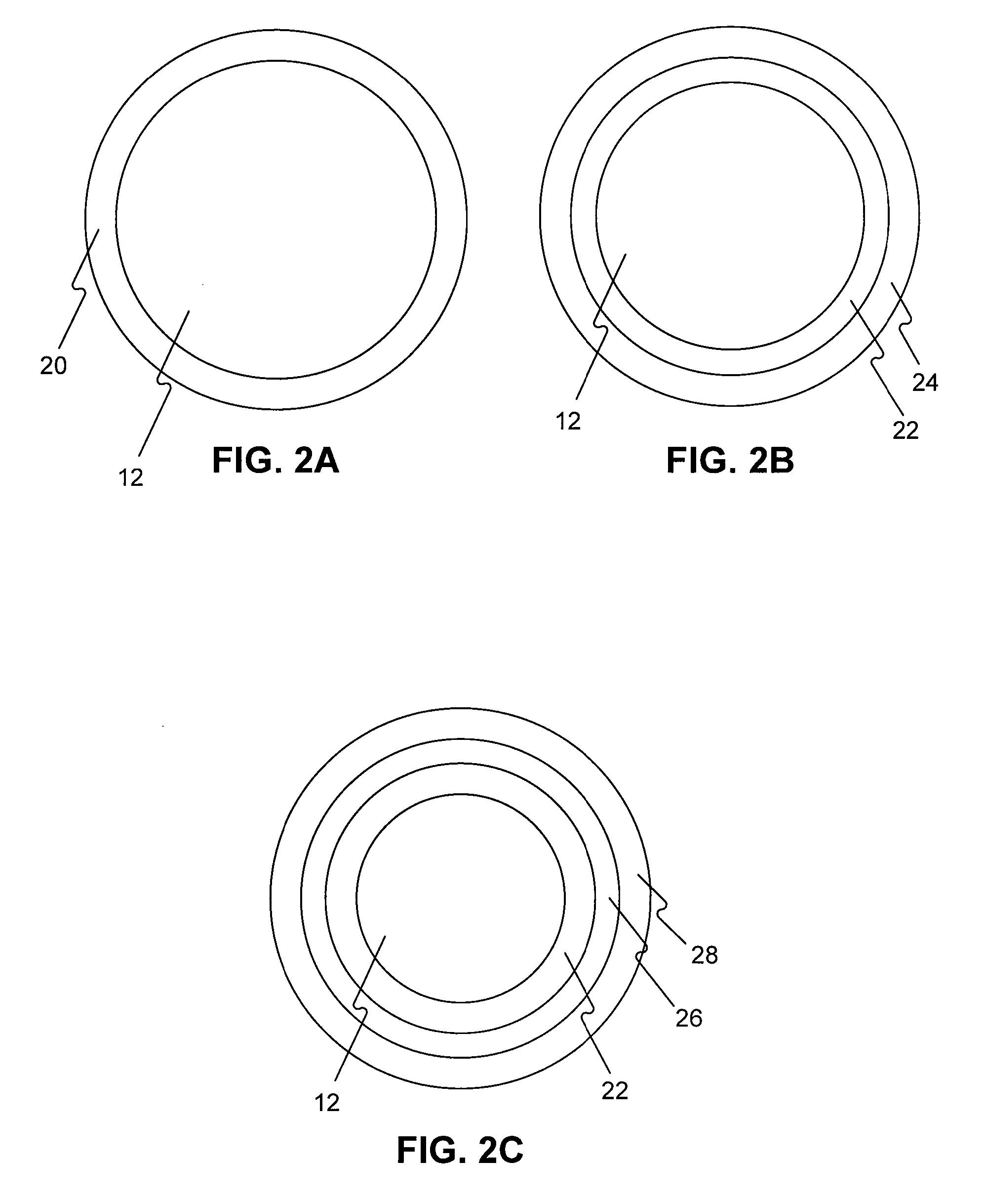
[0244]5 PTCA balloon catheters (3 mm in diameter and 20 mm in length) are folded with three wings under vacuum. The folded balloon under vacuum is sprayed or dipped in a solution (1-17) in example 1. The folded balloon is then dried, sprayed or dipped again, dried again, and sprayed or dipped again until sufficient amount of drug on the balloon (3 microgram per square mm) is obtained. The coated folded balloon is then rewrapped and sterilized for animal testing.

example 3

[0245]5 PTCA balloon catheters (3 mm in diameter and 20 mm in length) are folded with three wings under vacuum. The folded balloon under vacuum is sprayed or dipped in a solution (1-5) in example 1. The folded balloon is then dried, sprayed or dipped again in a solution (6-10), dried, and sprayed or dipped again until sufficient amount of drug on the balloon (3 microgram per square mm) is obtained. The coated folded balloon is then rewrapped and sterilized for animal testing.


PUM

PropertyMeasurementUnit
ethanol solubilityaaaaaaaaaa
total thicknessaaaaaaaaaa
particle size distributionaaaaaaaaaa

Abstract

The invention relates to a medical device for delivering a therapeutic agent to a tissue. The medical device has a layer overlying the exterior surface of the medical device. The layer contains a therapeutic agent and an additive. The additive has a hydrophilic part and a hydrophobic part and the therapeutic agent is not enclosed in micelles or encapsulated in particles or controlled release carriers.

Description

CROSS REFERENCE TO RELATED APPLICATIONS[0001]This application claims the benefit of priority under 35 U.S.C. § 119 of U.S. Provisional Application No. 60 / 860,084, filed on Nov. 20, 2006, U.S. Provisional Application No. 60 / 880,742, filed Jan. 17, 2007, U.S. Provisional Application No. 60 / 897,427, filed on Jan. 25, 2007, U.S. Provisional Application No. 60 / 903,529 filed on Feb. 26, 2007, U.S. Provisional Application No. 60 / 926,850 filed Apr. 30, 2007, U.S. Provisional Application No. 60 / 904,473 filed Mar. 2, 2007, U.S. Provisional Application No. 60 / 981,380 filed Oct. 19, 2007, and U.S. Provisional Application 60 / 981,384 filed Oct. 19, 2007, the disclosures of all of which are incorporated by reference herein.FIELD OF THE INVENTION[0002]Embodiments of the present invention relate to coated medical devices, and particularly to coated balloon catheters, and their use for rapidly delivering a therapeutic agent to particular tissue or body lumen. Embodiments of the present invention also...

Claims


Application Information

Patent Timeline
Patent Type & Authority Applications(United States)
IPC IPC(8): A61F2/02A61K31/337
CPCA61K31/337A61K31/436A61L29/16A61L29/18A61L31/16A61L31/18A61M25/10A61L2300/802A61L2300/416A61K2300/00A61K45/06A61P11/00A61P11/06A61P29/00A61P3/02A61P35/00A61P37/06A61P7/02A61P9/00A61L29/085A61L2300/216A61L2300/608A61L2420/06A61L2420/08A61M2025/105A61M2210/1025A61L29/08A61L2300/428
Inventor WANG, LIXIAO
Owner LUTONIX INC
Why Eureka
Try Eureka