

Drug releasing coatings for medical devices
a technology of medical devices and coatings, applied in the direction of catheters, dilators, surgery, etc., can solve the problems of fibrosis, thrombosis, fibrosis, etc., to prevent the loss of the lumen of the body, relieve stenosis, and reduce the risk of fibrosis
- Summary
- Abstract
- Description
- Claims
- Application Information
AI Technical Summary
Benefits of technology
Problems solved by technology
Method used
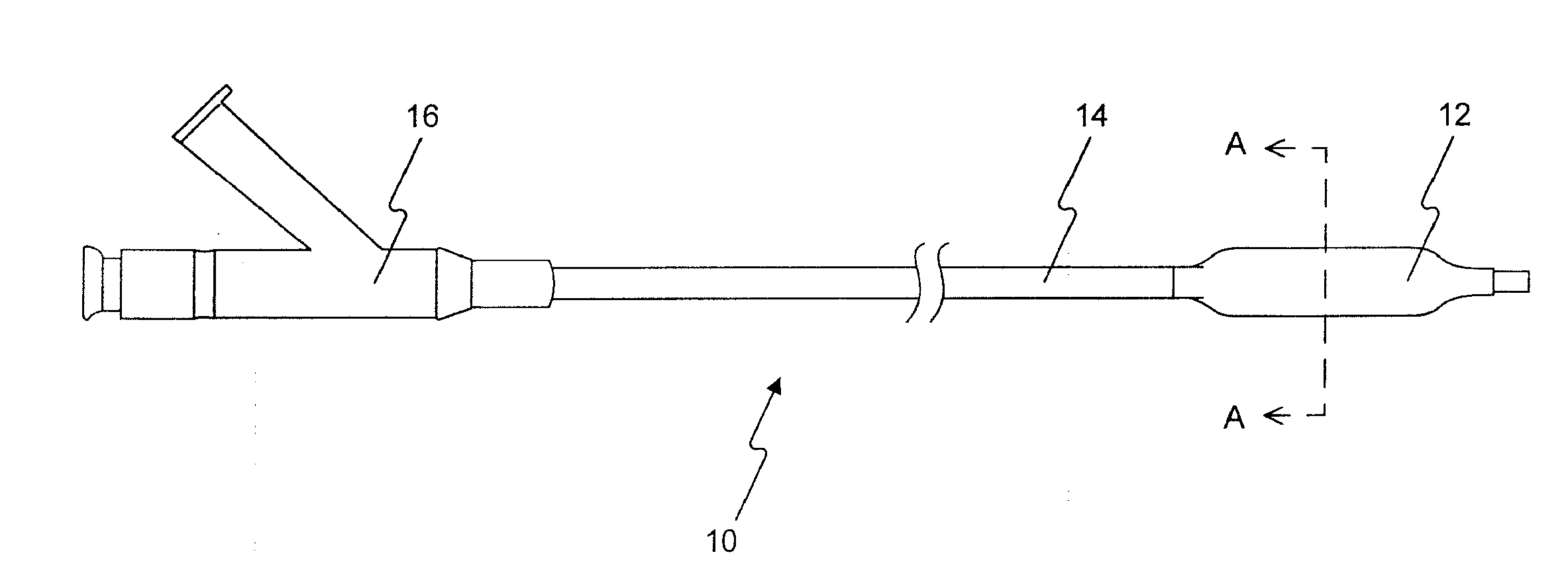
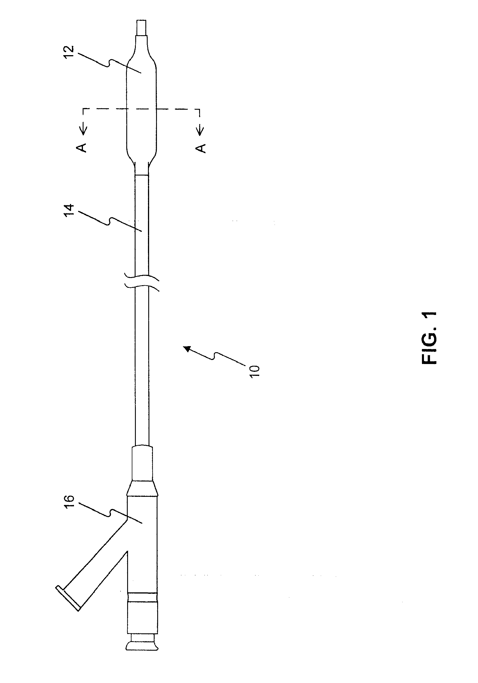
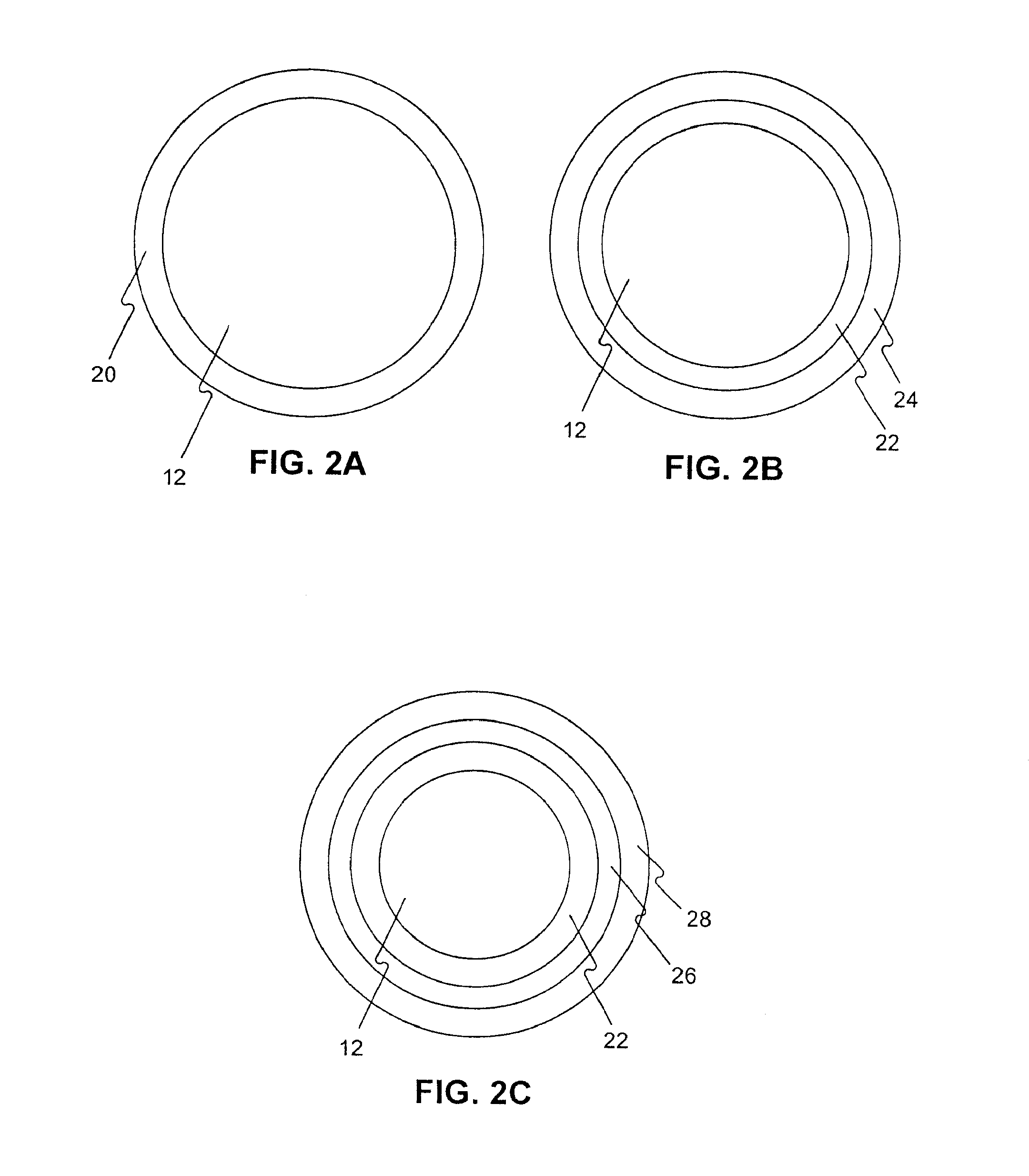
Image

Examples
example 1
[0242]Preparation of Coating Solutions:
[0243]Formulation 1—50-150 mg (0.06-0.18 mmole) paclitaxel, 2-6 ml acetone (or ethanol), 25-100 mg ascorbyl palmitate, 25-100 mg L-ascorbic acid and 0.5 ml ethanol were mixed.
[0244]Formulation 2—50-150 mg (0.05-0.16 mmole) rapamycin, 2-6 ml acetone (or ethanol), 50-200 mg polyglyceryl-10 oleate and 0.5 ml ethanol were wemixed.
[0245]Formulation 3—50-150 mg (0.06-0.18 mmole) paclitaxel, 2-6 ml acetone (or ethanol), 50-200 mg octoxynol-9 and 0.5 ml ethanol were mixed.
[0246]Formulation 4—50-150 mg (0.05-0.16 mmole) rapamycin, 2-6 ml acetone (or ethanol), 50-200 mg p-isononylphenoxypolyglycidol and 0.5 ml ethanol were mixed.
[0247]Formulation 5—50-150 mg (0.06-0.18 mmole) paclitaxel, 2-6 ml acetone (or ethanol), 50-200 mg Tyloxapol and 0.5 ml ethanol were mixed.
[0248]Formulation 6—50-150 mg (0.05-0.16 mmole) rapamycin in 2-6 ml acetone (or ethanol), 50-150 mg L-ascorbic acid in 1 ml water or ethanol, both, then were mixed.
[0249]Formulation 7—50-150 m...
example 2
[0271]5 PTCA balloon catheters (3 mm in diameter and 20 mm in length) were folded with three wings under vacuum. The folded balloon under vacuum was sprayed or dipped in a formulation (1-17) in example 1. The folded balloon was then dried, sprayed or dipped again, dried again, and sprayed or dipped again until sufficient amount of drug on the balloon (3 microgram per square mm) was obtained. The coated folded balloon was then rewrapped and sterilized for animal testing.
example 3
[0272]5 PTCA balloon catheters (3 mm in diameter and 20 mm in length) was folded with three wings under vacuum. The folded balloon under vacuum was sprayed or dipped in a formulation (1-5) in example 1. The folded balloon was then dried, sprayed or dipped again in a formulation (6-10), dried, and sprayed or dipped again until sufficient amount of drug on the balloon (3 microgram per square mm) was obtained. The coated folded balloon was then rewrapped and sterilized for animal testing.
PUM
| Property | Measurement | Unit |
|---|---|---|
| particle size distribution | aaaaa | aaaaa |
| melting point | aaaaa | aaaaa |
| water and ethanol solubility | aaaaa | aaaaa |
Abstract
Description
Claims
Application Information