
Oncolytic adenovirus

a technology of adenovirus and vector, applied in the field of adenovirus vector, can solve the problems of limited success, less efficacy than replicating, and limited success of the approach, so as to reduce the immunogenicity of the virus, the effect of maximizing the effect and reducing the effect of the effect of the virus

Inactive Publication Date: 2006-07-18
ONYX PHARMA INC
View PDF0 Cites 23 Cited by
  • Summary
  • Abstract
  • Description
  • Claims
  • Application Information

AI Technical Summary

Benefits of technology

[0102]Adenoviral therapy using the instant invention adenoviruses may be combined with other antineoplastic protocols, such as gene therapy. See, U.S. Pat. No. 5,648,478. As mentioned above, adenovirus constructs for use in the instant invention will exhibit specific cancer cell killing. Such constructs may also have prodrug activator genes, including thymidine kinase, cytosine deaminase, or others, that in the presence of the appropriate prodrug will enchance the antineoplastic effect of the invention E1a and / or E4 adenovirus vectors. See, U.S. Pat. No. 5,631,236.
[0103]Also, in the event that the instant invention adenoviral mutants elicit an immune response that dampens their effect in a host animal, they can be administered with an appropriate immunosuppressive drug to maximize their effect. Alternately, a variety of methods exist whereby the exterior protein coat of adenovirus can be modified to produce less immunogenic virus. See, PCT / US98 / 0503 where it is shown that a major immunogenic component of adenovirus' exterior coat, hexon protein, can be genetically engineered to be less immunogenic. This is done by creating a chimeric hexon protein by substituting for normal viral hexon protein epitopes a sequence of amino acids not normally found in hexon protein. Such adenoviral constructs are less immunogenic than the wild type virus.
[0104]Another aspect of the instant invention is the incorporation of heterologous genes with anti-neoplasia activity into the E1a and / or E4 shuttle vectors, preferably in the E1B, E3 regions of the virus, more preferably the E3B region, or in other regions of the virus where the heterologous gene exhibits a late expression pattern. Examples of such heterologous genes, or fragments thereof that encode biologically active peptides, include those that encode immunomodulatory proteins, and, as mentioned above, prodrug activators (i.e. cytosine deaminase, thymidine kinase, U.S. Pat. Nos. 5,358,866, and 5,677,178). Examples of the former would include interleukin 2, U.S. Pat. Nos. 4,738,927 or 5,641,665; interleukin 7, U.S. Pat. Nos. 4,965,195 or 5,328, 988; and interleukin 12, U.S. Pat. No. 5,457,038; tumor necrosis factor alpha, U.S. Pat. Nos. 4,677,063 or 5,773,582; interferon gamma, U.S. Pat. Nos. 4,727,138 or 4,762,791; or GM-CSF, U.S. Pat. Nos. 5,393,870 or 5,391,485. Additional immunomodulatory proteins further include macrophage inflammatory proteins, including MIP-3, (See, Well, T. N. and Peitsch, M C. J. Leukoc. Biol vol 61 (5): pages 545–50,1997), and cell suicide, or apoptosis inducing proteins, including BAD and BAX. See, Yang, E., et al. Cell, vol 80, pages 285–291 (1995); and Sandeep, R., et al Cell, vol. 91, pages 231–241 (1997). Monocyte chemotatic protein (MCP-3 alpha) may also be used. A preferred embodiment of a heterologous gene is a chimeric gene consisting of a gene that encodes a protein that traveres cell membranes, for example, VP22 or TAT, fused to a gene that encodes a protein that is preferably toxic to cancer but not normal cells. To increase the efficacy of the invention adenoviral E1A mutant constructs they may be modified to exhibit enhanced tropism for particular tumor cell types. For example, as shown in PCT / US98 / 04964 a protein on the exterior coat of adenovirus may be modified to display a chemical agent, preferably a polypeptide, that binds to a receptor present on tumor cells to a greater degree than normal cells. Also see, U.S. Pat. No. 5,770,442 and U.S. Pat. No. 5,712,136. The polypeptide can be antibody, and preferably is single chain antibody.Purification of Adenoviral Mutants
[0105]Adenovirus is routinely purified by a number of techniques including cesium chloride banding using an ultracentrifuge. However, for large scale production of adenovirus, methods which give larger yields than those readily obtainable by cesium chloride ultracentrifugation are desirable, and involve one or more chromatographic steps. The preferred method utilizes ion exchange chromatography. See, for example, PCT / US97 / 21504; and Huyghe et al., Human Gene Therapy, vol. 6: 1403–1416 (1996).Formulation
[0106]Adenovirus, including adenoviral mutants, may be formulated for therapeutic and diagnostic administration to a patient. For therapeutic or prophylactic uses, a sterile composition containing a pharmacologically effective dosage of adenovirus is administered to a human patient or veterinary non-human patient for treatment, for example, of a neoplastic condition. Generally, the composition will comprise about 103 to 1015 or more adenovirus particles in an aqueous suspension. A pharmaceutically acceptable carrier or excipient is often employed in such sterile compositions. A variety of aqueous solutions can be used, e.g., water, buffered water, 0.4% saline, 0.3% glycine and the like. These solutions are sterile and generally free of particulate matter other than the desired adenoviral vector. The compositions may contain pharmaceutically acceptable auxiliary substances as required to approximate physiological conditions such as pH adjusting and buffering agents, toxicity adjusting agents and the like, for example sodium acetate, sodium chloride, potassium chloride, calcium chloride, sodium lactate, etc. Excipients which enhance infection of cells by adenovirus may be included.
[0107]Adenoviruses of the invention, or the DNA contained therein, may also be delivered to neoplastic cells by liposome or immunoliposome delivery; such delivery may be selectively targeted to neoplastic cells on the basis of a cell surface property present on the neoplastic cell population (e.g., the presence of a cell surface protein which binds an immunoglobulin in an immunoliposome). Typically, an aqueous suspension containing the virions are encapsulated in liposomes or immunoliposomes. For example, a suspension of adenovirus virions can be encapsulated in micelles to form immunoliposomes by conventional methods (U.S. Pat. Nos. 5,043,164, 4,957,735, 4,925,661; Connor and Huang (1985) J. Cell Biol. 101: 582; Lasic DD (1992) Nature 355: 279; Novel Drug Delivery (eds. Prescott LF and Nimmo WS: Wiley, N.Y., 1989); Reddy et al. (1992) J. Immunol. 148: page 1585). Immunoliposomes comprising an antibody that binds specifically to a cancer cell antigen (e.g., CALLA, CEA) present on the cancer cells of the individual may be used to target virions, or virion DNA to those cells.

Problems solved by technology

Investigators initially used wild type viruses, and this approach met with some, albeit, limited success.
It has, however, met with limited success, and it appears to be less efficacious than replicating viruses.
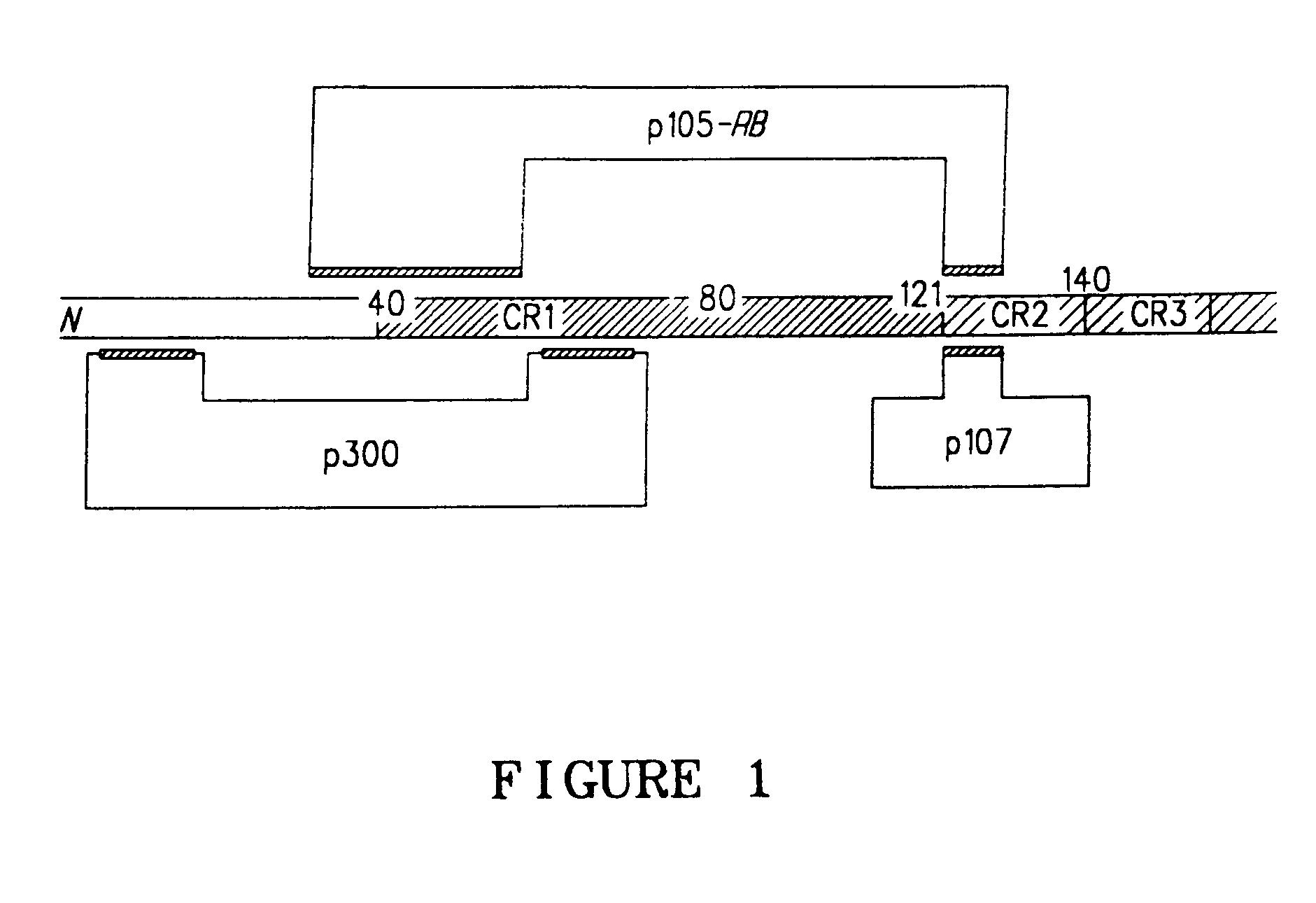
This results in an infection of the cell population with the E1a-RB(−) replication deficient adenovirus.

Method used


Image

  • Oncolytic adenovirus
  • Oncolytic adenovirus
  • Oncolytic adenovirus

Examples


example 1

E2F-1E4 Adenoviral Vector Construction.

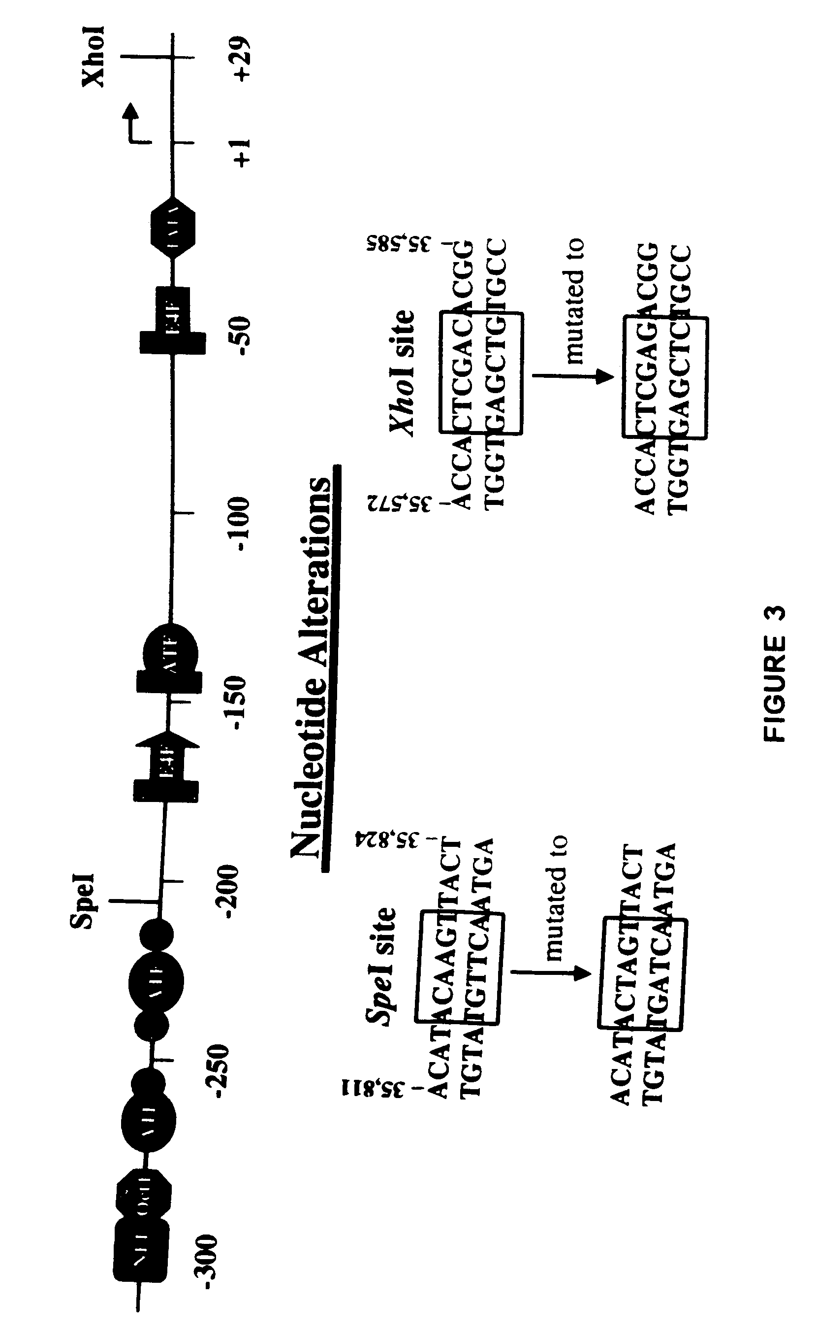
[0111]The recombinant plasmid pAd75–100 was obtained from Patrick Hearing and contains the Ad5 dl309 fragment from the EcoRI site at 75.9 map units (m.u.) to the right end of the viral genome at 100 m.u. (a BamHI linker is located at 100 m.u.) in pBR322 between the EcoRI and BamHI sites. This EcoRI to BamHI fragment was directly subcloned into Litmus 28 (New England Biolabs) to generate L28:p75–100. dl309. The wild type Ad5 E3 sequence that is missing in dl309 (Ad5 nucleotides 30,005 to 30,750) was restored by replacing the NotI to NdeI fragment in L28:p75–100. dl309 with a wild type NotI to NdeI fragment (Ad5 nucleotides 29,510-31,089) from pAd5-SN (described in U.S. Pat. Ser. No. 09 / 347,604 unpublished in-house vector). This plasmid was designated L28:p75–100.wt. In order to generate the promoter shuttle, a slightly smaller vector was generated to be the mutagenesis template. Plasmid pKSII+:p94–100 was constructed by directly subcloning the E...

example 2

E2F1-E4 Adenovirus Construction.

[0113]Ten micro grams of E2F1-E4PSV.309 were digested with EcoRI and BamHI, treated with calf-intestinal phosphatase, and gel purified. One micro gram of EcoRI digested dl922 / 47 TP-DNA was ligated to ˜5 micro grams of the purified fragment containing the wild type E2F-1 promoter driving the E4 region overnight at 16° C. Ligations were transfected into 293 cells using standard a CaPO4 transfection method. Briefly, the ligated DNA was mixed with 24 micro grams of salmon sperm DNA, 50 micro liters of 2.5M CalCl2, and adjusted to a final volume of 500 micro liters with H2O. This solution was added dropwise to 500 micro liters of Hepes-buffered saline solution, pH 7.05. After standing for 25 minutes, the precipitate was added dropwise to two 60 mm dishes of 293 cells which had been grown in DMEM supplemented with 10% fetal bovine serum (FBS) to 60–80% confluency. After 16 hours, the monolayer was washed one time with phosphate-buffered saline (minus calciu...

example 3

E2F1-E4 Viral Propagation and Confirmation.

[0114]Primary plaques were isolated with a pasteur pipette and propagated in a 6 well dish on 293 cells in 2 ml of DMEM supplemented with 2% FBS until the cytopathic effect (CPE) was complete. One-tenth (200 ml) of the viral supernatant was set aside for DNA analysis, while the remainder was stored at −80° C. in a cryovial. DNA was isolated using Qiagen's Blood Kit as per their recommendation. One-tenth of this material was screened by PCR for the presence of the desired mutations using the following primers: for dl922 / 47 (5′-GCTAGGATCCGAAGGGATTGACTTACTCACT-3′ and 5′-GCTAGAATTCCTCTTCATCCTCGTCGTCACT-3′) and for the E2F-1 promoter in the E4 region (5′-GGTGACGTAGGTTTTAGGGC-3′ and 5′-GCCATAACAGTCAGCCTTACC-3′). PCR was performed using Clontech's Advantage cDNA PCR kit in a Perkin Elmer 9600 machine using the following conditions: an initial denaturation at 98° C. for 5 min., followed by 30 cycles of denaturation at 98° C. for 1 min. and annealin...


PUM

PropertyMeasurementUnit
temperatureaaaaaaaaaa
pHaaaaaaaaaa
timeaaaaaaaaaa

Abstract

Viral vectors and methods of making such vectors are described that preferentially kill neoplastic but not normal cells, the preferred vector being an adenovirus that has the endogenous promoters in the E1A and / or E4 regions substituted with a tumor specific promoter which is preferably E2F responsive.

Description

FIELD OF THE INVENTION[0001]This application is a continuation-in-part of U.S. patent application Ser. No. 10 / 303,598 filed Nov. 25, 2002, which is a continuation-in-part of U.S. patent application Ser. No. 09 / 714,409 filed Nov. 14, 2000, which in turn claims priority from U.S. Provisional Application No. 60 / 165,638, filed Nov. 15, 1999.[0002]This invention relates to adenovirus vectors, and to methods for making and using such vectors. More particularly, it relates to improved adenovirus vectors containing mutations and substitutions in the promoters of the E1A and / or the E4 regions which confer substantial tumor cell specific oncolytic activity.BACKGROUND[0003]From the early part of this century, viruses have been used to treat cancer. The approach has been two-fold; first, to isolate or generate oncolytic viruses that selectively replicates in and kill neoplastic cells, while sparing normal cells. Investigators initially used wild type viruses, and this approach met with some, al...

Claims


Application Information

Patent Timeline
Patent Type & Authority Patents(United States)
IPC IPC(8): A61K48/00C12N15/00C12N15/63C12N15/86C12N5/00A61K35/76A61K35/761C12N15/861
CPCA61K35/76C07K14/005C12N7/00C12N15/86C12N9/78A61K35/13C12N2830/008C12N2710/10322C12N2710/10332C12N2710/10343A61K35/761A61P35/00A61P43/00
Inventor JOHNSON, LEISAFATTAEY, ALIHERMISTON, TERRYSHEN, JERRY YUQIAOLAQUERRE, SYLVIE
Owner ONYX PHARMA INC
Learn More