Method for investigating the fate of a test compound or the stateof a biological system by means of NMR of hyperpolarised NMR active nuclei

a technology of active nuclei and nmr, which is applied in the field of nmr spectroscopy, can solve the problems of increasing pressure on both the efficacy and safety evaluation process, increasing the sensitivity of nmr, and being unable to characterise the structure of metabolites fully and unambiguously, so as to enhance the corresponding nmr or mri signal, enhance the nuclear polarisation of a noble gas, and increase the sensitivity of analysis

Inactive Publication Date: 2013-02-12
GE HEALTHCARE LTD
View PDF17 Cites 2 Cited by
  • Summary
  • Abstract
  • Description
  • Claims
  • Application Information

AI Technical Summary

Benefits of technology

[0015]Although the NMR spectroscopy or imaging method of the present invention provides similar types of information about the fate of a test compound as conventional NMR or MRI, it offers potential advantages. Among these advantages are: a) increased sensitivity of analysis; and b) increased speed of acquisition of NMR spectra (or images). The analysis of drugs / metabolites or physiological compounds containing an NMR active nuclei may provide additional information previously only supplied by studying corresponding 14C-labelled compounds, whilst being free from the problems associated with radioactive isotopes.
[0016]Furthermore, in comparison to studies using fluorescent reagents and related labelling technology, for example, the current invention does not require the synthesis of an adduct comprising a reporter, e.g. a fluor, and the test compound in order to enable detection. Therefore the present invention offers the following advantages over conventional fluorescence based detection systems:
[0019]Similarly, although the NMR pattern profiling method of the present invention provides similar types of information about the state of a system when compared to existing NMR pattern profiling methods, it also offers potential advantages. Among those possible advantages are: a) increased sensitivity of analysis; and b) increased speed of acquisition of NMR spectra (or images). Increased detail in pattern profiles, may be realised as a consequence of increased sensitivity such that features become visible within spectra that would not be discernible from noise under conventional NMR. The sensitivity increase enables utilisation of 13C NMR as well as 1H NMR which collectively would provide additional information relative to 1H NMR profiles alone. Changes in patterns that are not visible in pattern profiles obtained using conventional NMR techniques may be observed using the method according to the present invention. These small changes may be particularly important for example when studying toxicity or carrying out quality assurance testing on cell cultures.

Problems solved by technology

This rapid evolution of potentially beneficial drugs has led to an increased pressure on both the efficacy and safety evaluation processes.
However, mass spectroscopy is, on its own, often not able to characterise the structure of metabolites fully and unambiguously.
Unfortunately, currently NMR is relatively insensitive.
In many cases, the relatively low sensitivity of NMR creates fundamental problems that affect the acquisition time needed in order to achieve a desired signal and the lower limit of detection (LOD) of analyte at a defined signal:noise ratio (e.g. 3:1).
In practical terms, the poor sensitivity of current NMR techniques limits their application in absorption, distribution, metabolism and excretion (ADME) studies.
During the early stages of drug development, the supply of candidate drug, and hence its metabolites, is limited and there is often not enough material available for analysis by NMR.
In addition, the concentration of metabolites produced by in vitro and in vivo screens is low and often well below the level needed for analysis by NMR.
It is not advisable to increase dosing because the routes of metabolism may change under such non-physiological conditions and the metabolites formed will be non-representative of these produced under standard patient treatment regimes.
This is very time consuming so, in practice, it often does not take place until late in the drug development phase.
This leads to many of the costly late phase candidate drug failures.
However, this is not feasible with the sensitivity of conventional NMR techniques.
The current situation with regard to Tox testing is arguably even more problematic than ADME.
Many candidate drugs are found to exhibit unacceptable toxicity late in clinical trials, and even occasionally post launch.
It is widely accepted within the pharmaceutical industry that current pre-clinical toxicological screens are inadequate.
Current in vitro screens are poorly predictive of the in vivo situation.
This is costly and time consuming.
Moreover, results from animal testing are not always predictive for humans.
However, since both methods ignore the dynamic metabolic status of the whole cell, tissue or organism, even in combination genomics and proteomics may not provide sufficient information about integrated cellular function in living systems to assess accurately the fate and toxicological profile of a drug candidate.
Currently, only the use of 1H NMR is practicable (due to insensitivity of detection for 13C using conventional NMR) and the information content of the analysis is limited by the chemical shift range of 1H.
However, its usefulness has been limited by the inability of current technology to detect and resolve differences in individual spectra due to the relatively low sensitivity of the NMR technique.
There is always a disadvantage with techniques such as fluorescent methods in that the additional chemical component may influence the measurement.

Method used


Image

  • Method for investigating the fate of a test compound or the stateof a biological system by means of NMR of hyperpolarised NMR active nuclei
  • Method for investigating the fate of a test compound or the stateof a biological system by means of NMR of hyperpolarised NMR active nuclei
  • Method for investigating the fate of a test compound or the stateof a biological system by means of NMR of hyperpolarised NMR active nuclei

Examples


example 1

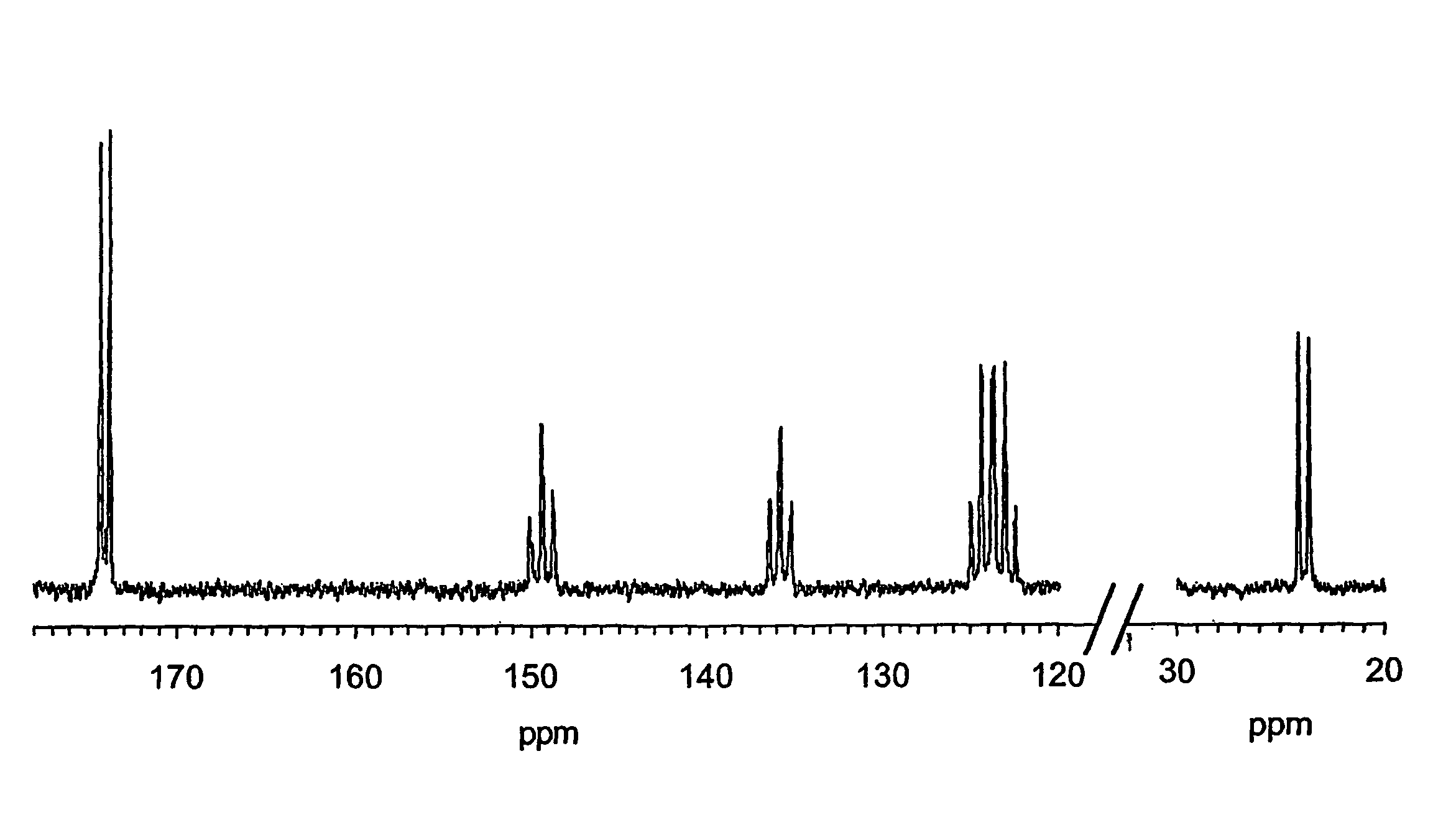
Study of (13C-carboxyl) Benzoic Acid Isolated From Rat Urine.

[0109](13C-carboxyl) Benzoic acid was purchased from SIGMA / Aldrich chemical company. A sample was added to rat urine at approximately 5 mg per ml and then isolated by solid phase extraction (SPE) and reversed phase high performance liquid chromatography (RP hplc, C18 Kromasil, 25×1 cm, 5 μm) using gradient elution with formic acid in water and formic acid in methanol. The isolated material was then dried and an aliquot re-analysed by RP hplc indicating a purity of 94%, with the main component co-eluting with carrier material, monitored by on-line UV detection (at 254 nm) and yielding the expected (M—H)− ion at 122 mass units by on-line electrospray ionisation mass spectrometry (EI-MS).

[0110]A sample of (13C-carboxyl) benzoic acid (89 μg, 0.72 μmol) obtained as described above was dissolved in sodium deutoxide in D2O (0.4% W / v, 7.5 ul, approximately 1.5 equivalents) and D2O (7.5 μl). Trityl radical dissolved in glycerol-D8 ...

example 2

Study of (13C-carboxyl) Hippuric Acid (Primary Benzoic Acid Metabolite) Isolated from Rat Urine After iv Administration of (13C-carboxyl) Benzoic Acid.

Embodiment Without a Holding Field Magnet

[0116](13C-carboxyl) Benzoic acid was administered intravenously at 10 mg / Kg (at 0 and 2 hrs) to 4 anaesthetised rats; urine was collected from canulated urethra. The major metabolite (13C-carboxy) hippuric acid was isolated by SPE and RP-hplc. The isolated material was then dried and an aliquot re-analysed by RP hplc indicating a purity of 99%, with the main component co-eluting with authentic carrier material, monitored by on-line UV detection (at 254 nm) and yielding the expected (M−H)−ion at 179 mass units by on-line electrospray ionisation mass spectrometry (EI-MS).

[0117]A sample of (13C-carboxyl) hippuric acid (463 μg, 2.57 μmol) obtained as described above was dissolved in sodium deutoxide in D2O (0.4% W / v, 17.5 μl, approximately 0.97 equivalents). Trityl radical dissolved in glycerol-D8...

example 3

Study of (13C-carboxyl) Hippuric Acid (Benzoic Acid Metabolite) Isolated from Rat Urine After iv Administration of (13C-carboxyl Benzoic Acid.

Embodiment Utilising a Holding Magnet

[0119]A sample of (13C-carboxyl) hippuric acid (463 μg, 2.57 μmol) obtained as described above was dissolved sodium deutoxide in D2O (0.4% W / v, 17.5 μl, approximately 0.97 equivalents). Trityl radical dissolved in glycerol D8 (26 μl, 25 mM) was added to the hippuric acid solution and the subsequent cocktail was mixed to homogeneity with a disposable plastic pipette. The sample was rapidly frozen, as small droplets, by dripping via a fine plastic pipette into a liquid nitrogen bath. The frozen sample drops were collected using small tweezers and placed in a liquid nitrogen cooled Kel-F cup and this was transferred for sample polarisation. The sample was polarised for 4 hours at a magnetic field of 3.354T and at a microwave frequency of 93.925 GHz. Microwave power was 100 mW and sample temperature was maintai...


PUM

PropertyMeasurementUnit
temperatureaaaaaaaaaa
magnetic fieldaaaaaaaaaa
temperatureaaaaaaaaaa

Abstract

The invention is concerned with Nuclear Magnetic Resonance (NMR) spectroscopy and Magnetic Resonance Imaging (MRI), particularly NMR spectroscopy. It provides hyperpolarization methods offering enhanced sensitivity of detection over conventional NMR for studying the fate of a test compound in a biological system. The methods are particularly suitable for studying metabolism and toxicity of drugs. The resulting NMR sensitivity increase is advantageous in two key aspects of NMR detection: test compounds can be detected at lower concentrations and substantial time saving can be achieved in cases where extensive averaging is conventionally employed to increase the signal to noise ratio of the corresponding NMR spectra. The methods can be used for studios that were not practical or not possible using conventional NMR.

Description

Technical Field[0001]This invention is concerned with Nuclear Magnetic Resonance (NMR) spectroscopy and Magnetic Resonance Imaging, particularly NMR spectroscopy. The spectra of NMR active nuclei vary depending on their environment. The present invention provides a method for obtaining information regarding the fate of a test compound, which may be exogenous, e.g. a drug, or an endogenous native compound, in a biological system by enhancing the nuclear polarisation of NMR active nuclei of the test compound (hereinafter termed ‘hyperpolarisation’) prior to NMR or MRI analysis. The invention also provides a method for carrying out NMR pattern profiling to obtain information on the status of a biological system.Definitions[0002]The term ‘test compound’ as used hereinafter, refers to a compound that may be exogenous or endogenous to the biological system in which it is to be studied and that is of physiological interest, e.g. a potential drug substance, which is not a noble gas nor a so...

Claims


Application Information

Patent Timeline
Patent Type & Authority Patents(United States)
IPC IPC(8): G01N1/00G01N24/12A61B5/055A61K49/10G01N24/08G01N33/483G01N33/50G01R33/28G01R33/465
CPCA61K49/10G01N33/5088G01R33/465G01R33/62Y10T436/24Y10T436/25G01R33/5601
Inventor COOK, NEILSANTOS, ALBIEBOSWORTH, NIGELWOLBER, JANLOOKER, MIKEKNOX, PETERARDENKJAER-LARSEN, JAN JENRIKGOLMAN, KLAES
Owner GE HEALTHCARE LTD
Why Eureka
Try Eureka