Method for preparing cornea lamina material

A cornea and lamellar technology, which is applied in the field of tissue engineering medical biomaterials, can solve the problems of disordered lamellar structure arrangement between collagen molecules, reduction of material bioactivity fusion ability, and low bioactivity of corneal lamellar materials, so as to reduce pollution risk, strong cell subculture ability, and controllable quality standards

Active Publication Date: 2012-07-11
SHAANXI RUISHENG BIOTECH
View PDF8 Cites 8 Cited by
  • Summary
  • Abstract
  • Description
  • Claims
  • Application Information

AI Technical Summary

Problems solved by technology

[0006] The current preparation methods of acellular corneal stroma all have certain safety risks and shortcomings of insufficient biological activity.
For example, 1) Chinese patent application (200410018107.5) discloses a cell-free heterogeneous corneal stroma and its preparation method and use. It uses ionic or non-ionic detergent combined with nuclease to remove cell components, and then dehydrates the stroma. Dry; due to the introduction of immunogenic nucleases, it is difficult to ensure complete elution in subsequent operations, and the remaining residues will increase immunogenicity; the bioactivity of the prepared corneal lamellar material is low
2) The decellularized corneal stroma and its preparation method disclosed in the Chinese patent application (200710027698.6) use ionic or non-ionic detergents to remove the cells to a certain extent, but the decellularization is not complete, and the collagen telopeptide The epitope structure still exists, and the resulting decellularized corneal stroma has high immunogenicity and safety risks
Similarly, the method for preparing the decellularized matrix disclosed in the Chinese patent application (200810026972.2) uses a combination of phospholipase and non-ionic detergent for decellularization, which improves th

Method used


Image

  • Method for preparing cornea lamina material
  • Method for preparing cornea lamina material
  • Method for preparing cornea lamina material

Examples


Embodiment 1

[0029] Treat the bovine eyeballs from the slaughterhouse, take the cornea-sclera complex and soak it in medical alcohol for 2 hours, then transfer it to 4°C normal saline for soaking, and set aside;

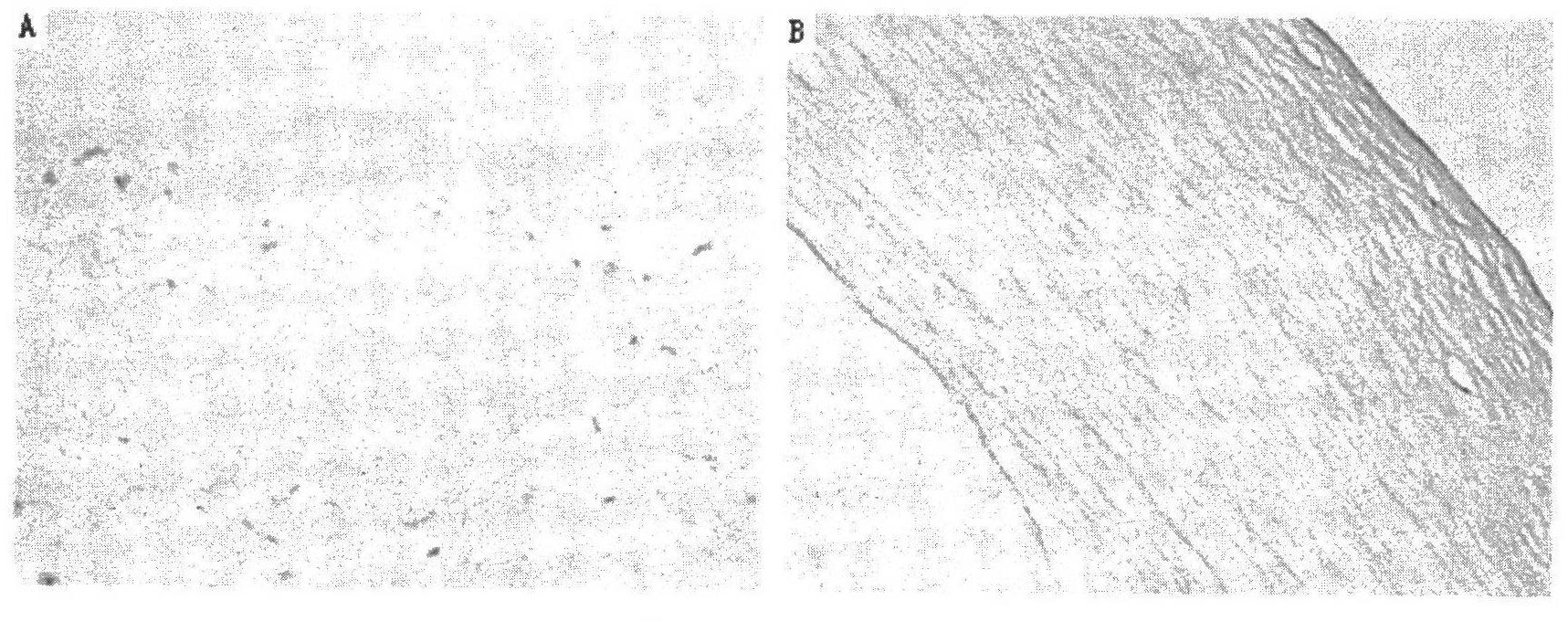
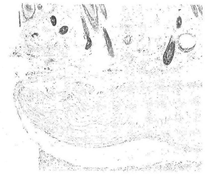
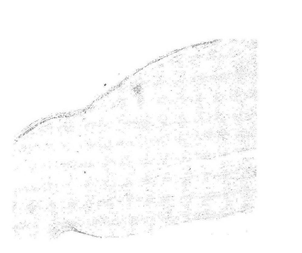
[0030] Step 1, decellularization treatment: put the cornea-sclera complex in 1mol / L sodium chloride aqueous solution containing 10ml / L Triton X-100, shake at 4°C for 5 hours; then place the cornea-sclera complex in In an aqueous solution containing 2ml / L of Triton X-100 and 0.1g / L of EDTA, shake at 4°C for 5 hours; repeat this step 6 times; hour; Obtain the decellularized cornea-sclera complex;

[0031] Step 2. Acquisition of amniotic epithelial stem cells: take primary amniotic epithelial stem cells and use primary cell culture medium to expand culture; the composition of primary cell culture medium is contained in commercial high-glucose DMEM culture medium, and EGF is 10ng / ml , human insulin 10mg / L, fetal bovine serum 150ml / L, L-glutamine 2×10 -3 mol / L, non-essential amino a...

Embodiment 2

[0038] Pig eyeballs from slaughterhouses were processed, and the cornea-sclera complex was soaked in medical alcohol for 2 hours, then soaked in 4°C normal saline, and set aside;

[0039] Step 1, decellularization treatment: put the cornea-sclera complex in 1mol / L sodium chloride aqueous solution containing 50ml / L Triton X-100, shake at 4°C for 5 hours; then place the cornea-sclera complex in In an aqueous solution containing 2ml / L Triton X-100 and 0.1g / L EDTA, shake at 4°C for 5 hours; repeat this step 8 times; obtain the decellularized cornea-sclera complex;

[0040] Step 2. Culture of amniotic epithelial stem cells: Take primary amniotic epithelial stem cells for culture; when the cell confluence reaches above 60%, digest with PBS buffer containing 4g / L trypsin for 6 minutes and collect the cell suspension, 600g Centrifuge for 5 minutes to obtain the cells; resuspend in culture medium A, and dilute with 1×10 4 / cm 2 The density of inoculated into the culture bottle and cu...


PUM

PropertyMeasurementUnit
Thicknessaaaaaaaaaa

Abstract

The invention discloses a method for preparing a cornea lamina material. The method comprises the following steps of: carrying out accellular processing on a cornea-sclera complex of a mammal eyeball, culturing an amnion epithelial stem cell, inducing and secreting TSP (Thrombospondin)-1 and co-culturing accellular cornea stroma and the amnion epithelial stem cell, sizing, packaging and sterilizing, and the like. According to an obtained cornea lamina transplanting material, the promotion and the balance among the biocompatibility, the stability, the transparency, the mechanical strength and the bioactivity are realized; the integrity of cornea collagen molecules and a lamina structure can be kept to the largest extent, and compared with that prepared with the traditional method, the accellular cornea stroma prepared according to the method disclosed by the invention is larger in tensile strength and has the moisture content and the transparency close to those of normal cornea; and various cell growth factors and the TSP-1 are gathered, and the cornea lamina material has special effects of inflammatory resistance and vascularization resistance, has the capability of greatly enhancing the bioactivity of the cornea lamina material and can be widely used for repairing complicated ocular surface coloboma such as inflammation and corneal ulcer grown in a vascularization way.

Description

technical field [0001] The invention belongs to the technical field of tissue engineering medical biomaterials, and in particular relates to a preparation method of a corneal lamellar material for transplantation. Background technique [0002] The cornea is divided into epithelial cell layer, Bowman's layer, stroma layer, Bowman's layer and endothelial cell layer according to the structure. Trauma, microbial infection, lesions and other factors will cause corneal function loss and vision loss, and severe corneal blindness will be formed. For corneal endothelial damage, penetrating keratoplasty is required; for corneal epithelium and stroma damage with intact endothelial cell layer, corneal lamellar transplantation can be used to repair. Corneal stroma injury is usually accompanied by corneal ulceration, complex inflammatory response, vascularization, leading to continuous degradation of corneal stroma. In the case of complex inflammation, it is difficult to obtain long-ter...

Claims


Application Information

Patent Timeline
IPC IPC(8): A61L27/36A61L27/54
Inventor 雷欣慧
Owner SHAANXI RUISHENG BIOTECH
Why Eureka
Try Eureka