Biphasic calcium phosphate porous bioceramic bone scaffold material and preparation method thereof and application
A biphasic calcium phosphate, bioceramic technology, applied in tissue regeneration, medical science, prosthesis and other directions, can solve the problems of single component, difficult to degrade biological activity, etc., and achieves low preparation cost, simple and feasible preparation method, and suitable porosity. Effect
- Summary
- Abstract
- Description
- Claims
- Application Information
AI Technical Summary
Problems solved by technology
Method used
Image
Examples
Embodiment 1
[0035] Embodiment 1, preparation of fishbone derived bone scaffold material
[0036] Raw material: salmon bone 100g, salmon, scientific name in English: Salmo salar, place of origin: Norway.
[0037] Removal of organic matter: Boil the fish bones in deionized water for 1 hour to remove visible organic matter, rinse with deionized water and boil again for 1 hour, repeat this step 3 times. Dry the cleaned fish bones in a constant temperature drying oven at 55°C for 24 hours to remove excess water.
[0038] High-temperature calcination: put the dried fish bones into a muffle furnace, control the heating rate at 5-10°C / min, control the high-temperature calcination temperature at 900°C, control the calcination time at 1 hour, and control the cooling rate at 10°C / min, and finally 47.8 g of calcined fish bones were obtained.
[0039] Grinding and sieving: the calcined bone was ground in an agate mortar, and sieved with a Taylor standard sieve of 35 mesh and 18 mesh wire mesh to obt...
Embodiment 2
[0042] Embodiment 2, composition, pore structure analysis and porosity determination of bone scaffold material
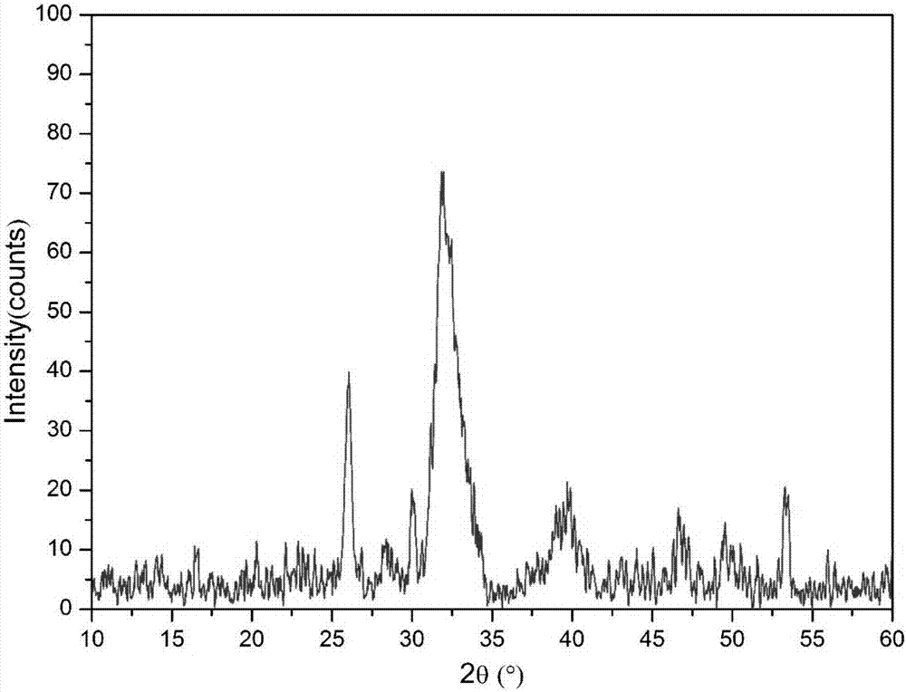
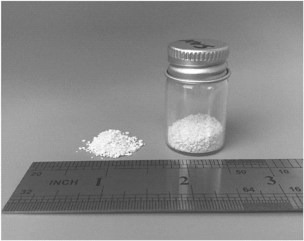
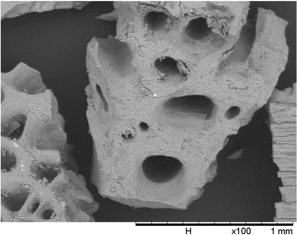
[0043] The bone scaffold material obtained in Example 1 is a white granular porous material, such as figure 2 shown. Using a scanning electron microscope to analyze its microstructure, it is found that the material has interconnected pore structures ranging in size from 50-300um, such as image 3 As shown, such a structure is conducive to cell ingrowth and nutrient transport. In order to understand the composition of the bone scaffold material, X-ray diffraction (XRD) and Fourier transform infrared spectroscopy (FTIR) were used to analyze the material.
[0044] The method for component analysis of the bone scaffold material in Example 1 by X-ray diffraction is as follows: the prepared granular bone scaffold material is repeatedly ground into a uniform fine powder in an agate mortar, pressed into tablets, tested on the machine, and set parameters It is a copper t...
Embodiment 3
[0048] Embodiment 3, the in vitro cytotoxicity experiment of bone scaffold material
[0049] According to the national standard GBT 16886.5-2003 method, the cytotoxicity was detected by using the material extract. The specific method is as follows:
[0050] Preparation of extract solution: add biphasic calcium phosphate porous bioceramic bone scaffold material (ratio of material mass to extraction medium capacity is 0.1g / mL) in DMEM medium containing 10% FBS and double antibody, at 37°C Continue soaking in a constant temperature shaker for 24 hours, then sterilize with a 0.22um filter, seal in a sterile bottle, and store in a refrigerator at 4°C for later use.
[0051] The CCK-8 method is used to detect the cytotoxicity of the material: take a 96-well plate, add 3000 cells to each well, and pre-culture for 24 hours. The DMEM medium of the double antibody was put into the incubator again for 1, 3, and 5 days. Before the test, add 10uL CCK-8 reagent to each well and incubate ...
PUM
| Property | Measurement | Unit |
|---|---|---|
| porosity | aaaaa | aaaaa |
Abstract
Description
Claims
Application Information
 - R&D
- Intellectual Property
- Life Sciences
- Materials
- Tech Scout
- Unparalleled Data Quality
- Higher Quality Content
- 60% Fewer Hallucinations
Browse by: Latest US Patents, China's latest patents, Technical Efficacy Thesaurus, Application Domain, Technology Topic, Popular Technical Reports.
© 2025 PatSnap. All rights reserved.Legal|Privacy policy|Modern Slavery Act Transparency Statement|Sitemap|About US| Contact US: help@patsnap.com


