Preservation of fetal nucleic acids in maternal plasma

a technology plasma, which is applied in the field of prenatal diagnosis of fetal abnormalities, can solve the problems of affecting only children, a large number of testing barriers, and carrying a substantial potential risk of pregnancy complications, so as to maintain the structural integrity of fetal nucleic acids

Active Publication Date: 2010-07-22
STRECK LLC
View PDF103 Cites 261 Cited by
  • Summary
  • Abstract
  • Description
  • Claims
  • Application Information

AI Technical Summary

Benefits of technology

[0013]One advantage of the present invention is the possibility for essentially simultaneous stabilizing of both the nucleated blood cells and cell-free nucleic acids. This helps to prevent cellular genomic nucleic acids (e.g., maternal cellular genomic nucleic acids) from being released into plasma, and further diluting the fetal nucleic acids (and associated biomarkers) of interest, while also maintaining the structural integrity of the fetal nucleic acids. An additional possible advantage of the present invention lies in its ability to maintain relative amounts of fetal nucleic acids. In vivo there is constant replenishment of the fetal nucleic acids to maintain a consistent amount of fetal nucleic acids but upon blood draw the fetal nucleic acid amounts will deteriorate without replenishment. The teachings of the present invention also contemplate the possibility to arrest the degradation of the fetal nucleic acids post-blood draw.
[0014]In a first aspect, the present invention contemplates a non-invasive prenatal screening method for the identification of fetal characteristics. The method includes the steps of: contacting a drawn maternal blood sample that includes a plurality of blood cells with a nucleic acid protective agent in an amount and time sufficient so that the blood cells are substantially prevented from (i) releasing genomic nucleic acids into the blood sample and from (ii) experiencing nuclease activity that degrades fetal nucleic acid; isolating fetal nucleic acids from the maternal blood sample; and analyzing the isolated fetal nucleic acids to identify a fetal characteristic.
[0015]The nucleic acid protective agent may include a formaldehyde releaser preservative agent such as one selected from the group consisting of: diazolidinyl urea, imidazolidinyl urea, dimethoylol-5,5-dimethylhydantoin, dimethylol urea, 2-bromo-2.-nitropropane-1,3-diol, oxazolidines, sodium hydroxymethyl glycinate, 5-hydroxymethoxymethyl-1-1aza-3,7-dioxabicyclo [3.3.0]octane, 5-hydroxymethyl-1-1aza-3,7dioxabicyclo[3.3.0]octane, 5-hydroxypoly[methyleneoxy]methyl-1-1 aza-3,7dioxabicyclo[3.3.0]octane, quaternary adamantine and any combination thereof. The concentration of the preservative agent prior to the contacting step may be between about 0.1 g/ml and about 3 g/ml. The concentration of the preservative agent prior to the contacting step may be between about 0.4 g/ml and about 0.8 g/ml. The concentration of the preservative agent prior to the contacting step may be a concentration at which cross-linking of nucleic acids and proteins is observed, as indicated by agarose gel electrophoresis. The amount of the preservative agent in a treated sample may be less than about 20 mg/ml of the blood sample.
[0016]The isolatin

Problems solved by technology

Further, hereditary diseases may only affect a child if both parents carry a necessary allele.
However, the difficulty in obtaining the genetic material of a fetus has presented a number of barriers to testing for the many known genetic markers for hereditary disease.
While providing reliable results, these procedures are often regarded as carrying a substantial potential risk of pregnancy complications due to their invasive nature.
Unfortunately, it has proved challenging to isolate fetal nucleic acids from maternal nucleic acids.
However, serum is generally not suitable for cell-free nucleic acid isolation since blood clotting processes release cellular nucleic acids which contaminate cell-free plasma DNA (see FIG. 1) as well as other deleterious substances that may destabilize the nucleated blood cells.
Unfortunately, this is frequently a tedious and time consuming multi-step process as it is important to use carefully controlled conditions to prevent cell breakage during centrifugation which will contaminate the cell-free nucleic acids with cellular nucleic acids released during breakage.
Once maternal cell lysis begins, the lysed maternal cells release additional nucleic acids which become mixed with the cell-free fetal nucleic acids and it becomes increasingly difficult to recover the fetal nucleic acids for testing.
Further, the amou

Method used


Image

  • Preservation of fetal nucleic acids in maternal plasma
  • Preservation of fetal nucleic acids in maternal plasma
  • Preservation of fetal nucleic acids in maternal plasma

Examples


example 1

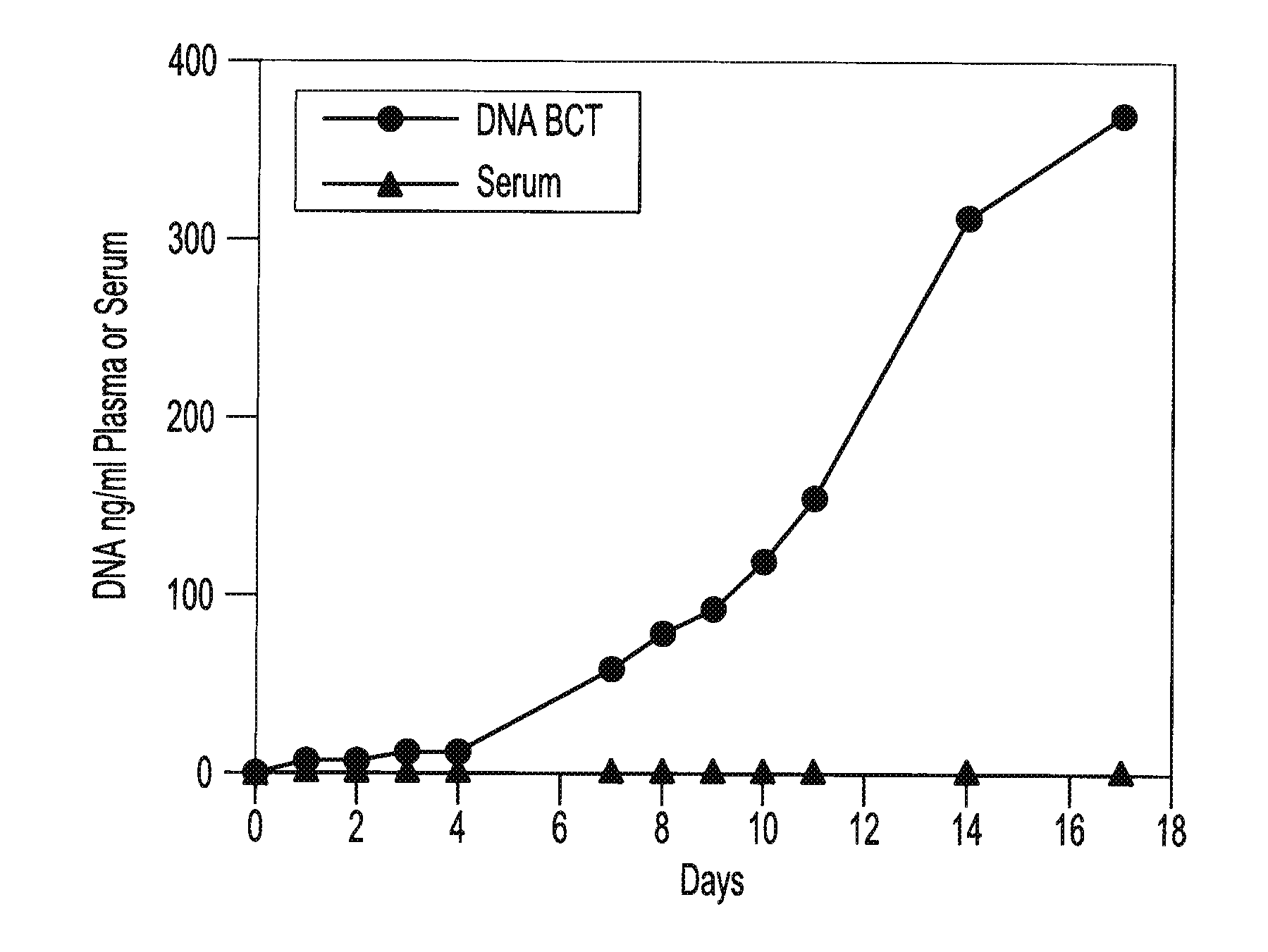
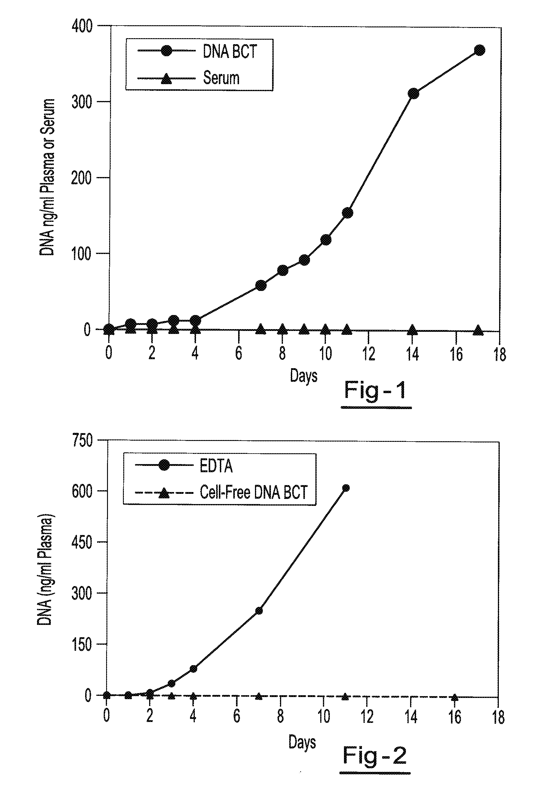
[0059]Blood samples are taken from a female donor and a male donor. The female blood sample is transferred into two tubes, tube A containing about 500 g / L IDU, about 80 g / L Tripotassium EDTA, and about 50 g / L glycine and tube B containing only the Tripotassium EDTA. Both tubes are stored at room temperature. White blood cells from the male blood sample are isolated and spiked into both tube A and tube B. 3 ml of blood are taken from each tube on day 0, day 1, day 2, day 3, day 4, day 7 and day 11. Each sample is centrifuged at room temperature at 800 g for 10 minutes and the upper plasma layer is transferred to a new tube and further centrifuged at 1500 g at room temperature for 10 minutes. The free circulating DNA in each tube is then purified using the NucleoSpin® Plasma XS kit available from Macherey-Nagel Inc., Bethlehem, Pa. The samples are then amplified by Real Time PCR amplification of a fragment of the Y-chromosome (using iQ SYBR Green Supermix reagents available from BIO-R...

example 2

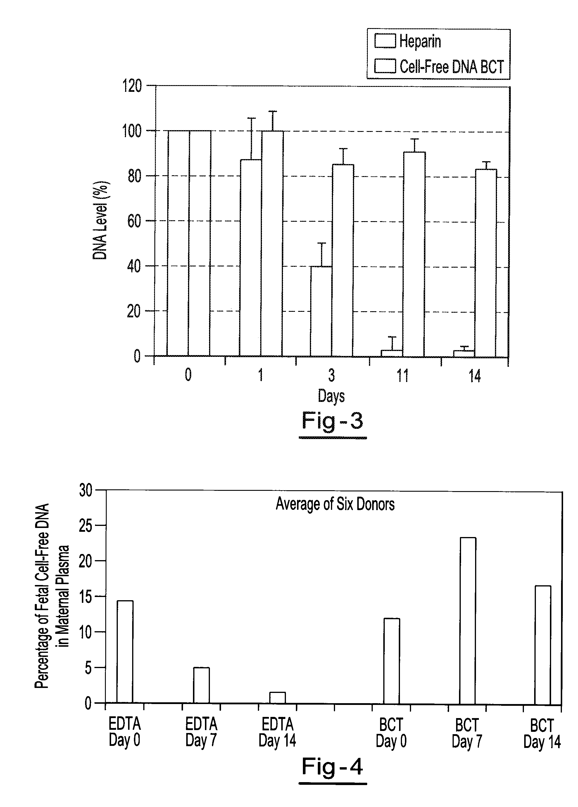
[0060]Blood samples from the same donor are drawn into two different types of blood collection tubes. One tube contains 500 g / L IDU, 81 g / L Tripotassium EDTA and 47 g / L glycine. The other tube contains only Heparin. All samples are centrifuged at 2100 g for 30 minutes at room temperature to separate the plasma. The plasma is then transferred to new tubes and non-human (lambda) DNA is then spiked into the plasma tubes. The spiked samples are then stored at room temperature for 0, 1, 2, 3, 4, 7, 11, and 14 days. Free circulating DNA is purified using the QIAamp® DNA Blood Mini Kit available from QIAGEN Inc. (Valencia, Calif.). DNA is extracted from each plasma sample. The samples are then amplified by Quantitative PCR (using iQ SYBR Green Supermix reagents available from BIO-RAD Laboratories (Hercules, Calif.)) to identify the amount of lambda DNA present. Results show a consistent relative percentage of lambda DNA presence at each measurement, indicating little if any decline in the ...

example 3

[0061]Two maternal blood samples from the same donor are drawn into two separate blood collection tubes. One tube contains about 500 g / L IDU, about 80 g / L Tripotassium EDTA, and about 50 g / L glycine. The other tube contains only the Tripotassium EDTA. Both tubes are stored at room temperature and 1 ml aliquots of blood are removed from each tube on day 0, day 7, and day 14 and plasma is separated. All samples are centrifuged at 800 g for 10 minutes at room temperature to separate the plasma. The plasma is then transferred into new tubes and centrifuged at 1500 g for 10 minutes at room temperature. Free circulating DNA is purified using the NucleoSpin® Plasma XS kit available from Macherey-Nagel Inc., Bethlehem, Pa. DNA is extracted from each plasma sample and eluted in 60 μl of elution buffer. An amount of 32 μl of eluted DNA is digested with 40 U of BstU 1 enzyme at 60° for 6 hours. The samples are then amplified by Real Time PCR (using TaqMan® RT PCR reagents available from Applie...


PUM

PropertyMeasurementUnit
Temperatureaaaaaaaaaa
Fractionaaaaaaaaaa

Abstract

A method for preserving and processing fetal nucleic acids located within maternal plasma is disclosed, wherein a sample of maternal blood containing fetal nucleic acids is treated to reduce both cell lysis of the maternal blood cells and deoxyribonuclease (DNase) and ribonuclease (RNase) activity within the fetal nucleic acids. The treatment of the sample aids in increasing the amount of fetal nucleic acids that can be identified and tested while maintaining the structure and integrity of the fetal nucleic acids.

Description

CLAIM OF PRIORITY[0001]This application claims the benefit of the filing date of U.S. Provisional Application Ser. Nos. 61 / 146,065, filed on Jan. 21, 2009 and 61 / 227,529, filed on Jul. 22, 2009, the entirety of the contents of these applications being hereby incorporated by reference for all purposes.FIELD OF THE INVENTION[0002]This invention relates to prenatal diagnosis of fetal abnormalities and more particularly to the preservation of fetal nucleic acids within a maternal blood sample.BACKGROUND OF THE INVENTION[0003]The demonstration by Leon et al. in 1977 that cell-free plasma DNA is elevated in cancer patients paved the way for the present day interest in cell-free plasma nucleic acid in disease diagnosis. Relatively recently, Lo et al. Lancet 350 (1997) 485-487 have identified the existence of circulating cell-free fetal nucleic acids in maternal plasma. Since this work, a number of studies have demonstrated that cell-free fetal nucleic acids present in maternal plasma can b...

Claims


Application Information

Patent Timeline
IPC IPC(8): C12Q1/68
CPCC12Q1/6806C12Q2527/125
Inventor FERNANDO, M. ROHANCHEN, KATE CHAO-WEI
Owner STRECK LLC
Why Eureka
Try Eureka