Platelet-derived growth factor compositions and methods for the treatment of osteochondral defects

a growth factor and platelet-derived technology, applied in the field of platelet-derived growth factor compositions and methods for the treatment of osteochondral defects, can solve the problems of increasing the recognition of pain and functional problems in patients, limited repair capabilities of cartilage in general, and very difficult healing of injury or trauma to articular cartilage, etc., to and increase cell number or cell growth

Inactive Publication Date: 2010-09-30
BIOMIMETIC THERAPEUTICS INC
View PDF100 Cites 50 Cited by
  • Summary
  • Abstract
  • Description
  • Claims
  • Application Information

AI Technical Summary

Benefits of technology

[0038]In some embodiments of the present invention, the cartilage phase is capable of increasing cell number or cell growth by about 100% to about 1000% (measured at about 2 days after cell seeding) in cells treated with PDGF in comparison to cells not treated with PDGF. In some embodiments, the cartilage phase is capable of increasing cell number or cell growth by about 100% (measured at about 2 days after cell seeding) in cells treated with PDGF in comparison to cells not treated with PDGF. In some embodiments, the cartilage phase is capable of increasing cell number or cell growth by about 200% (measured at about 2 days after cell seeding) in cells treated with PDGF in comparison to cells not treated with PDGF. In some embodiments, the cartilage phase is capable of increasing cell number or cell growth by about 300% (measured at about 2 days after cell seeding) in cells treated with PDGF in comparison to cells not treated with PDGF. In some embodiments, the cartilage phase is capable of increasing cell number or cell growth by about 400% (measured at about 2 days after cell seeding) in cells treated with PDGF in comparison to cells not treated with PDGF. In some embodiments, the cartilage phase is capable of increasing cell number or cell growth by about 600% (measured at about 2 days after cell seeding) in cells treated with PDGF in comparison to cells not treated with PDGF. In some embodiments, the cartilage phase is capable of increasing cell number or cell growth by about 800% (measured at about 2 days after cell seeding) in cells treated with PDGF in comparison to cells not treated with PDGF. In some embodiments, the cartilage phase is capable of increasing cell number or cell growth by about 1000% (measured at about 2 days after cell seeding) in cells treated with PDGF in comparison to cells not treated with PDGF.
[0039]In some embodiments of the present invention, both the osseous phase and the cartilage phase are capable of increasing cell number or cell growth in both phases by about 100% to about 1000% (measured at about 2 days after cell seeding) in cells treated with PDGF in comparison to cells not treated with PDGF. In some embodiments, both the osseous phase and the cartilage phase are capable of increasing cell number or cell growth in both phases by about 100% (measured at about 2 days after cell seeding) in cells treated with PDGF in comparison to cells not treated with PDGF. In some embodiments, both the osseous phase and the cartilage phase are capable of increasing cell number or cell growth in both phases by about 200% (measured at about 2 days after cell seeding) in cells treated with PDGF in comparison to cells not treated with PDGF. In some embodiments, both the osseous phase and the cartilage phase are capable of increasing cell number or cell growth in both phases by about 300% (measured at about 2 days after cell seeding) in cells treated with PDGF in comparison to cells not treated with PDGF. In some embodiments, both the osseous phase and the cartilage phase are capable of increasing cell number or cell growth in both phases by about 400% (measured at about 2 days after cell seeding) in cells treated with PDGF in comparison to cells not treated with PDGF. In some embodiments, both the osseous phase and the cartilage phase are capable of increasing cell number or cell growth in both phases by about 600% (measured at about 2 days after cell seeding) in cells treated with PDGF in comparison to cells not treated with PDGF. In some embodiments, both the osseous phase and the cartilage phase are capable of increasing cell number or cell growth in both phases by about 800% (measured at about 2 days after cell seeding) in cells treated with PDGF in comparison to cells not treated with PDGF. In some embodiments, both the osseous phase and the cartilage phase are capable of increasing cell number or cell growth by about 1000% (measured at about 2 days after cell seeding) in cells treated with PDGF in comparison to cells not treated with PDGF.

Problems solved by technology

Injury or trauma to the cartilage has been increasingly recognized as a cause of pain and functional problems in patients.
Cartilage in general has limited repair capabilities because chondrocytes are bound in lacunae and cannot migrate to damaged areas.
Further, in the case of articular cartilage damage, due to the absence of innervation and penetration by the vascular and lymphatic system and derivation of nutrition primarily through the synovial fluid and to some degree from the adjacent bone, injury or trauma to the articular cartilage is very difficult to heal, especially in the case of adult articular cartilage, which is mostly avascular and only 5% cellular.
While injury or trauma in chondral defects is only restricted in the cartilage itself without affecting the subchondral bone structures, injury or trauma in osteochondral defects affects both the cartilage and its underlying bone, and is very difficult to treat.
The compressive forces further impact underlying bone and cause injury to the blood supply and eventual necrosis.
However, each of these treatments has various drawbacks.
This treatment is complicated by the technical challenges of optimal plug positioning and tissue necrosis from the force required for harvesting the tissues.
Furthermore, patients often suffer from comorbidity of the harvest site and must remain in surgery for longer periods of time.
However, it has the main drawbacks of disease transmission risk and inferior result in comparison to the fresh autologous tissue grafting.
ACI has not been widely used due to its high cost (i.e., greater than $20,000 per procedure), necessity of two operations to harvest and implant the chondrocytes, increased operation time, localized morbidity at the harvest site, and inability to produce better outcomes than microfracture alone.
Blood and bone marrow (which contains stem cells) seep out of the fractures, creating a blood clot that releases cartilage-building cells.
The procedure is less effective in treating older or overweight patients, or cartilage damage that is larger than 2.5 cm.
Microfracture is also an incomplete fix for the osteochondral injury, because 1) an insufficient clot and quantity of cells are drawn into the defect to regenerate cartilage; 2) delamination / migration of the clot occurs after formation; and 3) Type I collagen found in fibrocartilage is generated, not the desirable Type II hyaline cartilage.

Method used


Image

  • Platelet-derived growth factor compositions and methods for the treatment of osteochondral defects
  • Platelet-derived growth factor compositions and methods for the treatment of osteochondral defects
  • Platelet-derived growth factor compositions and methods for the treatment of osteochondral defects

Examples


example 1

Evaluation of the Physical Characteristics of a Biphasic Plug for Application in Treatment of Osteochondral Defects

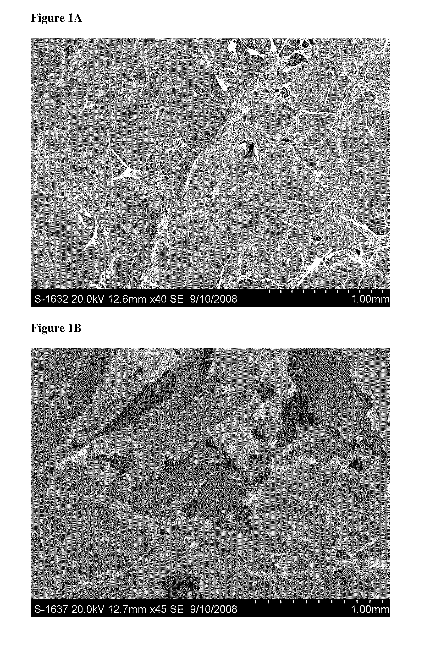
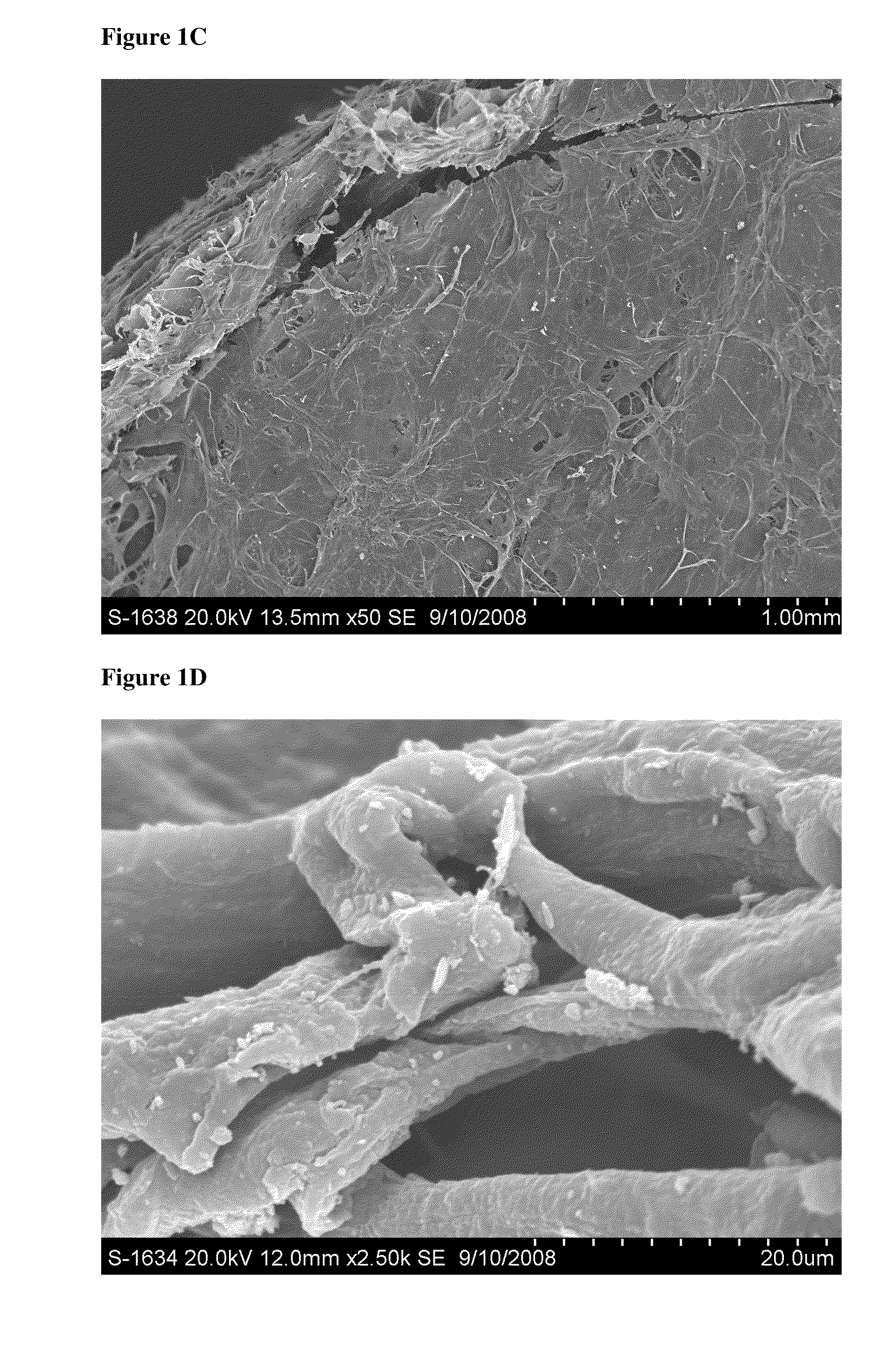
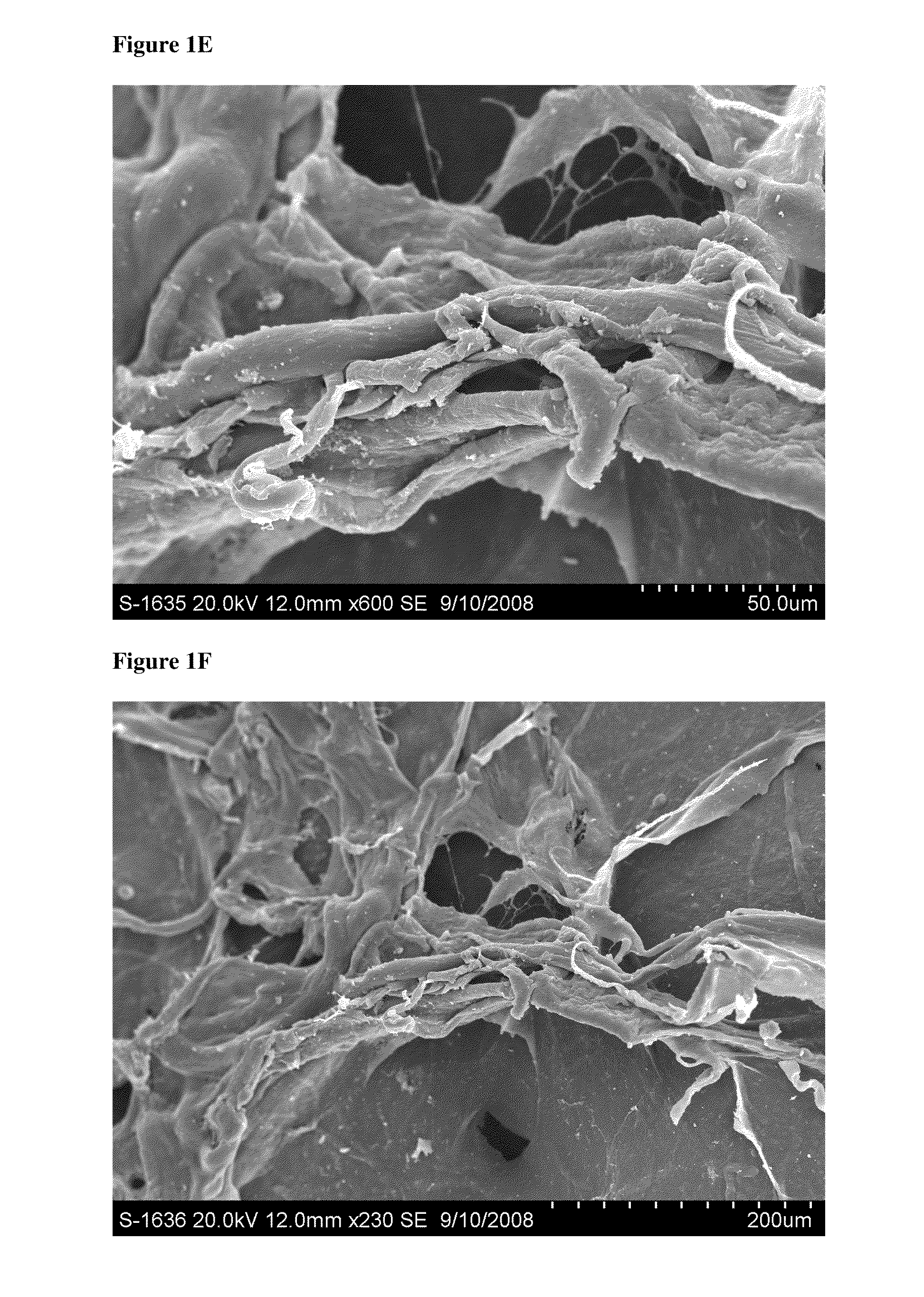
[0179]This study evaluated the surface topography, composition, and visualized porosity of a biphasic plug material from Orthomimetic's Chondromimetic using scanning electron microscopy.

[0180]In preparation, the plug (8.5 mm×8 mm) was placed in liquid nitrogen and vertically sectioned in two. The plug was placed in LN2 to maintain the structural integrity of the plug.

[0181]Once halved, the plug was then mounted with double sided adhesive tape to a 26 mm round sample mounting stub. The stub was then placed into the sputter coating apparatus. The sputter coating process bombards the sample to ensure thorough coating with gold particles to increase the electrical conductivity of the sample. Once the sputter coating process was completed, the sample was then ‘grounded’ with graphite glue to discourage charging when viewed in the electron microscope.

[0182]The samples were th...

example 2

Handling Characteristics of a Biphasic Plug

[0183]This study evaluated the handling characteristics of a biphasic plug (Orthomimetic's Chondromimetic Plug), both to evaluate the progress of hydration of the plug material in a buffer solution and to determine the effects of prolonged saturation of the plug material with elution buffer over time. Methylene Blue dye was used as a visual aid to document the hydration of the plug material.

[0184]For both the hydration and saturation study components, initial observations were noted, including: size (upper and lower phase), weight, texture, rigidity, and photographically.

[0185]For the hydration component of the study, a P200 pipette was used to add Methylene Blue dyed sodium acetate buffer to the plug material in increments of 50 μL. Aqueous Methylene Blue solution was made in 20 μl Methylene Blue and 5 mL sodium acetate buffer to make 1% x / v (volume / volume) Methylene Blue. Sodium acetate buffer (20 mM sodium acetate, pH 5.99) was made with...

example 3

[0192]Evaluation of the Release of rhPDGF-BB (Recombinant Human Platelet-Derived Growth Factor-BB) from a Biphasic Plug Material

[0193]This study evaluated the kinetics of rhPDGF-BB release from a biphasic matrix plug (Chondromimetic (Orthomimetics)) over time.

[0194]In sterile conditions, each plug was stabilized over a Sarstedt 15 ml conical polypropylene tube with a 27G½″ needle and syringe (plunger removed from syringe). Each of Orthomimetic's Chondromimetic matrix plug (×3; 8.5 mm×8 mm) was loaded with 450 μL rhPDGF-BB (e.g., 0.3 mg / ml or 1.0 mg / ml), and then allowed to sit at room temperature saturated within conical tube for ten minutes (see FIG. 3).

[0195]Following incubation, 9 ml elution buffer (1% L-glutamine, 1% Pen-Strep, 2% FBS (heat inactivated, Gamma-irradiated), 2.5% HEPES) was placed into 15 ml conical tubes, numbered 1-6. Conical tubes numbered 1-3 contained loaded sample plugs, tubes numbered 4-6 served as controls, where 450 μL rhPDGF-BB was added directly to the e...


PUM

PropertyMeasurementUnit
concentrationaaaaaaaaaa
weightaaaaaaaaaa

Abstract

The present invention provides compositions and methods for treating an osteochondral defect. In one embodiment, provided is a composition for treating an osteochondral defect comprising a biphasic biocompatible matrix and platelet derived growth factor (PDGF), wherein the biphasic biocompatible matrix comprises a scaffolding material and wherein the scaffolding material forms a porous structure comprising an osseous phase and a cartilage phase. In another embodiment, also provided is a method for treating an osteochondral defect in an individual comprising administering to the individual an effective amount of a composition comprising a biphasic biocompatible matrix and PDGF to at least one site of the osteochondral defect, wherein the biphasic biocompatible matrix comprises a scaffolding material and wherein the scaffolding material forms a porous structure comprising an osseous phase and a cartilage phase.

Description

CROSS-REFERENCE TO RELATED APPLICATIONS[0001]This application claims the benefit under 35 U.S.C. §119(e) of U.S. Provisional Patent Application No. 61 / 209,520, filed Mar. 5, 2009, and U.S. Provisional Patent Application No. 61 / 164,259, filed Mar. 27, 2009, this application is also a continuation-in-part of PCT Application No. ______ filed on Mar. 5, 2010, Attorney Docket No. 597792001540, titled “Platelet-Derived Growth Factor Compositions And Methods For The Treatment Of Osteochondral Defects”; the entireties of which are herein incorporated by reference.TECHNICAL FIELD[0002]This invention relates to compositions and methods for treating an injury or a defect in a cartilage and a bone, particularly to the treatment of osteochondral defects in a cartilage and a bone adjacent to the cartilage in an individual by administering compositions to the individual comprising a biphasic biocompatible matrix in combination with platelet-derived growth factor (PDGF) to at least one site of the ...

Claims


Application Information

Patent Timeline
Patent Type & Authority Applications(United States)
IPC IPC(8): A61K9/00A61K38/18A61P19/00
CPCA61K38/1858A61L27/227A61L27/46A61L27/54A61L27/56A61L2300/414A61P19/00A61P19/08A61K38/36A61K9/70A61K9/0002
Inventor KESTLER, HANS K.NICKOLS, JOSHUAWISNER-LYNCH, LESLIE A.RODEN, COLLEEN M.LIU, YANCHUN
Owner BIOMIMETIC THERAPEUTICS INC
Why Eureka
Try Eureka