
Use of mycoplasma bovis antigen

a technology of mycoplasma and antigen, applied in the field of use of mycoplasma bovis antigen, can solve the problems of significant economic losses worldwide, organisms persisting in unsanitary, warm, moist environments, and i>mycoplasma /i> is difficult to treat, so as to achieve treatment and/or prophylaxis of infections, and cattle treatment and/or prophylaxis

Active Publication Date: 2012-04-19
BOEHRINGER LNGELHEIM VETMEDICA GMBH
View PDF3 Cites 9 Cited by
  • Summary
  • Abstract
  • Description
  • Claims
  • Application Information

AI Technical Summary

Benefits of technology

[0039]The present invention relates to combination vaccines and/or the combined use of immunogenic compositions for the treatment and/or prophylaxis of cattle against microbiological infections, wherein the infections are caused by M. bovis and at least one further cattle relevant pathogen. The combination vaccine as described herein comprises at least one M. bovis antigen, preferably the attenuated, avirulent M. bovis as provided herewith and one further immunologically active component effective for the treatment and/or prophylaxis of infections caused by a further pathogen of cattle. The combined use or the method of co-administration of two or more antigens of pathogens affecting cattle comprises administering a first immunogenic composition comprising a M. bovis antigen, preferably the attenuated, avirulent M. bovis, as provided herewith, and at least one further immunologically active component effective for the treatment and/or prophylaxis of infections caused by a further pathogen of cattle.
[0040]According to a further aspect, the present invention relates to a combination vaccine and/or the combined use of immunogenic compositions for the treatment and/or prophylaxis of cattle against infections of the respiratory and/or reproductive system. The combination vaccine comprises the attenuated, avirulent M. bovis as described herein and at least one further immunologically active component effective for the treatment and/or prophylaxis of infections caused by Bovine Viral Diarrhea virus (BVDV), Bovine Herpes virus (BHV), Bovine Respiratory Syncytial Virus (BRSV), Parainfluenza

Problems solved by technology

Mycoplasma bovis (M. bovis) is considered to be one of the more pathogenic species of Mycoplasma and causes significant economic losses worldwide.
In general, Mycoplasmas are difficult to treat since they lack a cell wall or membrane, which tends to make them resistant to several classes of commonly used broad-spectrum antibiotic treatments.
The organism persists in unsanitary, warm, moist environments.
The prior art is deficient in that killed M. bovis is not as effective or efficient in lessening the severity of clinical symptoms associated with a Mycoplasma bovis infection.
Passively derived immunity does not appear to prevent BRSV infections but will reduce the severity of disease.
The viral infection alone is not life-threatening but predisposes cattle to secondary bacterial pneumonia, which may result in death.
Vaccines are not available for prevention of these viral respiratory diseases.
Under normal conditions, M. haemolytica generally remains confined to the upper respiratory tract, in particular the tonsillar crypts, and is difficult to culture from healthy cattle.
The interaction between the virulence factors of the bacteria and host defenses results in tissue damage and development of pneumonia.
The animal will become unthrifty in appearance if the pneumonia becomes chronic, which is usually associated with the formation of pulmonary abscesses.
It appears to be identical to Histophilus ovis and Haemophilus agni, etiologic agents of ovine septicemia, mastitis, and epididymitis; however, transmission of H. somnus between sheep and cattle has not been demonstrated.
Reproductive problems may not necessarily be preceded by bacteremia, but the pathogenesis is poorly defined.
Occasionally, animals are found dead, indicating a rapidly fatal course.
Experimental infections usually result in unapparent to mild signs of respiratory disease.
However, it has been shown that C. fetus fetus can also be a significant cause of the classic infertility syndrome usually attributed to Campylobacter fetus venerealis.
Campylobacter spp are very labile and are destroyed quickly by heating, drying, and exposure to the atmosphere.
Because the organisms survive in surface waters for extended periods, the disease is often waterborne.
However, Leptospira hardjo infections usually do not cause hemolytic anemia, which makes diagnosis more difficult.
In older cattle, signs vary greatly and diagnosis is more difficult.
Brucella abortus Infection spreads rapidly and causes many abortions in unvaccinated herds.
Cattle may ingest contaminated feed and water, or lick contaminated genitals of other animals.
Exposure to direct sunlight kills the organisms within a few hours.
Infections may also cause stillborn or weak calves, retained placentas, and reduced milk yield.
Long-standing infections may result in arthritic joints in some cattle.
Diseases thus produced are a constant threat to successful livestock production in many parts of the world.
Lesions develop without any history of wounds, although bruising or excessive exercise may precipitate some cases.
At first, the swelling is small, hot, and painful.
As the disease rapidly progresses, the swelling enlarges, there is crepitation on palpation, and the skin becomes cold and insensitive as the blood supply to the area diminishes.
In some cattle, the lesions are restricted to the myocardium and the diaphragm, with no reliable ante mortem evidence of the localized lesion.
Clostridium novyi has been suspected but not yet confirmed as a cause of sudden death in cattle and pigs fed high-level grain diets, and in which pre-existing lesions of the liver were not detectable.
Affected animals tend to lag behind the flock, assume sternal recumbency, and die within a few hours.
Differentiation from acute fascioliasis may be difficult, but peracute deaths of animals that show typical lesions on necropsy should arouse suspicion of infectious necrotic hepatitis.
Wounds caused by accident, castration, docking, unsanitary vaccination, and parturition may become infected.
Similarity to blackleg is marked, and differentiation made on necropsy is unreliable; laboratory confirmation is the only certain procedure.
Infection with C. perfringens types A, B and C causes severe enteritis, dysentery, toxemia, and high mortality in young calves.
The disease has been suspected in well-nourished beef calves nursing high-producing cows grazing lush pasture and in sudden death syndrome in feedlot cattle; however, supportive laboratory evidence in the latter is lacking.
Infection with Salmonella spp can produce diarrhea in animals of all ages, especially those that are stressed, closely stocked, or exposed to a heavily contaminated feed or water supply.
In older animals, the disease is manifested by dysentery and toxemia, and mortality can be significant.
Calves infected with pathogenic E. coli can develop severe diarrhea causing fatal dehydration, or fatal septicemic infections.
The organism is quite resistant and can survive on pasture for more than 1 year, but sunlight, alkaline soils, and drying reduce its survival rate.
This may take months to years to develop and is usually paralleled by a decline in cell-mediated immunity and a rise in ineffective serum antibody.
Diseased animals required prolonged treatment and can spread infection to both animals and humans.
Reduced weight-gains, reduced milk yields, and weight loss accompany many infections in cattle.
Although T. foetus can survive the process used for freezing semen, it is killed by drying or high temperatures.
However, immunity is not long lasting and reinfection does occur.
This results in repeat breeding and a prolonged calving season.
Calves may be aborted, stillborn, born underweight, weak, or paralyzed, or they may become paralyzed within 4 weeks of birth.
This situation of endemic stability can be upset by either a natural (eg, climatic) or artificial (eg, acaricide treatment) reduction in tick numbers to levels where tick transmission of Babesia to calves is insufficient to ensure all are infected during this critical early period.

Method used


Image

  • Use of mycoplasma bovis antigen
  • Use of mycoplasma bovis antigen
  • Use of mycoplasma bovis antigen

Examples


example 1

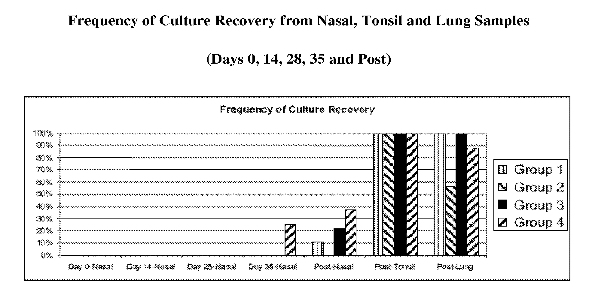
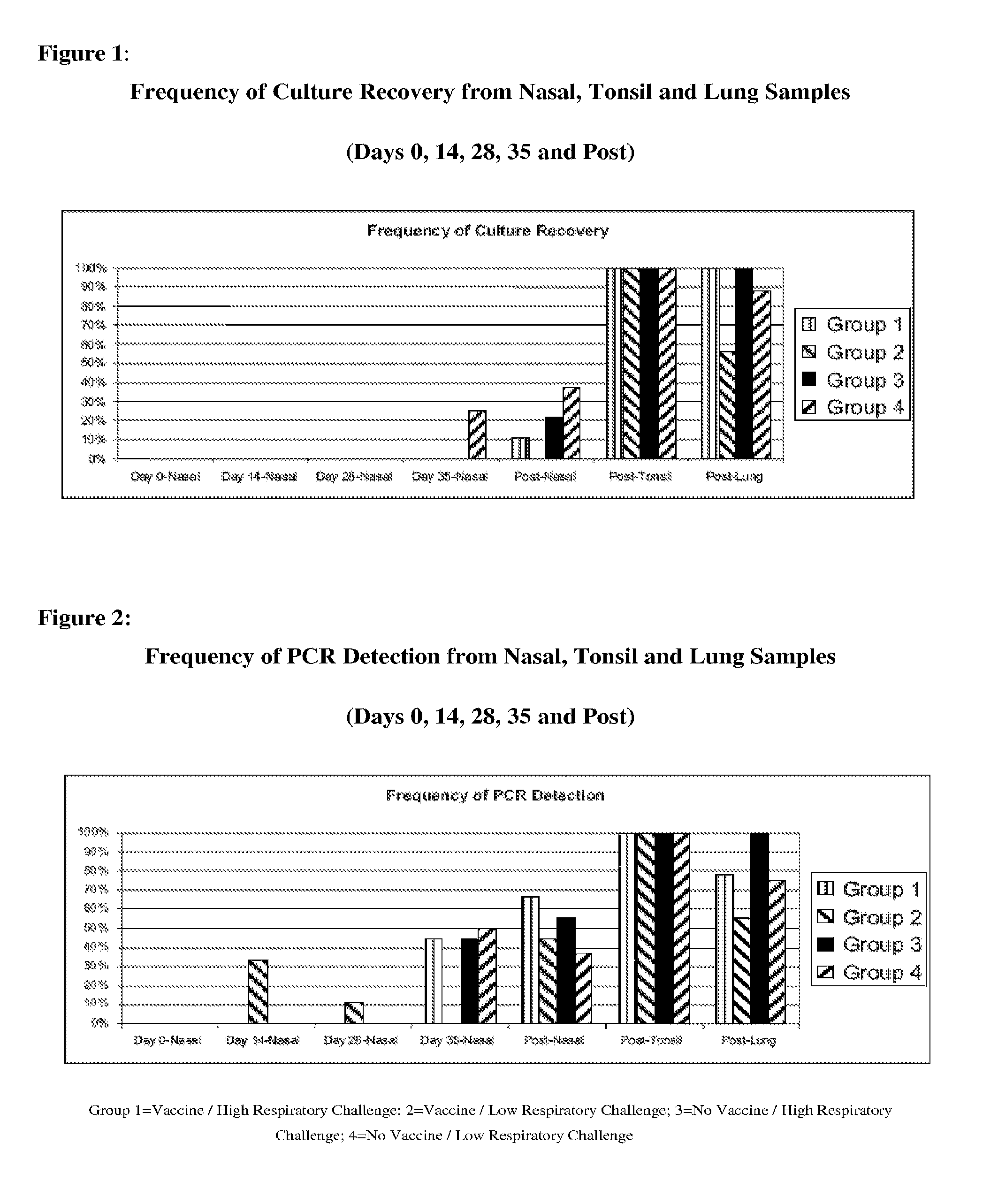
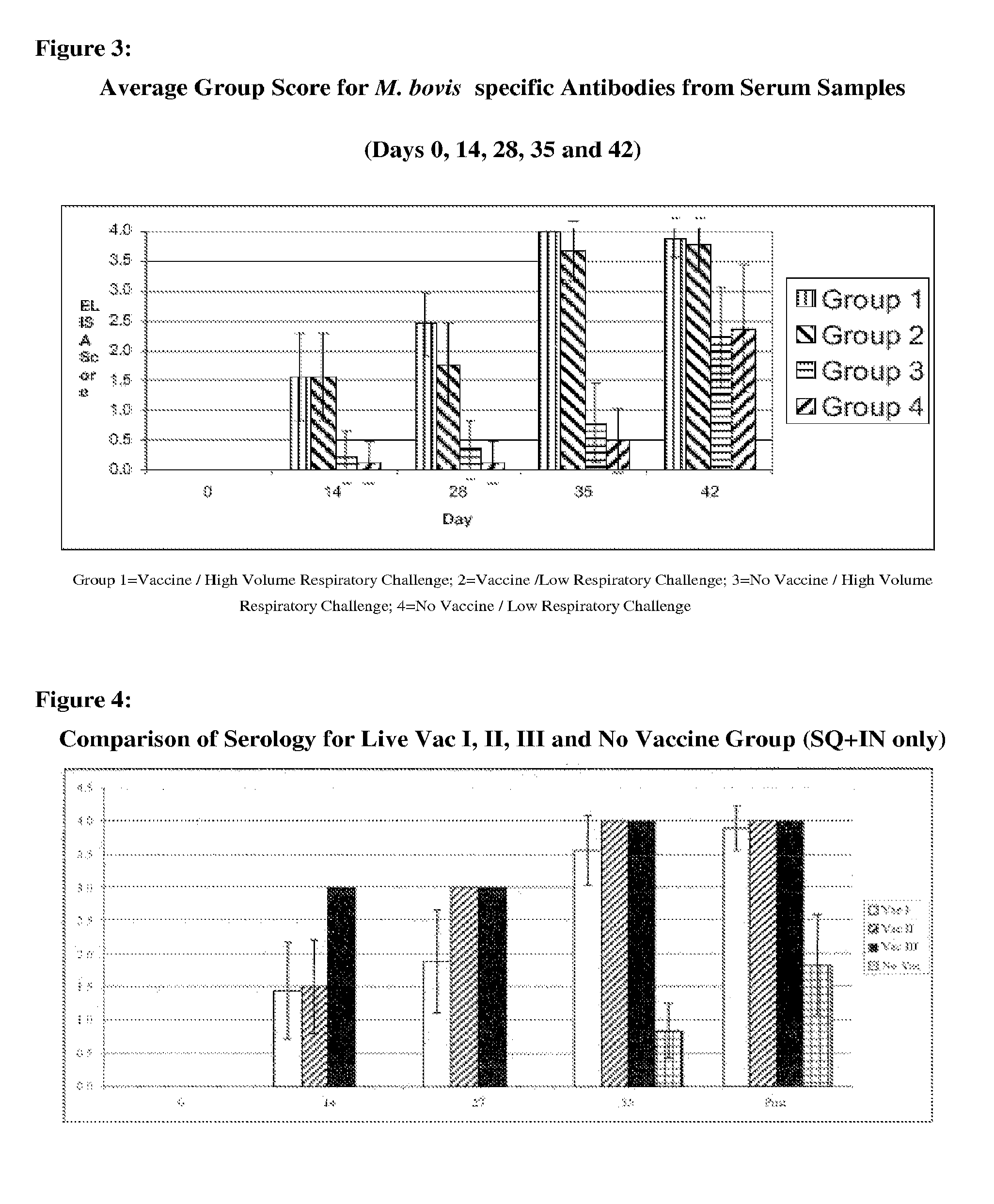
[0211]This Example assessed the efficacy of an experimental live M. bovis vaccine using two different challenge models in a target species.

Materials and Methods

[0212]Thirty-five colostrum deprived (CD) Holstein calves ranging in age from 4-8 weeks of age were used. All animals met the inclusion criteria, namely that they tested negative for M. bovis and were in good health at the time of the challenge. The calves were first randomly assigned to 1 of 4 groups. Groups 1-3 each contained 9 calves, and Group 4 contained 8 calves. Group 1 and 2 calves were vaccinated with Live Vac 1, which is a culture of M. bovis isolate 052823 passaged 106 times (05-2823-1A-3A P106+) (ATCC Designation No. PTA-8694) while Group 3 and 4 calves were vaccinated with media only. The vaccine isolate for Groups 1 and 2 was obtained from naturally occurring disease outbreak and then serially passaged 106 times in M. bovis appropriate media. The culture was grown 24±2 hours at 37° C. after inoculation with an a...

example 2

[0230]The purpose of this investigation was to determine the safety and efficacy of three live vaccine candidates.

Materials and Methods

[0231]The calves used for the study were 6±2 weeks of age and were divided into 6 groups. As shown in Table 7, group 1 was comprised of 10 animals, which were vaccinated with M. bovis Live Vaccine I on day 0 (D0) and D14 of the study. Group 1 was vaccinated with 2 ml subcutaneously and 2 ml intranasally on D0 and D14. Group 2 was comprised of 10 animals and was vaccinated with 2 ml of M. bovis Live Vaccine I subcutaneously on D0 and D14 of the study. Group 3 was comprised of 9 calves vaccinated with M. bovis Live Vaccine I on D0 and D14. Group 3 was vaccinated with 2 ml intranasally. Group 4 was a control and was not administered any vaccine. Group 5 was comprised of 2 calves that were vaccinated with M. bovis Live II vaccine. Group 5 was vaccinated with 2 ml subcutaneously and 2 ml intranasally on D0 and D14. Group 6 was comprised of two calves that...

example 3

[0247]This example describes the DNA fingerprinting process used to differentiate M. bovis strains by isolating, amplifying and detecting DNA.

Materials and Methods

[0248]Mycoplasma sp. isolates were used in the studies. Isolates were obtained from in-house sources or field isolates obtained from infected animals. Isolates were grown using a combination of Mycoplasma-selective agar and broth for 1-7 days. To isolate DNA, broth cultures were spun and pelleted. DNA from the pellet was then extracted (using the Qiagen DNeasy Tissue Kit and resuspended in molecular grade water). Genomic DNA was quantitated using Picogreen (Invitrogen). Primers were designed based on the known insertion sequences (transposable elements) present in the bacterial genome (Mycoplasma bovis). Outwardly facing primers were manually selected from the element ends (excluding the terminal repeat regions) at a Tm of 55-58 C. PCR reactions were then carried out using a multiplex PCR master mix (Qiagen Multiplex PCR K...


PUM

PropertyMeasurementUnit
Timeaaaaaaaaaa
Volumeaaaaaaaaaa
Volumeaaaaaaaaaa

Abstract

The present invention relates to combination vaccines and / or the combined use of immunogenic compositions for the treatment and / or prophylaxis of cattle against microbiological infections, wherein the infections are caused by M. bovis and at least one further cattle relevant pathogen. The combination vaccine as described herein comprises at least one M. bovis antigen, preferably the attenuated, avirulent M. bovis as provided herewith and one or more further immunologically active components effective for the treatment and / or prophylaxis of infections caused by a further pathogen of cattle.

Description

SEQUENCE LISTING[0001]This application contains a sequence listing in paper format and in computer readable format, the teachings and content of which are hereby incorporated by reference. The sequence listing is identical to that incorporated in WO2008 / 030619.BACKGROUND[0002]Mycoplasma bovis (M. bovis) is considered to be one of the more pathogenic species of Mycoplasma and causes significant economic losses worldwide. Mycoplasmas cause severe clinical signs in cattle of all ages. M. bovis is the most frequent Mycoplasma pathogen found to cause pneumonia, mastitis, and arthritis in cattle and its etiological role has also been associated with otitis, keratoconjunctivitis, synovitis, and reproductive disorders in cows and bulls. In general, Mycoplasmas are difficult to treat since they lack a cell wall or membrane, which tends to make them resistant to several classes of commonly used broad-spectrum antibiotic treatments. Mycoplasmas differ from viruses in that Mycoplasmas are large...

Claims


Application Information

Patent Timeline
IPC IPC(8): A61K39/295A61K39/116A61P37/04A61P31/22A61P31/12A61P31/14A61P31/20A61K39/02A61P31/04
CPCA61K39/0241A61K2039/522A61K2039/54C12R1/35A61K2039/545A61K2039/552C12N1/36A61K2039/543A61P1/04A61P31/00A61P31/04A61P31/12A61P31/14A61P31/20A61P31/22A61P37/04C12R2001/35C12N1/205
Inventor BECK, MICHAELHAYES, PHILLIP WAYNEKNITTEL, JEFFREY P.
Owner BOEHRINGER LNGELHEIM VETMEDICA GMBH
Learn More