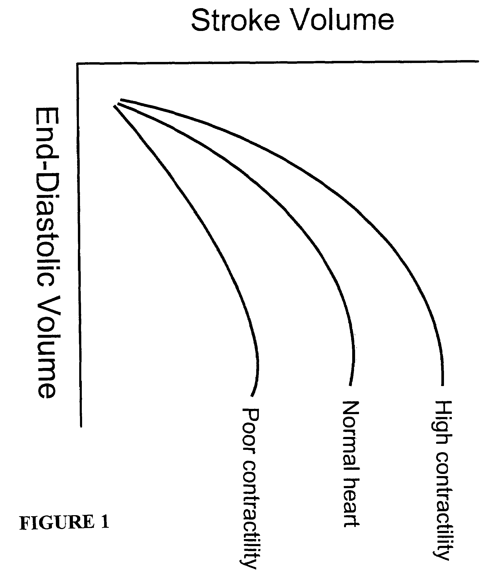
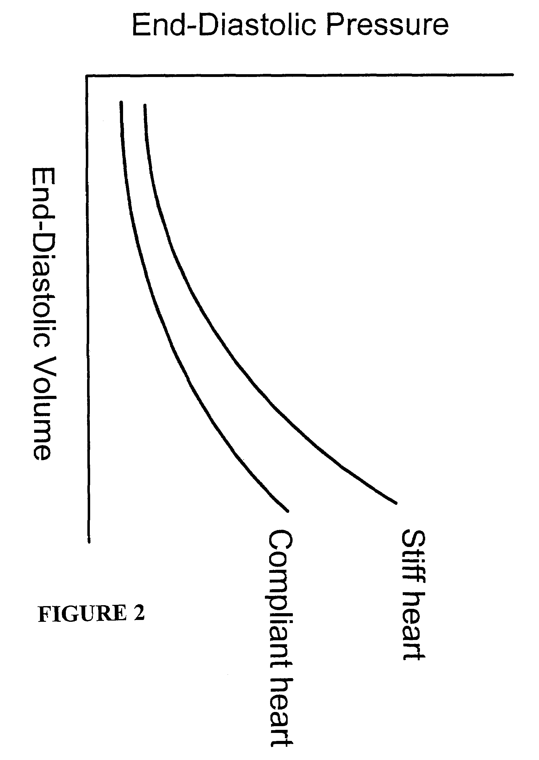
[0047]The
clinical diagnosis of diastolic dysfunction is relatively easy if a
pulmonary artery catheter is placed. If the patient has normal left
ventricular function, yet the pulmonary
artery catheter reveals a high
left atrial pressure, then the diagnosis is confirmed. However, most clinicians are unwilling to place a pulmonary
artery catheter, so a great deal of effort has been made to estimate
left atrial pressure using echocardiography. At present, all the techniques that have been proposed have met with very limited success. Certainly there is no
consensus on how to diagnose diastolic dysfunction with echocardiography alone. The ability to make determinations of cardiac properties and parameters noninvasively, such as cardiac tissue stiffness and contractility, however, makes the diagnosis of diastolic dysfunction trivially easy (and non-invasive) because wall stiffness in late
diastole reflects
left atrial pressure.
[0055]Methods and systems of the present invention provide determinations of
strain rate as the rate of change of strain, measured directly using non-invasive
ultrasound techniques over time.
Strain rate is measured, not as bulk movement of
myocardial tissue but, rather, as relative movements of selected target sites within
myocardial tissue. And, because the time of peak strain rate is evanescent, improvements in accuracy of peak strain measurements provided using methods and systems of the present invention, reduce the amount of necessary
time averaging of the
signal, and improve the
cycling rate of the measurement. Both passive and active
modes of the present invention may be implemented to determine strain in myocardial tissue. Moreover, the improved accuracy, non-invasiveness and cost-effective attributes of methods and systems of the present invention permit use of strain and strain rate measurements for monitoring myocardial contractility and tissue properties, as well as diagnosis of myocardial dysfunction.
[0057]Tissue
Doppler ultrasound techniques have been used to detect
myocardial ischemia, primarily in experimental situations that involve severe
ischemia and consequent impairment of systolic dysfunction.
Strain rate patterns change dramatically with the onset of
ischemia, characterized by a delayed onset of (contraction) strain rate, decreased peak systolic strain rate and strain, post-systolic shortening, and decreased peak strain rate during early ventricular filling. See, e.g., Pislaru C, Anagnostopoulos P D, Seward J B et al.: Higher myocardial strain rates during isovolumic relaxation phase than during ejection characterize acutely
ischemic myocardium, J Am Coll Cardiol 2002; 40:1487–1494. The magnitude of
infarction can be determined as well when the myocardium is exposed to dobutamine. Transmural
infarction (total infarction) is identified by an almost complete absence of strain rate or integrated strain over the
cardiac cycle, whereas incomplete infarction demonstrates reduced strain and strain rate at rest, and progressive post-systolic increases in strain (post-systolic shortening) in response to dobutamine. See, e.g., Weidemann F, Dommke C, Bijnens B et al.: Defining the transmurality of a chronic
myocardial infarction by ultrasonic strain-rate imaging, Circulation 2003;107:883–888. The use of dobutamine stress, in combination with strain
rate analysis, appears to be the best method to assess how much myocardial tissue remains alive after a
myocardial infarction. See, e.g., Hoffmann R, Altiok E, Nowak B et al:
Strain rate measurement by doppler echocardiography allows improved assessment of myocardial viability in patients with depressed left
ventricular function. J Am Coll Cardiol 2002; 39:443–449. Using PET scanning to define the degree of viable tissue in areas of myocardium that had suffered infarction, viable tissue demonstrated increases in strain rate to dobutamine whereas non-viable tissue did not. Prediction of viability from strain rate was better than standard 2-D echo analysis of
wall motion and better than examination of tissue velocities alone.
[0068]The concept that the tension wave can be accurately determined over time, especially during isovolumic relaxation and early
diastole, has the potential to characterize uniquely a wide range of disorders. Constrictive and restrictive pericariditis and cardiomyopathies have distinctive patterns of pressure in the ventricular chamber. If tension follows the same pattern as pressure, then tension measurements could easily accomplish what currently requires invasive measurement. Graded
myocardial ischemia, as opposed to abrupt
total occlusion of coronary
artery blood flow, may first present as diastolic dysfunction and therefore precede ECG changes and even changes in
systolic function. Thus, if a
system can monitor both diastolic and
systolic function, that
system has the most chance of detecting
ischemia early and provide the physician a greater opportunity for intervention before the condition worsens. As with all applications, assessment of
diastolic function may be accomplished using either the passive or active ultrasound
modes of the present invention, or both
modes simultaneously or alternately.
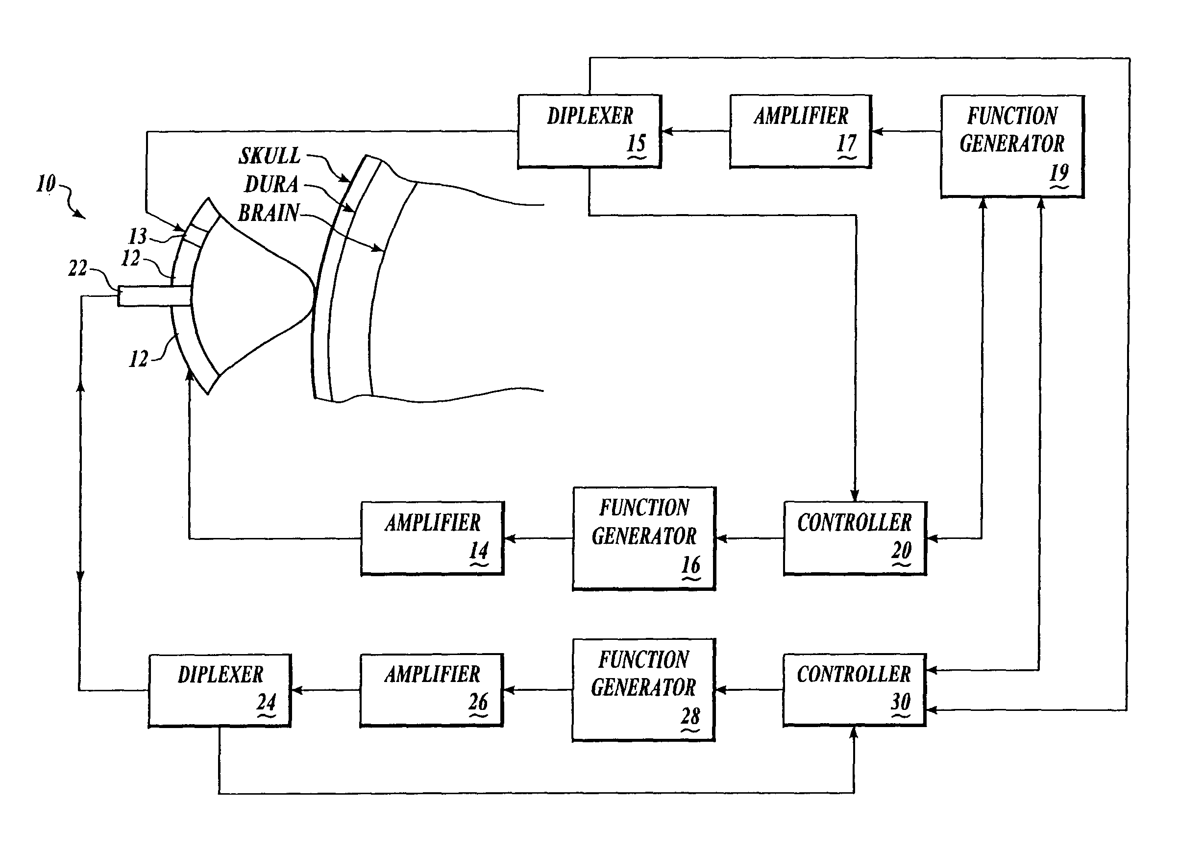
[0072]Single or multiple interrogation signals administered from different places and / or at different times may insonify single or
multiple target tissue sites. Intrinsic and / or induced acoustic properties of the insonated
target tissue may be assessed, by acquiring scatter or emission data, simultaneously and / or sequentially. One of the advantages of the methods and systems of the present invention is that
target tissue sites may be volumetrically small, and
spatially resolved, to provide data from localized tissue sites with a high degree of spatial resolution. In this way, localized differences in tissue properties may be identified and associated with a spatial location within the interrogated tissue. According to one embodiment, tissue sites of varying size and / or location are assessed simultaneously or sequentially. For most applications, the use of acoustic source(s) and / or
transducer(s) capable of interrogating and detecting
target tissue sites having a volume of from 1 mm3 to 100 cm3 are suitable.
[0085]Following the (optional) environmental assessment, an acoustic force is applied by an acoustic
transducer, at a predetermined frequency, to displace targeted cardiac tissue at a targeted location. The deformation may be produced at any desired location within cardiac tissue, depending on the focus (foci) of the ultrasonic
transducer(s) producing the
acoustic radiation force. In some systems, variable foci ultrasonic transducers are provided, and a diagnostic procedure is carried out using a plurality of target tissue sites. According to one embodiment for assessment of cardiac output, the focus (foci) of the ultrasonic transducer(s) is preferably provided in proximity to the surface or a small distance below the surface of a
ventricle wall, to maximize the tissue displacement induced by the
radiation pressure that arises from the impedance mismatch between cardiac tissue and fluid.

