

Methods for the diagnosis and treatment of bone disorders
a bone disorder and bone disease technology, applied in the field of metabonomics, can solve the problems of fracture, bone pain, bone deformity and abnormalities of calcium and phosphate homeostasis, and weak mechanical strength of scurvy tissue, and achieve the effect of reducing the risk of fracture, and improving the quality of li
- Summary
- Abstract
- Description
- Claims
- Application Information
AI Technical Summary
Benefits of technology
Problems solved by technology
Method used
Image

Examples
example 1
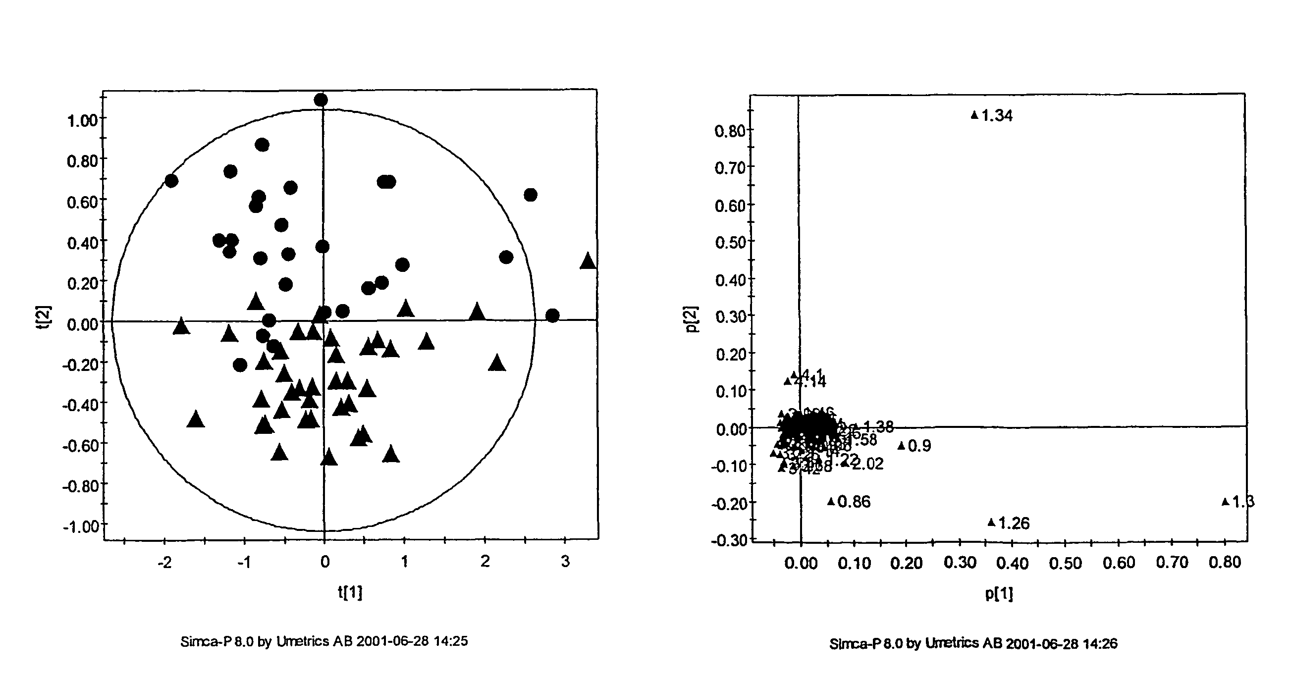
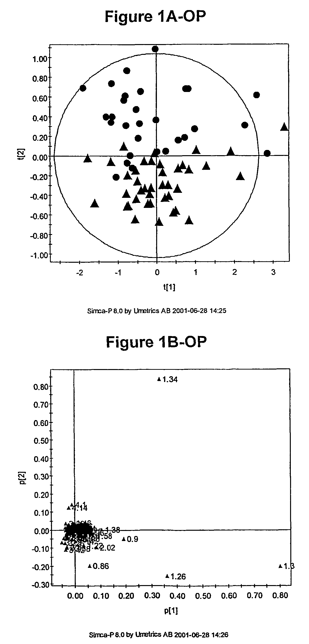
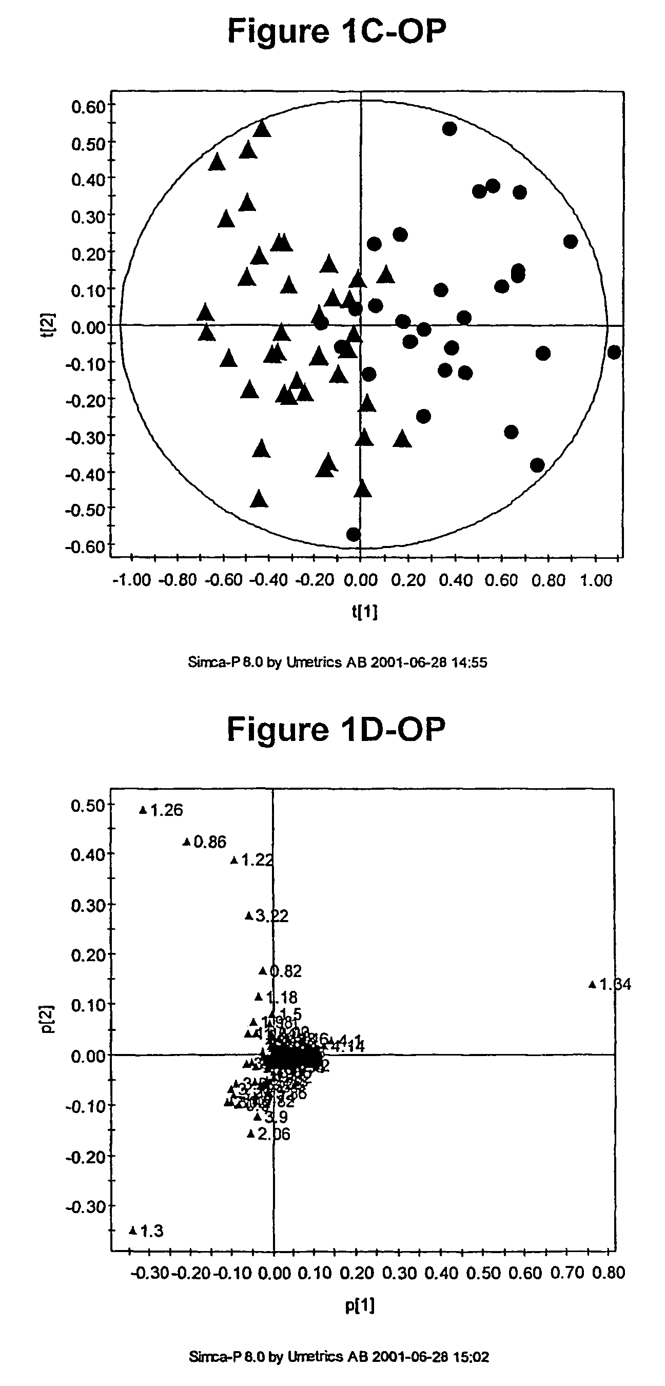
[0789]As discussed above, the inventors have developed novel methods (which employ multivariate statistical analysis and pattern recognition (PR) techniques, and optionally data filtering techniques) of analysing data (e.g., NMR spectra) from a test population which yield accurate mathematical models which may subsequently be used to classify a test sample or subject, and / or in diagnosis.
[0790]These techniques have been applied to the analysis of blood serum in the context of osteoporosis. The metabonomic analysis can distinguish between individuals with and without osteoporosis. Novel diagnostic biomarkers for osteoporosis have been identified, and methods for associated diagnosis have been developed.
[0791]Briefly, metabonomic methods were applied to blood serum sample for subjects in an osteoporosis study. Biomarkers, including free proline, were identified as being diagnostic for osteoporosis. Subsequently, proline levels were used to classify (e.g., diagnose) patient...
example 2
[0826]As discussed above, peak assignment in the NMR study described above suggested that proline is particularly significant for distinguishing subjects with osteoporosis from subjects with normal bone mineral density.
[0827]This finding was confirmed using a novel high-throughput microtitre format assay for proline (also developed by the inventors) to the same serum samples used in the NMR study (subjects diagnosed with osteoarthritis were excluded from the data analysis). The independent biochemical assay data confirms that the differences in the NMR spectra attributed to proline are in fact due to proline.
[0828]Without wishing to be bound to any particular theory, the inventors postulate that low serum proline is associated with low bone mineral density through a causal link whereby proline deficiency slightly but significantly decreases the rate of synthesis of collagen, the key structural protein in bone.
[0829]The data are summarised in the following ...
example 3
Further Application of Isatin Assay
[0833]In an independent study of 865 women with OP (using the same WHO definition of osteoporosis as in the first study) and 612 women with normal bone mineral density, serum proline was found to be lower among women with osteoporosis, specifically, 211±4 μM in OP versus 252±3 μM in control subjects (p<0.001 Student's unpaired t-test).
[0834]Further analyses of these cohorts indicate that serum proline concentration is correlated with bone mineral density, even among healthy controls (r=0.271; p<0.0001 Spearman's rank correlation coefficient, versus lumbar spine bone mineral density from DEXA scan).
[0835]A similar correlation was seen with femoral neck bone mineral density (r=0.202; p<0.0001).
[0836]However, low serum proline was not significantly associated with the presence of clinical fracture (213±16 μM in those with fracture compared to 229±8 μM in those with OP defined by low bone mineral density but no fracture).
PUM
| Property | Measurement | Unit |
|---|---|---|
| time | aaaaa | aaaaa |
| time | aaaaa | aaaaa |
| time | aaaaa | aaaaa |
Abstract
Description
Claims
Application Information