
Self-expanding valve for the venous system

a venous system and self-expanding technology, applied in the field of self-expanding valves, can solve the problems of varicose veins, blood stagnation, damage to more distal valves, etc., and achieve the effects of removing solubilized cell membranes

Inactive Publication Date: 2009-03-05
QUIJANO RODOLFO C +1
View PDF11 Cites 46 Cited by
  • Summary
  • Abstract
  • Description
  • Claims
  • Application Information

AI Technical Summary

Benefits of technology

[0019]The geometry of the free margin or coapting edge of the valve can be approximated with a similar catenary equation. Thus, in this manner the shape of the venous valve is defined and experiments suggest that the valve meets the desired specifications in flow control, allowing quasi-laminar flow to pass through the valve, minimizing turbulence that is deleterious and leads to thrombus formation, and providing ample coaptation to ensure the competency of the valve under conditions of diameter changes in the agger or better described as dilatation of the “annulus” of the venous valve.
[0020]It is one object of the present invention to provide a graft sheet material for use in a venous valve device, wherein the graft sheet material is formed from a segment of connective tissue protein or collagen, and the segment is decellularized and crosslinked with a crosslinking agent resulting in reasonably acceptable cytotoxicity and reduced enzymatic degradation.
[0021]In some aspects, there is provided a biological tissue material or tissue sheet material comprising a process of removing cellular material and lipid from a natural tissue and crosslinking the natural tissue with a crosslinking agent or with ultraviolet irradiation, the tissue material being characterized by reduced antigenicity, reduced immunogenicity and reduced enzymatic degradation upon placement inside or on a patient's body, wherein porosity of the natural tissue is optionally increased. In one aspect, the increase of porosity is adapted for promoting tissue regeneration, when in need. In a preferred embodiment, the natural tissue or tissue sheet material is selected from a group consisting of bovine pericardium, equine pericardium, porcine pericardium, ovine pericardium, caprine pericardium, kangaroo pericardium, fascia lata, dura mater and the like. In still another embodiment, the crosslinked decellularized natural tissue material is loaded with at least one growth factor, at least one bioactive agent, or stem cells / regenerative cells.
[0024]The process for the production of a decellularized tissue or tissue sheet may further comprise dehydrating the decellularized tissue. Alternately, the dehydrating is carried out by soaking the decellularized tissue in glycerol or in glycerol-alcohol mixture (for example, 80% glycerol-20% ethanol). Alternately, the process may further comprise lyophilizing (freeze-drying) the decellularized tissue or tissue patch / sheet in a sterile environment, preferably removing all or substantial amount of the crosslinking agent. Thus, for its use, a reconstitution with specially formulated solutions or simple sterile de-ionized water or saline may suffice to return the material to its flexible, durable, strong, viable state.

Problems solved by technology

If the perforating valves become incompetent, the reverse pressure is transmitted directly to the superficial venous system, reversing the flow, damaging more distal valves, and eventually leading to varicose veins.
The continuous pressure on this area can cause stagnation of blood, and venous stasis ulcers can develop.
Thinning of the dermis ensues associated with poor blood supply that makes the skin very susceptible to trauma.
The smallest scratch will rupture the skin that has little normal blood flow, and the rupture becomes an ulcer that is unsightly, ill-smelling, painful and difficult to heal.
Venous ulcers are notoriously slow to heal; one study showed that 50% of ulcers had been open for one year or more.
An ulcer may heal by various applications of unguents and salves, bandaging and repeated cleaning, thus reverting to the third stage, but it can also progress and give rise to worsening conditions that may necessitate amputation of the limb.
Also it is estimated that all this has as root cause, the dysfunction or destruction of one or more of the valves along the veins of the leg.
This condition if not corrected can be fatal.
These procedures result in some amelioration of symptoms, but recurrence is seen often.
The same experience was not proved successful in the human.
Prosthetic venous valve replacements made of Gore-Tex® PTFE, polyurethane materials have been tried but unfortunately results invariably were suboptimal with thrombus developed quickly, leaflets hardened and incompetence and regurgitation returned.
The infection rate of these materials was also subject of great concern.
The tissue preserving and crosslinking agents used to date, have brought on various serious adverse events to the health of the patients during their use.
The most used fixative and sterilant, glutaraldehyde, has provided excellent preservation of xenogeneic collagen, however, depending on the process conditions used, the residuals present in the tissue even after extensive rinsing prior to implantation, present still undesirable toxicity levels, are irritant to human live tissue, and can induce thrombus formation (clots), hemolysis, and fibrin and protein deposition on the tissue implanted, often precipitating the failure of the device.

Method used


Image

  • Self-expanding valve for the venous system
  • Self-expanding valve for the venous system
  • Self-expanding valve for the venous system

Examples


Embodiment Construction

[0053]The following detailed description is of the best presently contemplated modes of carrying out the invention. This description is not to be taken in a limiting sense, but is made merely for the purposes of illustrating general principles of embodiments of the invention.

[0054]A “tissue” or “tissue material” refers to a material of biological tissue origin that might be decellularized and crosslinked to form or used in a medical device. A tissue sheet, such as a pericardial sheet, is in a sub-group of tissue material (including sheet form and non-sheet form).

[0055]An “implant” refers to a medical device which is inserted into, or grafted onto, bodily tissue to remain for a period of time, such as an extended-release drug delivery device, tissue valve, replacement venous valve, drug-eluting stent, vascular graft, wound healing or skin graft, orthopedic prosthesis, such as bone, ligament, tendon, cartilage, and muscle.

[0056]A “decellularization process” is meant to indicate the pr...


PUM


Abstract

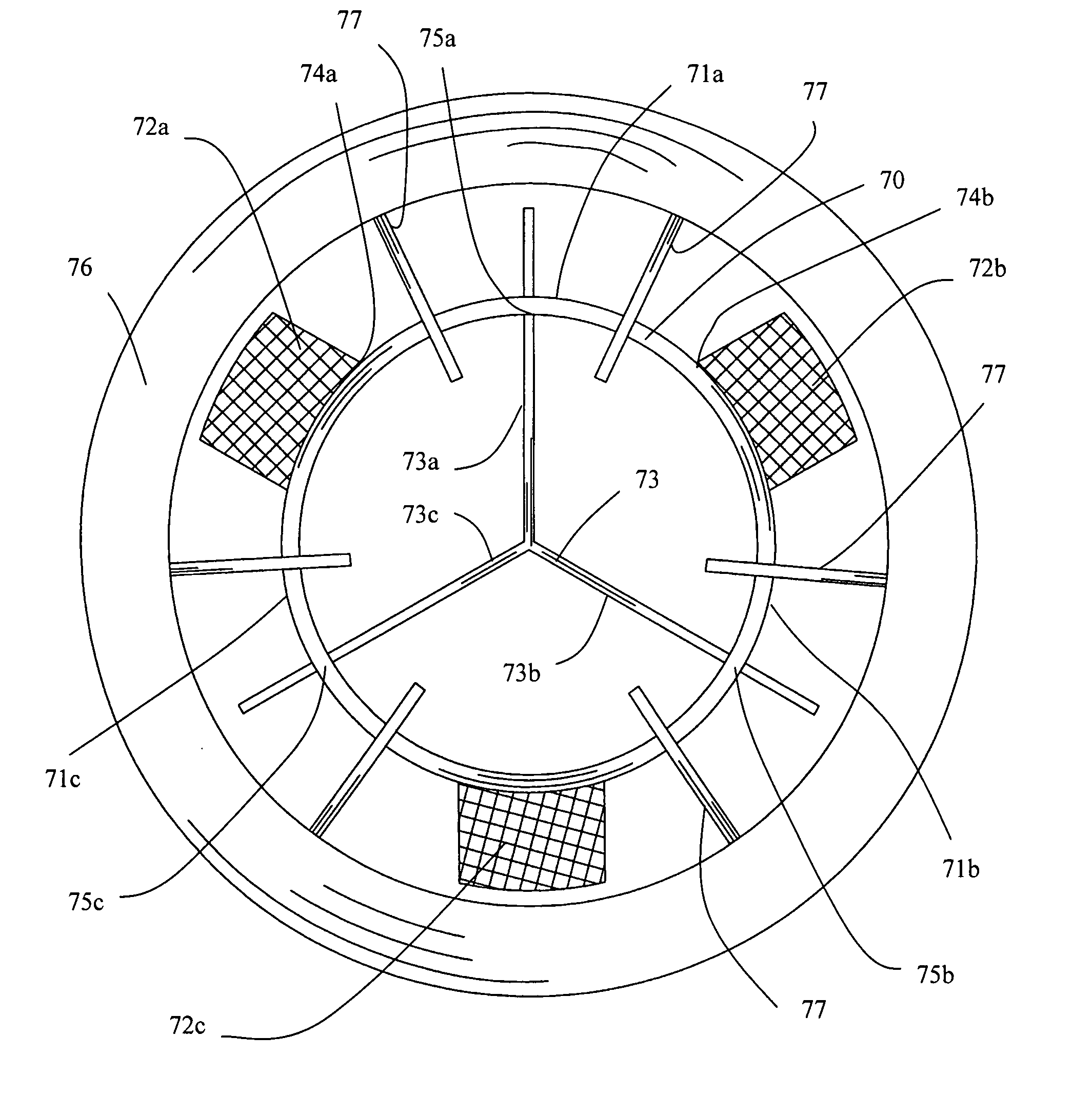
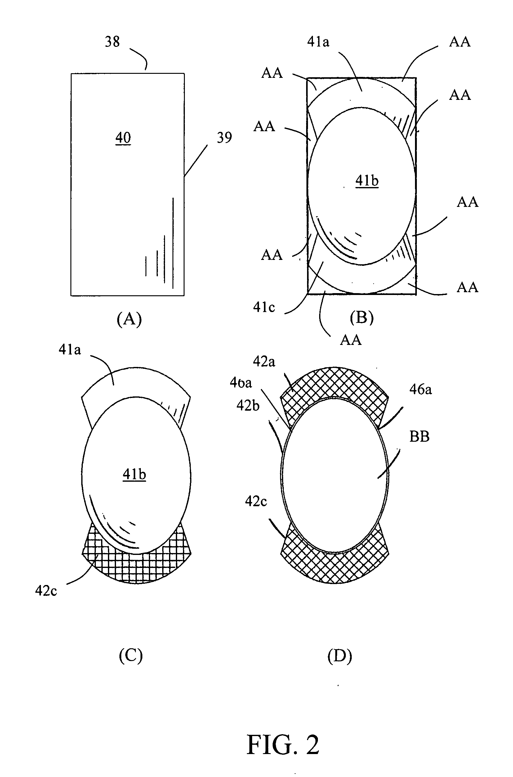
A venous valve device and method of formation are described to provide antegrade blood flow in the deep venous vessels of the leg or in other venous vessels of the body having incompetent or irreversibly dysfunctional valves. The venous valve device is made of a sheet of biocompatible material, comprising a longitudinal wire-frame structure that is a continuous seamless wire loop and plural anchoring mesh-lattice wing members spaced apart and connected to the base of the wire-frame structure.

Description

FIELD OF THE INVENTION[0001]The present invention generally relates to a self-expanding valve to control the flow of blood in any section of the venous system. More particularly, the present invention relates to a medical device having shape-memory alloys frame with cross-linked decellularized pericardial tissue or polymer or Nitinol membrane as a replacement venous valve.BACKGROUND OF THE INVENTION[0002]Various disease conditions of the venous system, from the foot to the groin, can be attributed to partial or total dysfunction of unidirectional valves that are normally found in specific regions of the deep, perforating or superficial veins of the leg. The human leg has both a superficial vein system and a deep vein system that are in fluid communication to each other through a series of perforating veins, each with a perforator vein valve. The superficial short and long saphenous veins carry blood at low pressure and have valves to prevent reversal blood flow, thus maintaining flo...

Claims


Application Information

Patent Timeline
Patent Type & Authority Applications(United States)
IPC IPC(8): A61F2/06
CPCA61F2/01A61F2/2418A61F2230/0095A61F2220/0058A61F2230/0026A61F2/2475A61F2/2415
Inventor QUIJANO, RODOLFO C.TU, HOSHENG
Owner QUIJANO RODOLFO C
Learn More