As will be explained in detail further this kind of environment is far from the
natural environment for a cell meaning that e.g. results of many experiments may misrepresent the natural response of the cell to a particular
drug candidate.
Unfortunately, to date, most of the assay techniques are not particularly successful for the study of these mechanisms.
Firstly, the relative large (>100 μm) size of the standard glass capillaries limits the physiological analogies to the proximal microvascular regions.
Secondly, such studies can only be utilised to study single end-points and cannot be utilised to examine cell choices in migration.
In this regard, studies relating to the effects of chemokines have largely been limited to cellular arrest on adhesion
receptor ligands and have not been extended to the study of cell
crawling.
Although these methods provide semi-quantitative information regarding a
cell type's affinity for a particular substratum, there is no simple method for quantitative characterisation of binding or methods enabling a prolonged study of cell rolling, the ensuing capture by the substratum and subsequent attachment.
Furthermore, direct studies of changes in
cell morphology,
cell growth and biochemical changes cannot be provided easily with these techniques since, determining the
kinetics of attachment and resulting morphological changes requires multiple replicated experiments being analysed at different times.
U.S. Pat. No. 5,998,160 (Berens et al) describes a static assay which, unfortunately, does not have any consideration of cell flow and rolling.
One of the major disadvantages of this and the previous adhesion assays is the geometrical design (
microscope slides and multiple well chambers), which does not at all resemble the
in vivo situation.
It is suggested that one of the disadvantages of the assays described in those specifications is that the
biological process of transmigration through the micropores is difficult to observe due to the geometrical configuration of the apparatus involved.
This obviously leads to difficulties due to optical aberrations.
In effect, the study of the cells morphology changes while transmigrating across the membrane and their subsequent cytoskeletal changes reverting to their former state is a process which is difficult to monitor and
record due to limitations with current techniques. in addition, although it is possible to after experiment parameters following the
initiation of the experiment, such as the introduction of a second. chemoattractant, recombinant or cell-derived, at some specified time after commencing the experiment, it is not possible to distinguish separate effects from each said chemoattractant.
The
pharmaceutical industry has major problems in the
drug screening process and while
high throughput screening (HTS) has been extremely successful in the
elimination of the large majority of unsuitable
drug candidates, it has not progressed significantly beyorid that and usually, after a successful HTS assay, a pharmaceutical company may still have some 10,000 possible drug candidates requiring assessment.
There are other disadvantages such as the transport and subsequent reaction of the drug following its injection into the animal.
Probably the most important
disadvantage is that it does not in any way test in a real situation, drug
efficacy.
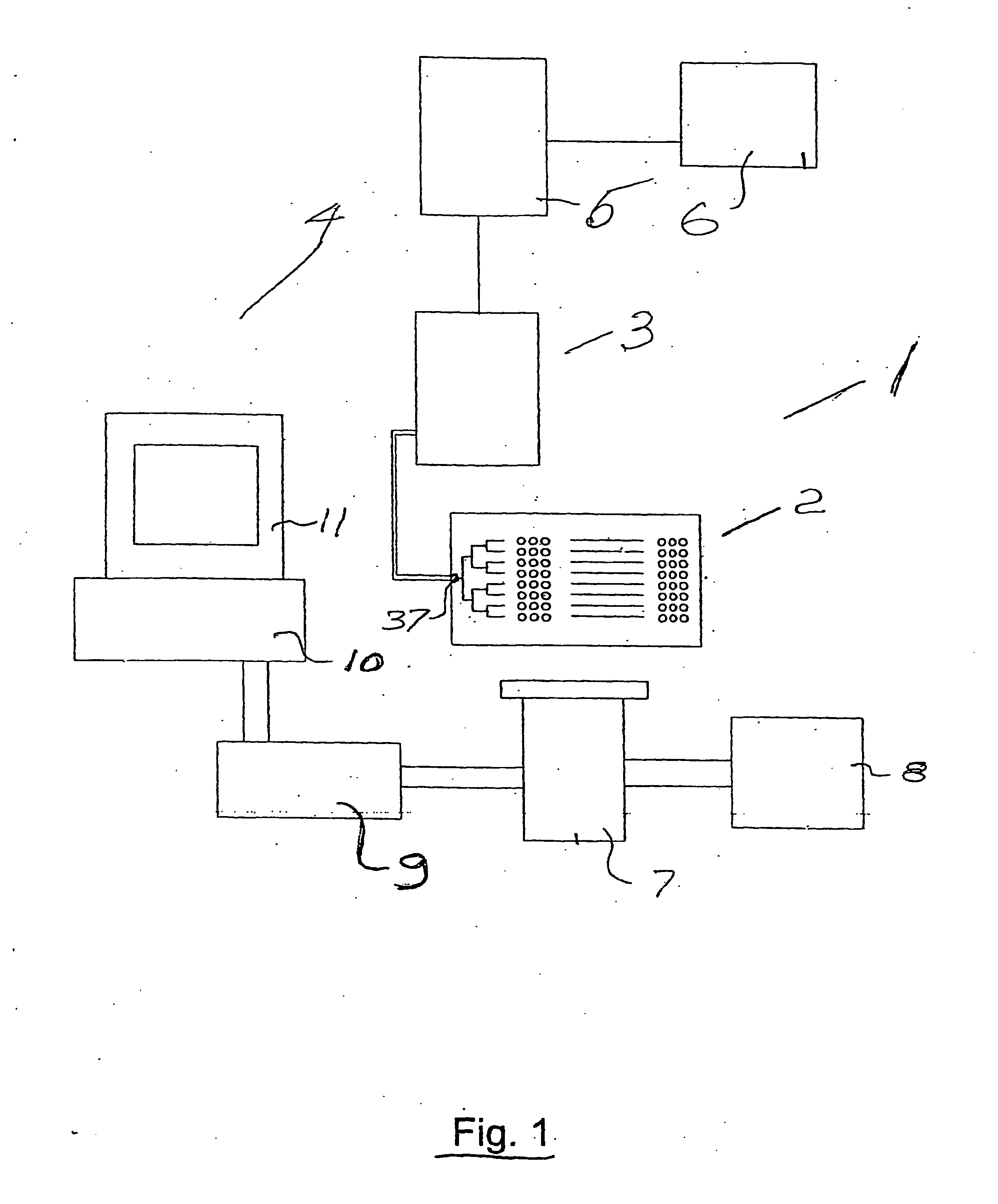
Our investigations to date have not revealed any techniques for performing assays to test the interaction of a large number of chosen compounds with living cells while the cells or compounds mimic the
in vivo situation of
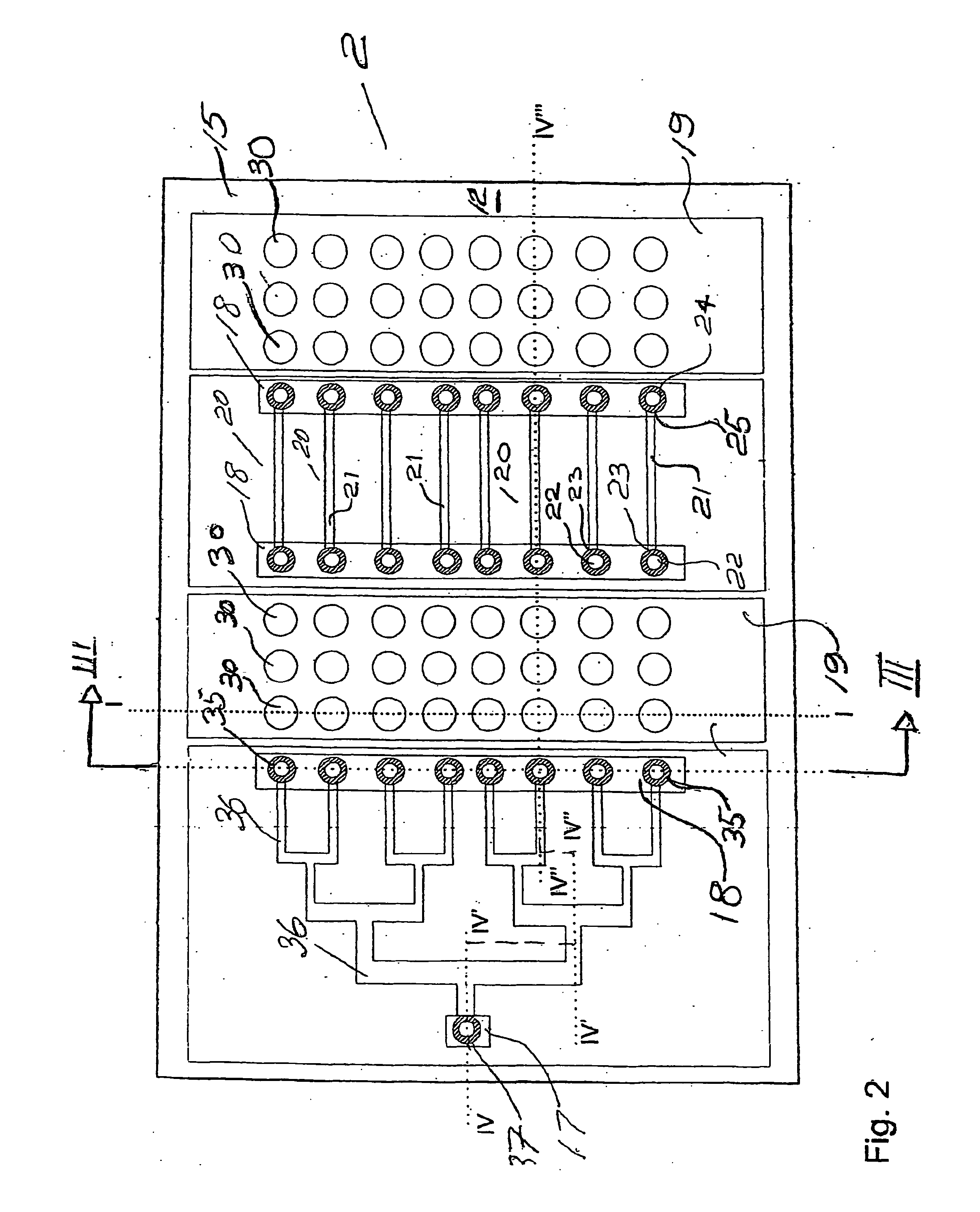
continuous flow.
The
disadvantage of the parallel flow chamber is that it requires significant volumes of the sample for the tests, typically in the range of 100-400 microlitres with dead volumes in the order of millilitres.
The size of the parallel flow chamber is also too large to allow performing a number of experiments in parallel and in a typical configuration only one experiment is performed at a time.
As a result parallel flow chambers failed to become one of key tools in a pharmaceutical company unlike the instruments supporting the
high throughput screening applications.
Arguably, the most fundamental reason why the
cell based assays are currently not performed in the biochip format under the conditions of
continuous flow is that there is no adequate pump that can deliver the low flow rates required. liquid flow in a typical parallel flow chamber is maintained using a
syringe pump meaning that the typical flow rate in the range of 10-100 microlitres / min is achieved.
Therefore, the flow rate of a
syringe pump is far too large for the control of the biochip.
However, although the electroosmotic pump can
handle homogeneous liquids such as
DNA solutions, it is not effective for maintaining the flow of cell suspensions.
The cell suspensions when pumped with electroosmotic pump tend to block the channels and the reproducible pumping rate is difficult to achieve.
There is a further
disadvantage in performing tests using parallel flow chamber.
Therefore, as the biochemical processes are determined by this ratio, the result of the experiment in the parallel flow chamber may misrepresent the result of the in-vivo test.
Although the miniaturisation of the assay devices gives many advantages, the delivery of
low volume samples into the biochip still remains a problem.
Such a handling of the sample is unsuitable for the biochip implementation, when significant
sample volume reduction is required.
Simply scaling down of the parallel flow chamber also brings additional difficulties.
The relatively
large sample volume required to operate the syringe pump hinders further miniaturisation of the parallel flow chamber.
One disadvantage of this method is that the one sample liquid cannot be easily replaced in the microwell reservoir without
contamination with a previously used sample.
Another disadvantage is that during the experiment, which may run up to several hours, e.g. cell culturing, is that such a
low volume of the sample liquid may evaporate from an open microwell reservoir.
Despite a major effort in developing pumping systems for a microchannel structure, the problem still remains.
Many conventionally used pumping systems are operating with significantly bigger volumes of liquids, therefore they cannot provide pumping accuracy or in some cases adequate pumping speed when it comes to establishing flows inside the microstructures with a microchannel
diameter from 5 to 100 μm.
Unfortunately, such an operation results in a large pressure surge which alters the volumetric flow rate.
This invention, however, is directed towards relatively large flow rates of the order of microlitres per minute and would be useful for
drug infusion but would not be particularly suitable for microchannel structures and the like, where the flow rates are, as mentioned already, substantially less.
Unfortunately, heretofore, such syringe and positive displacement pumps have been relatively inefficient at delivering fluid flow at rates of the order of nanolitres per minute, which flow rate is required to transport liquids in microchannel structures.
Generally, the limitation on the flow rate is the movement accuracy of the various mechanical parts of the syringe pump such as the
stepper motor,
plunger, valves, and so on.
Thus, general sealing surface wear makes it impossible to achieve accuracy for shorter displacements.
A further disadvantage of the syringe pump when used for pumping liquids in microchannel structures, is that it cannot deliver a sufficiently low pumping speed for many applications of the structures.
Indeed, one can readily appreciate that at this speed,
visual observation is difficult and further-would not allow for the manipulation or sensing of biological samples.
Thus, heretofore, positive displacements pumps and in particular, syringe pumps, while very attractive for their simplicity, have not as of yet been useful for these applications.
By analysing the state of the art literature, one can conclude that gas / air bubbles are considered generally detrimental to pump performance.
As they are compressible, the accuracy of the volume dispensing is compromised by the presence of such bubbles.
These cations are not in sufficient density to neutralize all the negative charges, therefore a second layer of cations forms.
Since ions are in solution, they drag the whole
buffer solution with them and cause electroosmotic flow.
This makes the control of the
zeta potential and therefore electroosmotic flow control a complicated task.
In some cases this alteration can cause a reverse of electroosmotic flow or its complete cancellation.
Transport of particles in electroosmotic pumping systems is also difficult, due to the fact that during transport they can acquire an electrical charge and can be moved by the
electric field, which in some cases causes the flow to reverse.
When a static pressure is opposed to the electroosmotic flow, the resulting flow can produce a turbulence, which doesn't allow controllable mixing of fluids and biological samples and decreases the speed of electroosmotic flow.
However, when the channel is long, a large
voltage needs to be applied to it, which may be impractical for highly integrated structures.
However, this requires careful placement and alignment of a plurality of electrodes along the channel.
One of common problems that is usually encountered in these two types of liquid pumping
system is the appearance of gas bubbles, which are easily obtained during pumping as a result of
electrolysis.
Pumping of liquids by pumps based on electroosmosis and electrohydrodynamic phenomena relies on the electrical contact throughout the liquid, which disappears in the presence of bubbles rendering pumping by these methods difficult.
Therefore, appearance of gas bubbles inside microfluidic structures poses major problems for such pumps.
Procedures for priming these pumps is complicated and requires a
vacuum pump or special injection devices, to prevent appearance of bubbles in the micropumps main pumping chamber.
Therefore, it is also impractical to use micropumps as a part of disposable microfluidic biochips.
The fluids can only be transported in one direction and no reversed flow is possible.
Control of the flow rate in the individual channels is not possible dynamically, but only by designing a specific geometry of the microchannel structures.
Therefore mixing is only possible with predefined ratios.
This would require an additional increase in the rotation to pump them and therefore would lead to non-reliable experiments particularly in the case of sequentially executed experiments.
Despite several types of pump methods proposed for pumping liquids in the microchannel structures, there is no simple solution, which can be used in many of the applications utilizing microchannel structures.
All methods have some disadvantages, which are more or less significant for different applications.
For example, it's not practical to use micromachined pumps in applications of disposable biochips.
Integration micromachined pumps with a disposable device would increase the cost of it.
In the same example electroosmotic pumps cannot provide a great degree of reliability.
It seems to be impractical when every disposable
chip needs to be treated before an experiment in order to successfully control electroosmotic velocity.
Such bubbles are commonly detrimental to pump's performance.

