However, the methods of determining
cell death and proliferation currently in use all suffer from important limitations.
Some of these limitations make the assays impractical for use in HTS, and also limit their utility in traditional research environments.
However, these processes are slow and lack sensitivity.
These enzymes are typically present in low quantities in most cells, and they do not lend themselves to simple activity assays, making the process of determining cell death cumbersome and insensitive.
Aside from the problems of handling and
waste disposal of radioactive materials, these assays also suffer from various artifacts, and are tedious because of the pretreatment and
recovery steps required.
These assays are useful for examining individual cells, but for quantification of overall
cytotoxicity they are inefficient because each cell must be counted individually, either by laborious microscopic analysis or by very expensive and time-consuming
flow cytometry.
Moreover, some
modes of death (such as complement-mediated
lysis) are not easily assessed by this method, because the
dead cell remains intact for a limited period of time, after which it can no longer be counted because it has disintegrated.
The methods are generally slow and tedious, and thus are not suitable for high-
throughput screening applications.
However, while these methods are useful for qualitative definition of the mode of death, they have no advantages over the ATP-release
assay in quantitative determinations of
cytotoxicity or proliferation.
Proliferation assays are also in common use as indirect
cytotoxicity assays, but there are serious drawbacks with this approach; these are discussed below in connection with the ATP-release
assay.
Metabolically active cells reduce the dyes at rates much greater than quiescent cells; the readout may therefore be a poor reflection of the
cell number.
Metabolism-based assays are not suitable for measurement of
cellular cytotoxicity (for example, the activities of cytotoxic T lymphocytes), or any other assay
system in which live cells other than the target cells are present, because these other cells will yield a substantial and often ill-defined background
signal.
Although they have not been thoroughly characterized with respect to their effects on
cell metabolism, it is known that various agents, such as antioxidants, can interfere with performance of the dyes.
This method is quite accurate, but is extremely tedious and quite expensive.
The labor-intensive aspect of this method is exacerbated by the fact that multiple dilutions of each sample must usually be plated in order to ensure that at least one plate will yield a countable number of colonies.
Although strictly speaking this is a cytotoxicity assay, in that ATP released by dead cells is measured, it is rarely used as a direct cytotoxicity assay, because of the very
short lifetime of
extracellular ATP.
The
ATP content of cells is subject to strong metabolic fluctuations, which will cause artifacts.
Finally, in cytotoxicity mode, the assay suffers from very important drawbacks that are common to all proliferation assays used in this mode.
This leads to the second problem, which is that a direct readout is almost always preferable to a
signal that depends on subtracting two large numbers, as the user must do to use a
proliferation assay to measure cytotoxicity.
Another very important difficulty is a time-consuming problem with this approach which does not involve the actual assay step.
However, if the user is measuring live cells in order to derive the cytotoxicity
signal, then the user must wait much longer, until the cytotoxic effect has had
sufficient time to cause a detectable difference between the
test sample and the control.
Furthermore, the required time interval is not known in advance, and if the experiment is stopped too soon, it must be repeated (or abandoned, since the user will not know whether a result showing no difference between test and control is due to the lack of an effect or insufficient time to show an effect).
This is a serious drawback to the use of any
proliferation assay for cytotoxicity work, including the ATP-release assay.
This method is homogeneous, but requires a 15-minute incubation, and a further 10-minute “dark-adjustment” period before the luminance read; it is therefore too slow for high-efficiency HTS.
This assay method is not suitable for use with other types of cells in general, since most cells do not express
alkaline phosphatase in sufficient quantity.
Moreover, it involves the use of a substrate whose general effects
on cells have not been characterized.
In terms of sensitivity, this assay represents an advance over conventional release assays; however, the disadvantages of this approach are serious.
First,
stable transfection itself is a labor-intensive and expensive procedure; yet this must be done for every target cell line of interest if the method of Schafer et al, is to be used.
Stable transfection does not always work, and, if it does, may alter the metabolic characteristics of the target cell and thereby severely complicate interpretation of the results of the experiment.
The method may not be applicable to cell types outside of these that may be transfected in this manner: expression systems would be different, and the enzymes might be produced in insufficient quantities, in inactive form, or not at all.
Moreover, the assay is not homogeneous.
This in itself is a very serious drawback in the high-
throughput screening environment, since it adds a complex step to the procedure.
Finally, according to the authors,
luciferase had a half-life of approximately 30 minutes under the conditions used, and this was found to be inadequate for quantification of cell death in prolonged assays.
The luminance signal continued to increase with time, a feature which allowed the user to decide when an acceptable signal had been achieved “
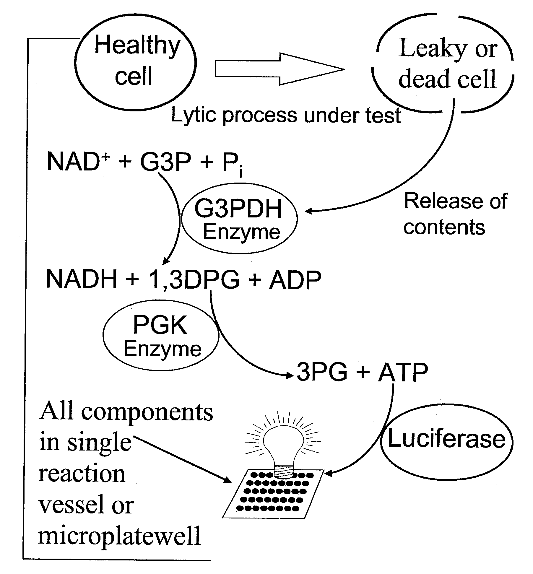
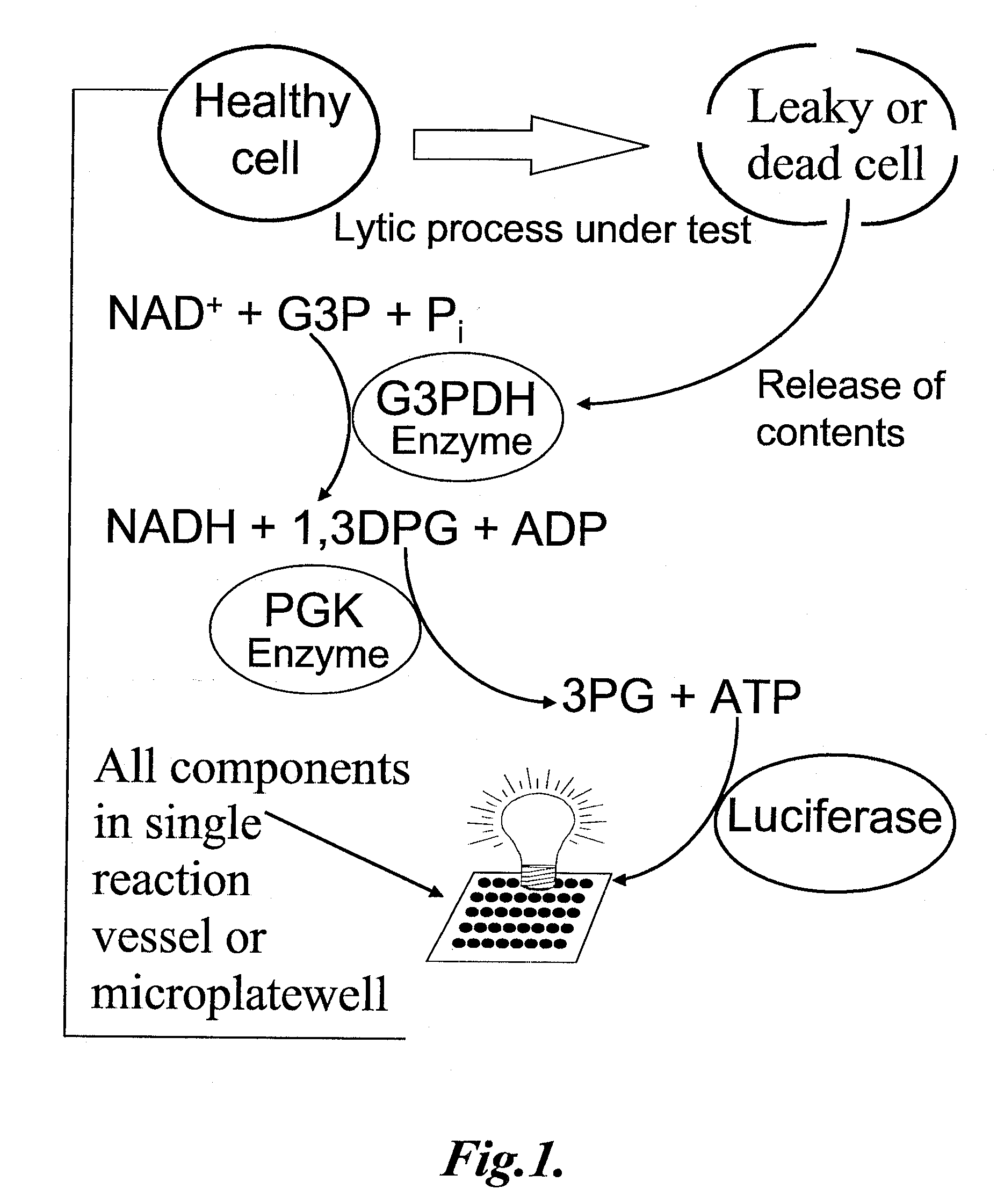
on the fly.” Nevertheless, the GPL assay had its own disadvantages which prevented it from being commercially viable.
It was cumbersome to execute, in that it involved four transfer steps (cocktail to reaction vessel, sample to reaction vessel,
luciferase to luminance vessel, aliquot of reaction to luciferase) and two incubations prior to the actual read.
Moreover, because the assay cocktail was not compatible with live cells, tests involving
bacteria, erythrocytes, or other non-adherent cells or microbes were still more tedious, because the live cells had to be separated from the supernatant by
centrifugation prior to the assay.
These features contributed to the unsuitability of the GPL assay for use in high-throughput screening, especially the necessity of several transfers and the separation of the cells from the supernatant.
It was also of limited utility for research use because of its complexity of operation.
As mentioned above, an important
disadvantage shared by most cytotoxicity and proliferation assays currently available is that they do not permit measurement of both cytotoxicity and proliferation in a
single sample.
In summary, the cytotoxicity and proliferation assays currently available are far from ideal.
The traditional release assays suffer from poor sensitivity and speed,
Metabolism-based assays are slow, inaccurate with respect to actual
cell number, and subject to serious artifacts.
CFU assays are too slow and tedious for routine use.
ATP-release assays are destructive, one-time assays of moderate sensitivity, and they have numerous important drawbacks as cytotoxicity assays.
Although the published coupled luminescent assay (CGPL) is superior to the other cytotoxicity and proliferation assays in many ways, it nevertheless is cumbersome and impractical for use in high-throughput screening or research environments because of the
processing, numerous transfer steps, and lack of a dual cytotoxicity / proliferation mode.
However, assay methods in current use for phosphatases are burdened with a number of drawbacks, including poor throughput or sensitivity, the use of radioactivity, and difficulty of interpretation due to the use of unnatural substrates and / or
reaction conditions.
Poor throughput and / or sensitivity are often due to the nature of the assay; for example, assays utilizing antibodies against phosphorylated target molecules generally require extended incubations, assays making use of electrophoretic separations are too slow to allow the throughput desired, and assays using radioactivity are inherently inconvenient and also suffer from poor throughput.
However, these assays, which generally make use of antibodies or other ligands directed against phosphorylated target molecules for detection of
phosphatase activity, generally require long incubation times for ligand-target association that significantly reduce the value of these assays in high-throughput screening.
These assays also typically involve multiple additions of antibodies or other ligands, and / or wash steps, as well as the design, synthesis, and subsequent ongoing cost of
fluorophore-containing biomolecules or synthetic compounds.
Finally, many FP assays, and other assays which rely on detection of a phosphorylated target molecule, suffer from an additional
disadvantage in that the
phosphatase activity yields a negative signal, i.e., a decrease in the phosphorylated molecule which is the target of detection.
For one thing, several kinds of artifacts can give rise to a negative signal, including
protease contamination or unexpected denaturation of a critical
protein.
Moreover, a negative signal is usually limited in its
dynamic range by its very nature.
Although this method is still in use in research, it is extremely inconvenient, involving the expense of the
label itself, the difficulty and expense of creating or'
purchasing the labeled compound, a separation step, and the danger and tedium of dealing with the radioactive products.
(2001) Anal. Biochem. 298:241), which is quite slow and involves multiple reaction steps, making it unsuitable for high-throughput applications.
(1998) Japanese
Patent Application Number 10121688), but involves multiple mixing steps and the use of immobilized enzymes with flow cells in a portable sampling device, making it unsuitable for a high-throughput screening environment.
In any case this method has never been shown to be compatible with phosphatase activities.
The use of these highly unnatural substrates in high-throughput
screening procedures poses a different set of problems, especially problems of interpretation.
This is even more likely to be the case if the unnatural substrate has a substantially higher Km (Michaelis constant) for the enzyme than the natural substrate, since competitive inhibitors identified in such a
system may successfully compete for the weakly binding unnatural substrate, but may be ineffective against the strongly binding, natural substrate.
Similarly, important inhibitors may not be identified by such a
system, especially if the substrate is smaller, more labile than, or kinetically distinct from the natural substrate.
For example, p-nitrophenylphosphate is a commercially important substrate for
alkaline phosphatase, because it is very labile and yields a colorimetric result, but its use in inhibitor screening applications could lead to
false rejection of good inhibitors.
An inhibitor might be strong enough to exhibit useful inhibition of the natural reaction, but not strong enough to prevent most of this very labile ester from being hydrolyzed.
This could lead to rejection of valuable “hits” in a screening situation.
In short, when the reaction being studied is not the same as the natural reaction that is the desired target, there is a substantial risk that the information gathered will not be biologically useful or relevant.
These methods work only with alkaline phosphatases, and are not readily extensible to other phosphatases, since a new substrate and / or reaction series might have to be designed and synthesized for each phosphatase.
In many or most cases this may be impossible or prohibitively expensive.
Moreover, the methods are not rapid, homogeneous assays, for example, the assay recently reported by Olesen et al. involves 3-4 transfers and at least 2 separate incubations, over a period of at least 30 minutes.
This would make it most inconvenient for a high-throughput setting.
Another serious drawback of these approaches, discussed above, is the use of unnatural substrates.
While it is interesting that
protein phosphatase 2A hydrolyzes this highly unnatural substrate, the rate of
hydrolysis was so poor that the
detection limit was more than 1000-fold worse than by fluorimetric methods (however, these fluorimetric methods also required one hour, involved multiple steps, and required highly unnatural substrates).
While it is unknown whether this work can be transferred to other
protein phosphatases, it is clear that such hypothetical methods, if possible, would likely be insensitive, very slow, and non-homogeneous, and would also make use of unnatural substrates, with all the disadvantages discussed above.
However, current methods of detecting and / or quantifying cAMP have important drawbacks.
Traditional methods involve laborious preparations of extracts and / or radioactive tracers, with many attendant disadvantages.
The “Hit-Hunter” kit offered by Applied Biosystems and DiscoverX is sensitive to concentrations of cAMP in the nanomolar range, but the assay system is extremely complicated, involving
complementation of a proteolytically cleaved galactosidase enzyme by a cAMP-complexed fragment that is usually bound to a
specific antibody, but is released when free cAMP is present.
Thus it may be hard to draw quantitative conclusions about cAMP concentration in this system.
This method is conceptually and biochemically complex, involving components that are expensive to prepare, and like most techniques involving
antibody association or dissociation, it is relatively slow.
This led to a considerable background signal.
However, pyruvate orthophosphate dikinase is not commercially available, and is a complex enzyme that is difficult to
handle successfully.
Purification of the enzyme from natural sources is very laborious, as described in U.S. Pat. No. 5,891,659.
For example,
nitrate is an important component of many fertilizers, and frequently appears as an undesirable contaminant in
groundwater or runoff water from agricultural operations.
However, current methods of detecting
nitrate have important drawbacks.
Colorimetric and
mass-
spectroscopy methods generally require returning the sample to a central laboratory, and therefore have excessive turn-around times. Although some personal and portable instruments exist, these are generally expensive, with limited sensitivity, and some are quite heavy (TL-200 from Timberline Instruments for
ammonia detection, for example weighs 15 kg).
Hach provides the OptiQuant UV
Nitrate Analyzer, which is a continuous method, but is of limited sensitivity, and is very expensive.
The Nico2000 is much more reasonably priced, but has a limit of detection of about 0.3 parts per million, or roughly 5 μM, which is inadequate for many applications.
This electrochemical instrument is highly subject to interference from
chloride and
bicarbonate ions.
Nitrate reductase has been used to reduce
nitrate to
nitrite, followed by calorimetric detection via the Griess reaction; however, for detection of NOS activity, it is necessary to add NADPH, and this interferes with the Griess reaction.
Cayman provides a
Nitrate /
Nitrite Colorimetric
Assay Kit, in which the enzyme
lactate dehydrogenase is provided to consume excess NADPH, but this kit involves multiple steps, and is still subject to the other disadvantages of the Griess reaction.
The Griess method involves the use of dangerous chemicals and requires several steps.
Molecular Probes provides fluorescent methods for detection of
nitrite, but these methods are not suitable for
specific detection of nitrate without at least one additional step.
Methods of measuring LDH activity are also relatively straightforward, although they have generally been slow and unsuitable for high-throughput applications.
If the activity of ACHE is blocked, the body is unable to switch off signals to muscles and other organs, resulting in convulsions and death.
Unfortunately, current methods of assessing ACHE activity and detecting ACHE inhibitors have serious drawbacks.
This assay is too slow and inadequately sensitive for use in high-throughput applications.
Real-time systems can be engineered to detect specific molecules or defined sets of molecules (such as
nerve gases) by
mass spectrometry or other methods based on molecular weight, but these methods are very expensive, require a high degree of expertise to establish, and suffer in any case from the severe limitation that they are limited to detection of particular structures.
If a substance not present in the data-base is encountered, the system has no way of detecting it reliably.
(2004)
Assay and
Drug Development Technologies 2:373-382), but the equipment is very expensive, and the “high throughput” is 4-5 seconds per sample, which cannot compare with the capabilities of coupled luminescent technology-several hundred samples in the
cycle time of a luminometer, which can be as little as 2 seconds.


