Valved stent for chronic venous insufficiency

a valve and venous insufficiency technology, applied in the field of valved stents, can solve the problems of affecting the normal retrograde flow of blood, failure of valves, and malfunction of valves, and achieve the effect of minimizing turbulen

Inactive Publication Date: 2009-10-08
QUIJANO RODOLFO C +1
View PDF0 Cites 34 Cited by
  • Summary
  • Abstract
  • Description
  • Claims
  • Application Information

AI Technical Summary

Benefits of technology

[0018]The invention herein disclosed would overcome major issues and complications that the heretofore proposed devices for the correction of chronic venous insufficiency. It is one object of the invention to provide a valved stent having a specially designed lattice structure, fabricated from one piece tube of temperature sensitive shape memory alloy, incorporating within a very specifically designed geometrical membrane of biocompatible material of specific thickness that will approximate quite closely the configuration of human and animal venous valves. It is very important that the coupling of the specially aggregated lattice members and the membrane shape that is obtained by consideration of the course of some of the members that form the lattice of the stent when fully expanded be accomplished in a very orderly manner for each size or diameter well as length of valve assembled as well as the configuration of valve assembled. Thus, in this manner the shape of the venous valve is defined and experiments suggest that the valve meets the desired specifications in flow control, allowing quasi-laminar flow to pass through the valve, minimizing turbulence that is deleterious and leads to thrombus formation, and providing ample coaptation to ensure the competency of the valve under conditions of diameter changes in the agger or better described as dilatation of the “annulus” of the venous valve.

Problems solved by technology

The veins are very flexible such that if limbs are elevated, all the blood can leave the vein and they collapse into thin ribbon-like flat shape that does not allow suction or siphoning of blood along its length.
These varicosities are often unsightly, and patients complain of aching, tiredness, restless legs, bocturnal cramps and itching, Such symptoms develop because of venous hypertension and are present in about half of the population, although physicians believe there is no relation to varicosities.
There is no evidence at present that demonstrate that early varicose vein surgery will prevent these complications from developing.
Valves may have failed due to thrombosis or by their leaflets becoming stretched or shrunk becoming unable to coapt or appose to each other to provide closure that impedes retrograde flow of blood normally.
Inactivity due to arthritis or paralysis can cause the malfunction of these valves.
The varicose vein may become adherent to the thin overlying superficial skin, and stretched by pressure the dark venous blood will be noticeable through the skin, and be vulnerable to hemorrhage by minor trauma.
Thinning of the dermis ensues associated with poor blood supply that makes the skin very susceptible to trauma.
The smallest scratch will rupture the skin that has little normal blood flow, and the rupture becomes an ulcer that is unsightly, ill-smelling, painful and difficult to heal.
Venous ulcers are notoriously slow to heal; one study showed that 50% of ulcers had been open for one year or more.
An ulcer may heal by various applications of unguents and salves, bandaging and repeated cleaning, thus reverting to the third stage, but it can also progress and give rise to worsening conditions that may necessitate amputation of the limb.
Although a great variety of treatments have been tried for centuries to correct venous insufficiency and mostly the end stages of tissue damage and ulceration, no treatment has ever provided reliable and lasting improvement of the condition.
For centuries, salves and unguents, bandaging and other external applications of a host of medications were the practice, but that ameliorated symptoms only temporarily.
These procedures require extreme expertise, and few physicians are so well trained; pose a great risk of thrombosis and thromboembolism, a frequent event when a vein is dissected or reanastomosed, and an event that can prove to be fatal at times if it progresses to embolize the thrombus in the lung.
The trauma of surgery in venous disease is well known to steeply increase the risk of thrombosis, thromboembolism and pulmonary embolism.
The frame unable to maintain its programmed diameter in the longitudinal direction, namely not having sufficient radial strength to maintain the diameter of the vein as needed along the vein course, narrowed the inflow orifice causing hemodynamic detriment that precipitated thrombosis and failure.
Both of these methods used in the arterial system where pressures and flows are distinctly higher suffer from total proper function, failing often because of peri-valvular reflux, due to incomplete seal by the stent.
Inventions of devices and methods for least invasive treatment of venous insufficiency to eliminate need of open surgical treatment are not as plentiful as those used for correction of arterial problems.
Both the stent and the material fail to provide the function of a venous valve, the stent configuration does not provide stability within a vessel to maintain it parallel to the flow of blood, as it tilts and disturbs the normal flow of venous flow, and allows reflux, allows peripheral or peri-stent valvular leakage or reflux.
The membrane that is not fixed or cross-linked and is configured in an isosceles triangular fashion rather that the geometry of a venous valve, does not provide for the proper fluid vector fields to minimize thrombosis and to be able to withstand the stresses and strains found in the limb venous system.

Method used


Image

  • Valved stent for chronic venous insufficiency
  • Valved stent for chronic venous insufficiency
  • Valved stent for chronic venous insufficiency

Examples


example no.1

EXAMPLE NO. 1

[0066]Tissue Sheet Preparation via Decellularization

[0067]In one embodiment of the present invention, porcine pericardia procured from a slaughterhouse are used as raw materials. In the laboratory, the pericardia are first gently rinsed with fresh saline to remove excess blood on tissue. The cleaned pericardium before delipidation process is herein coded specimen-A. The procedure used to delipid the porcine pericardia is described below: A portion of the trimmed pericardia is immersed in a hypotonic tris buffer (pH 8.0) containing a protease inhibitor (phenylmethyl-sulfonyl fluoride, 0.35 mg / L) for 24 hours at 4° C. under constant stirring. Subsequently, they are immersed in a 1% solution of Triton X-100 (octylphenoxypolyethoxyethanol; Sigma Chemical, St. Louis, Mo., USA) in tris-buffered salt solution with protease inhibition for 24 hours at 4° C. under constant stirring. Samples then are thoroughly rinsed in Hanks' physiological solution and treated with a diluted cho...

example no.2

EXAMPLE NO. 2

[0070]Tissue Sheet Preparation via Crosslinking

[0071]The decellularized tissue (specimen-B) of porcine pericardia are fixed with various crosslinking agent. The first specimen is fixed in 0.625% aqueous glutaraldehyde (Merck KGaA, Darmstadt, Germany) as reference. The second specimen is fixed in genipin (Challenge Bioproducts, Taiwan) solution at 37° C. for 3 days. The third specimen is fixed in 4% epoxy solution (ethylene glycol diglycidyl ether) at 37° C. for 3 days. The aqueous glutaraldehyde, and genipin used are buffered with PBS (0.01M, pH 7.4). The aqueous epoxy solution was buffered with sodium carbonate / sodium bicarbonate (0.21M / 0.02M, pH 10.5). The amount of solution used in each fixation was approximately 200 mL for a 10 cm×10 cm porcine pericardium. Subsequently, the fixed decellularized specimens are sterilized in a graded series of ethanol solutions with a gradual increase in concentration from 20 to 75% over a period of 4 hours. Finally, the specimens are...

example no.3

EXAMPLE NO. 3

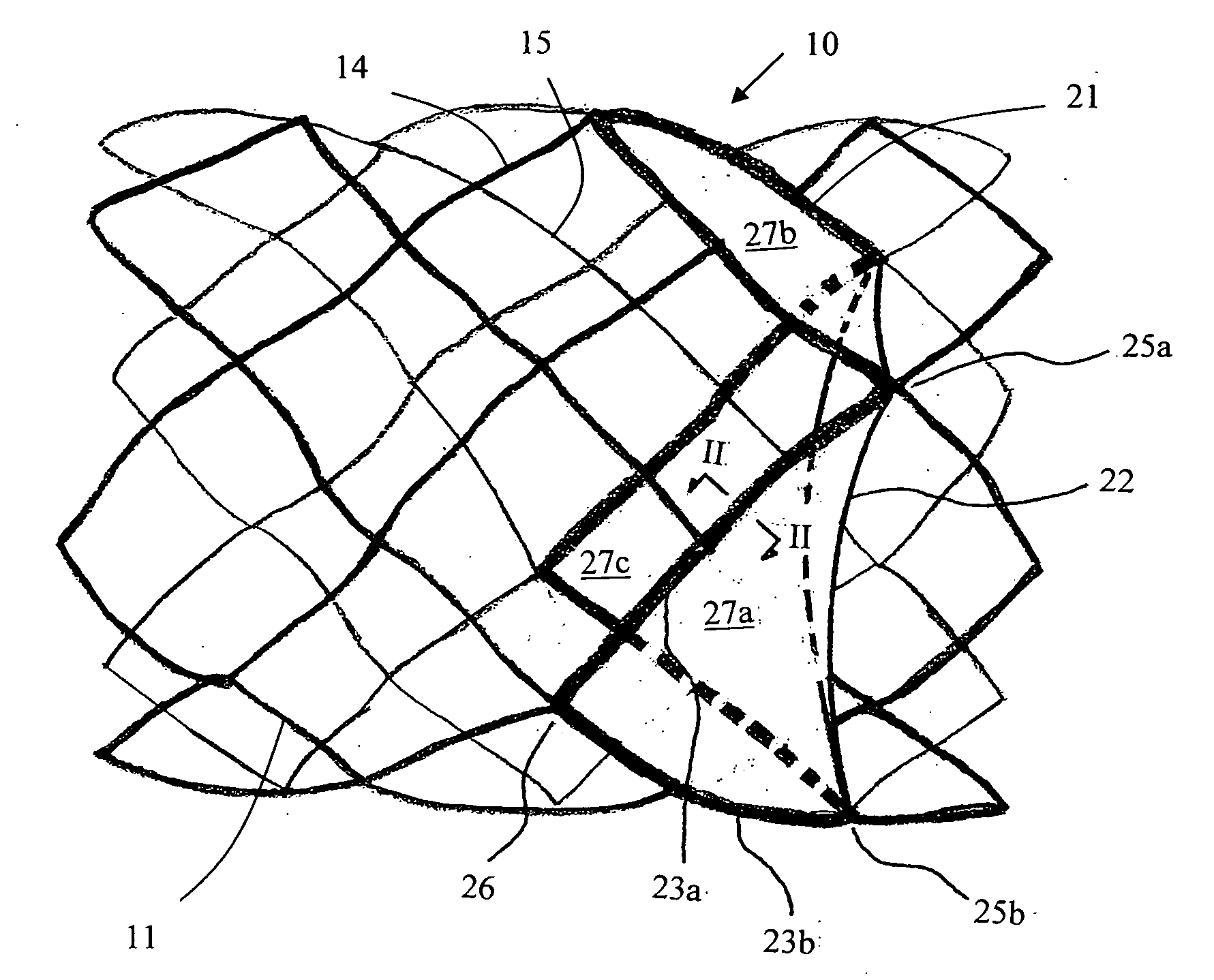
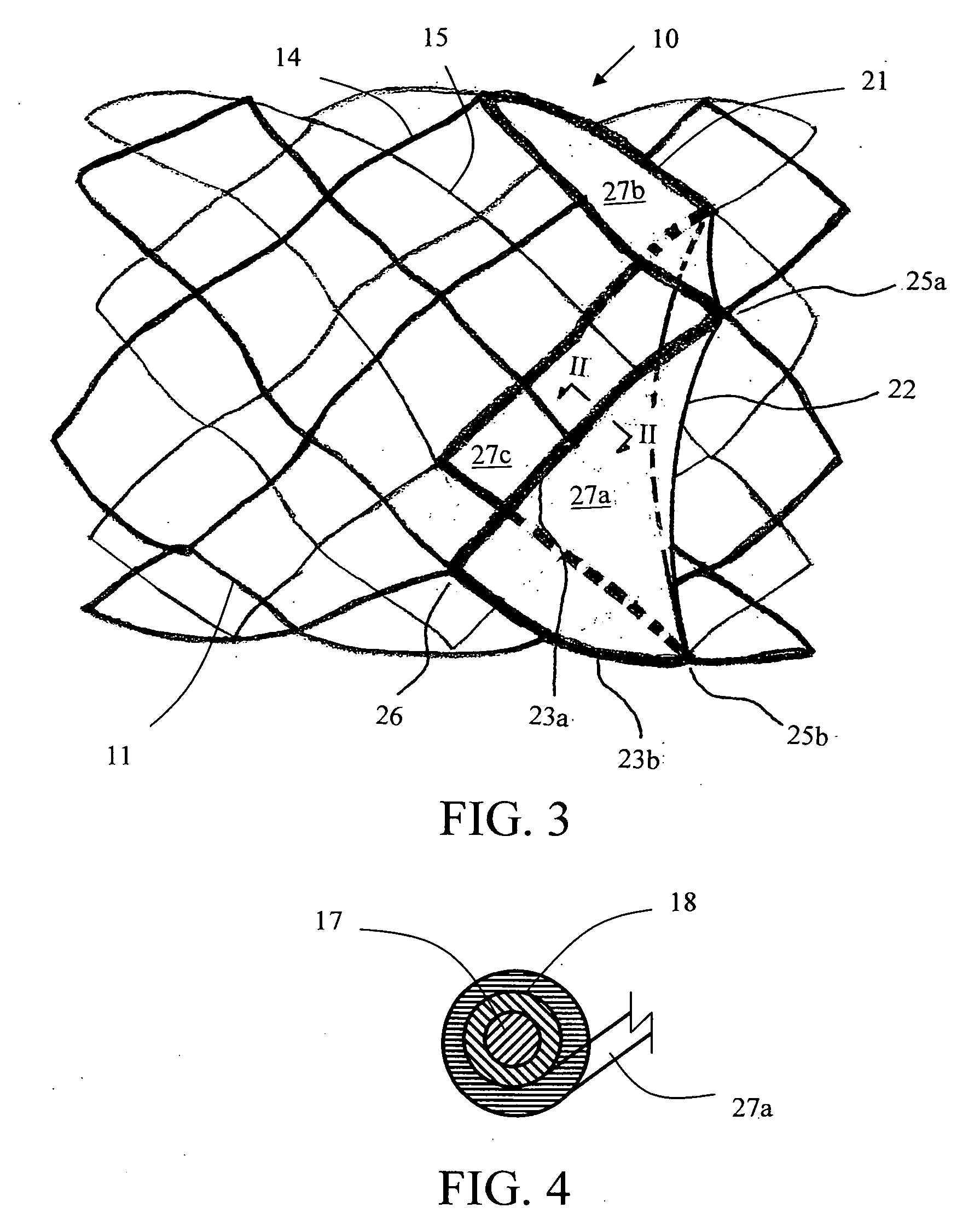
[0092]Tissue Sheet Trimming as a Leaflet

[0093]The valved stent of FIG. 5 shows two margins of attachment (23a, 23b) being sized and configured in a concave shape, the concave-shaped margins of attachment are trimmed or cut from a flat membrane sheet according to illustration of FIG. 8. FIG. 8A shows a trimmed 3-D membrane sheet that is bordered by a free margin (from point 25a to point 25b via point 24), a first margin of attachment (23a) (from point 25a to point 26 following the corresponding spline strut section), and a second margin of attachment (23b) (from point 26 to point 25b following the corresponding spline strut section). At a first specific axial distance upstream from the free margin level (line AA), say H1, the length of chord (line BB) is measured or calculated between the two points on each of the right-spiral and left-spiral splines at that H1 level. There is an angle, θ, between either the right-spiral spline (also known as lattice or strut segment) or...


PUM


Abstract

The invention discloses a valved stent and process of manufacture for treating chronic venous insufficiency having the geometry of the supporting frame and its coupling to the membrane of a specific geometry that provides the valvular mechanism for optimal function. The membrane may comprise a decellularized pericardial tissue via chemical treatment with cholic acid or bile salts and crosslinked.

Description

FIELD OF THE INVENTION[0001]This invention discloses a valved stent for the replacement of absent or destroyed or correction of incompetent venous valves. More specifically, the invention discloses the geometry of the supporting frame and its coupling to the membrane of specific geometry that provides the valvular mechanism for optimal function.BACKGROUND OF THE INVENTION[0002]Being somewhat different in structure than the arteries, veins are specifically designed to allow blood flow in one direction only, toward the heart, and this is only possible by the presence of numerous valves found along the lumen of most veins. Only in the vena cava, the iliac veins and the portal system of veins in the liver valves absent. The valves form an essential part of the pumping system that is returning blood to the heart from the lower limbs against the force of gravity and thus protect the peripheral tissues from the retrograde pressures of the column of blood in the veins when the person is upr...

Claims


Application Information

Patent Timeline
Patent Type & Authority Applications(United States)
IPC IPC(8): A61F2/06
CPCA61F2/2418A61F2/2475A61F2230/0054A61F2220/0075A61F2220/005
Inventor QUIJANO, RODOLFO C.TU, HOSHENG
Owner QUIJANO RODOLFO C
Why Eureka
Try Eureka