Tissue Engineered Cartilage, Method of Making Same, Therapeutic and Cosmetic Surgical Applications Using Same

a technology cartilage, which is applied in the field of tissue engineered cartilage, can solve the problems of less resistance of cartilage generated by seeding a hydrogel or preformed three-dimensional polymeric scaffold to compressive force, joint pain and loss of mobility, and despecification of seeded chondocytes, etc., and achieve the effect of uniform diameter

Inactive Publication Date: 2010-03-11
US DEPT OF HEALTH & HUMAN SERVICES
View PDF0 Cites 26 Cited by
  • Summary
  • Abstract
  • Description
  • Claims
  • Application Information

AI Technical Summary

Benefits of technology

[0012]In other aspects, the nanofibrous polymer supports comprise electrospun polyester polymers which have been approved for use in surgical applications by the FDA or equivalent regulatory agency. In general, electrospinning is a process of producing nanofibers or microfibers of a polymer in which a high voltage electric field is applied to a solution of the polymer. The drawn nanofibers are collected in on a target covering one of the electrodes. By ...

Problems solved by technology

Cartilage defects resulting from aging, joint injury, and developmental disorders cause joint pain and loss of mobility.
Thus, cartilage generated by seeding a hydrogel or preformed three dimensional polymeric scaffold are less resistant to compressive force than nat...

Method used


Image

  • Tissue Engineered Cartilage, Method of Making Same, Therapeutic and Cosmetic Surgical Applications Using Same
  • Tissue Engineered Cartilage, Method of Making Same, Therapeutic and Cosmetic Surgical Applications Using Same
  • Tissue Engineered Cartilage, Method of Making Same, Therapeutic and Cosmetic Surgical Applications Using Same

Examples


example

Isolation and Culture of Bone Marrow-Derived hMSCs

[0146]With approval from the Institutional Review Board of Thomas Jefferson University, bone marrow-derived hMSCs were obtained from the femoral heads of patients undergoing total hip arthroplasty, and processed as previously described (Noth U, et al. J Orthop Res 2002;20:1060-9; Haynesworth S E, et al. Bone 1992;13:81-8; and Wang M L, et al. J Orthop Res 2002;20:1175-84). Briefly, whole bone marrow was curetted from the exposed cutting plane of the femoral neck, washed extensively in Dulbecco's Modified Eagle's medium (DMEM; BioWhittaker, Walkersville, Md.), separated from contaminating trabecular bone fragments and other tissues using a 20-gauge needle attached to a 10-cc syringe, and cultured in DMEM, 10% fetal bovine serum (FBS; Premium Select, Atlanta Biologicals, Atlanta, Ga.) from selected lots (Caterson E J, et al. Mol Biotechnol 2002;20:245-56), and antibiotics (50 μg / mL streptomycin, 50 IU / mL of penicillin; Cellgro, Herndon...

example 2

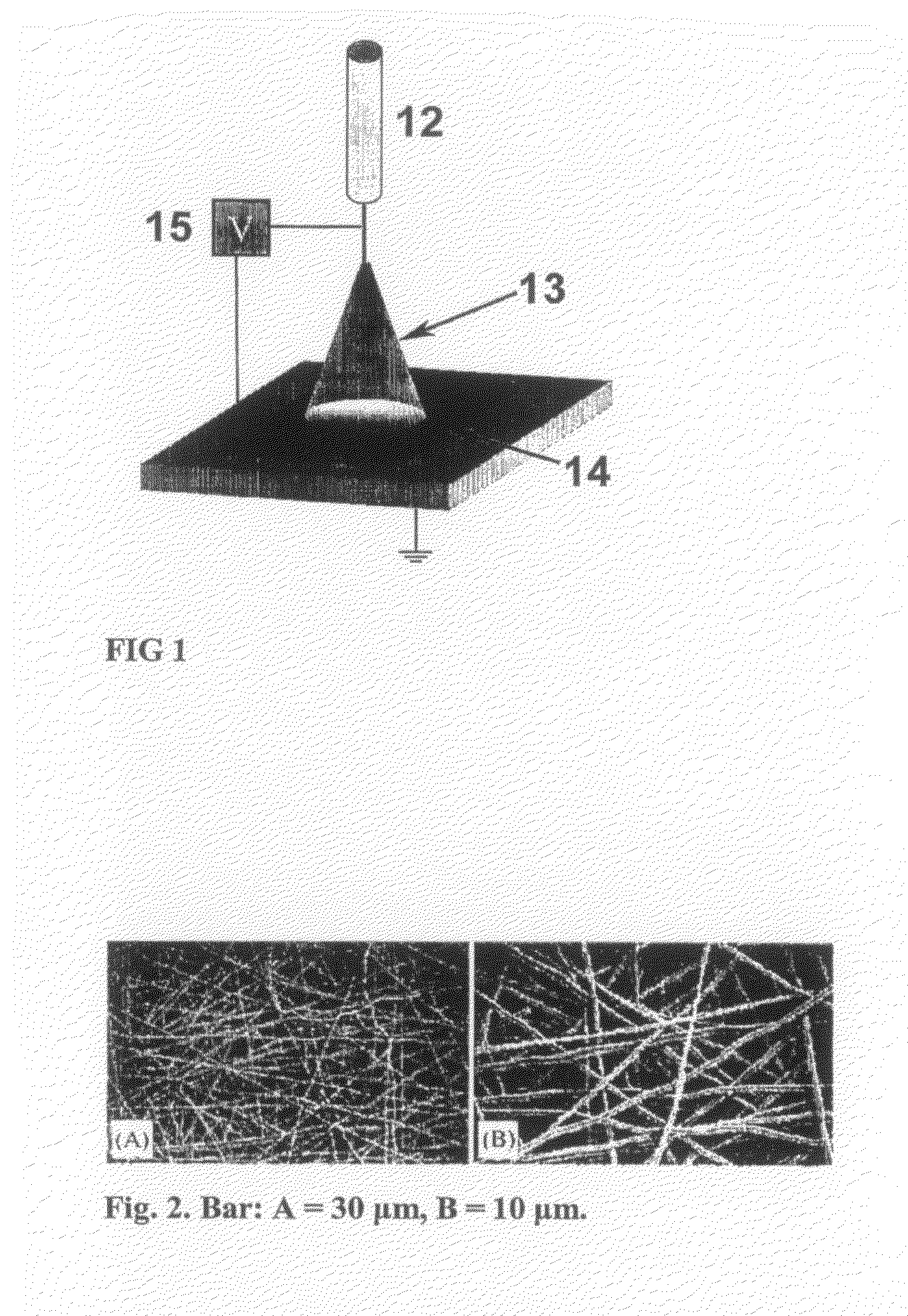
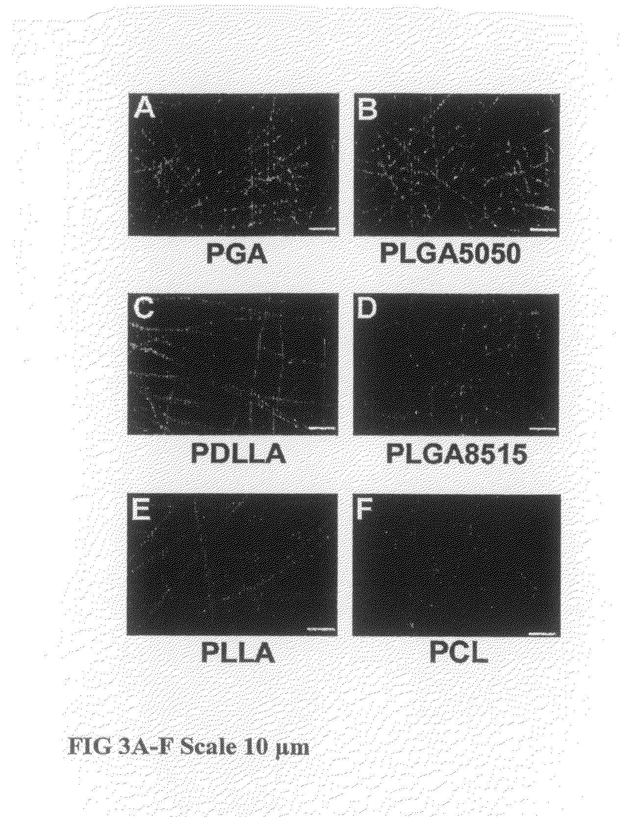
Fabrication of Electrospun Nanofibrous PCL Scaffolds

[0147]Nanofibrous scaffolds were fabricated according to an electrospinning process described previously (Li W J, et al. J Biomed Mater Res 2003;67A:1105-14). Briefly, PCL polymer was dissolved in an organic solvent mixture (1:1) of tetrahydrofuran (THF; Fisher, Pittsburgh, Pa.) and N,N, dimethylformamide (DMF; Fisher, Pittsburgh, Pa.) at a final concentration of 0.14 g / mL. The polymer solution was delivered through the electrospinning apparatus at a constant flow rate of 0.4 mL / h under an applied 0.6 kV / cm charge density, resulting in a 144 cm2 mat with an approximate thickness of 1 mm. To remove residual organic solvent, the non-woven polymer mat was placed within a vacuum chamber for 48 h, and subsequently stored in a desiccator. Prior to cell seeding, squares measuring 10 mm×10 mm×1 mm were fashioned from the electrospun mat, sterilized by ultraviolet irradiation for 30 min per side in a laminar flow hood, and pre-wetted for 24...

example 3

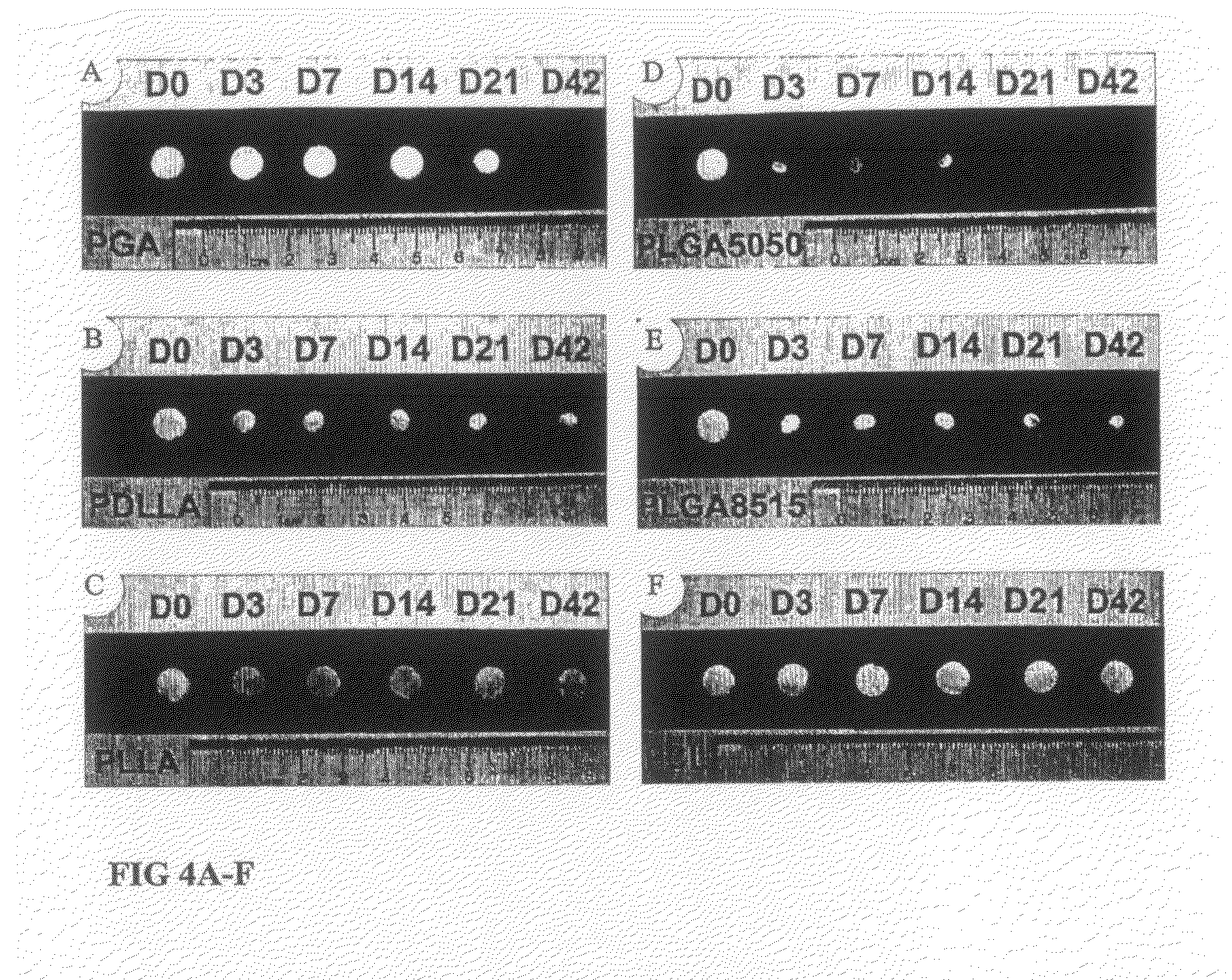
Seeding and Differentiation of hMSCs on PCL Scaffolds

[0148]Pre-processed nanofibrous PCL scaffolds were placed in 24-well tissue culture plates (Corning Glass Works, Corning, N.Y.) coated with 0.3% poly(2-hydroxyethyl methacrylate) (poly HEMA; Polysciences, Warrington, Pa.) to prevent normal cell attachment to tissue culture polystyrene. Cellular scaffolds were incubated at 37° C. for 4 h to allow MSCs to diffuse into and adhere to the scaffold before the addition of 2 mL of culture medium to each well. During the 4 h incubation, 20 μL of serum containing culture medium was applied every 30 min to each cellular scaffold to prevent the constructs from drying. For chondrogenic differentiation studies, 4×10⁵ hMSCs were seeded per PCL scaffold and maintained in a chemically defined medium containing serum-free DMEM, 50 μg / mL ascorbate, 0.1 μM dexamethasone, 40 μg / mL L-proline, 100 μg / mL sodium pyruvate, ITS-plus (Collaborative Biomedical Products, Cambridge, Mass.), antibiotics, and 10 ...


PUM

PropertyMeasurementUnit
Pressureaaaaaaaaaa
Diameteraaaaaaaaaa
Diameteraaaaaaaaaa

Abstract

Cartilage has been constructed using biodegradable electrospun polymeric scaffolds seeded with chondrocytes or adult mesenchymal stem cells. More particularly engineered cartilage has been prepared where the cartilage has a biodegradable and biocompatible nanofibrous polymer support prepared by electrospinning and a plurality of chondocytes or mesenchymal stem cells dispersed in the pores of the support. The tissue engineered cartilages of the invention possess compressive strength properties similar to natural cartilage. Methods of preparing engineered tissues, including tissue engineered cartilages, are provided in which an electrospun nanofibrous polymer support is provided, the support is treated with a cell solution and the polymer-cell mixture cultured in a rotating bioreactor to generate the cartilage. The invention provides for the use of the tissue engineered cartilages in the treatment of cartilage degenerative diseases, reconstructive surgery, and cosmetic surgery.

Description

[0001]The present application claims the benefit of U.S. provisional application No. 60 / 690,988, filed Jun. 15, 2005, which is incorporated herein by reference in its entirety.STATEMENT REGARDING FEDERALLY SPONSORED RESEARCH OR DEVELOPMENT[0002]Research supporting this application was carried out by the United States of America as represented by the Secretary, Department of Health and Human Services.FIELD OF INVENTION[0003]The present invention relates to tissue engineered cartilage comprising a nanofibrous biocompatible polymer support having chondocytes dispersed therein, which has compressive strength equal to natural cartilage, methods of fabricating tissue engineered cartilage by culturing a mixture of stem cells or chondocytes and a electrospun nanofibrous polymer substrate in a suitable bioreactor and methods of treatment comprising implantation of tissue engineered cartilage into a patient.BACKGROUND OF THE INVENTION[0004]Diseased or damaged cartilage has often been replaced...

Claims


Application Information

Patent Timeline
IPC IPC(8): A61L27/38
CPCA61L27/18A61L27/3817A61L27/3852A61L27/3895A61L27/56A61Q90/00A61L2400/12A61L2430/06C08L67/04A61P19/04
Inventor LI, WAN-JU
Owner US DEPT OF HEALTH & HUMAN SERVICES
Why Eureka
Try Eureka