Soft tissue repair material and method for preparing same

A technology for repairing materials and soft tissues, applied in biochemical equipment and methods, tissue regeneration, tissue culture and other directions, can solve the problems of reduced biological activity and the risk of in vivo transplantation of biologically active reagents, and achieves simple methods, low prices, and a wide range of materials. Effect

Active Publication Date: 2018-11-27
SHANDONG EYE INST
View PDF5 Cites 4 Cited by
  • Summary
  • Abstract
  • Description
  • Claims
  • Application Information

AI Technical Summary

Problems solved by technology

Although the decellularization effect has been achieved on the small intestinal submucosa tissue, a large amount of organic solvents such as peracetic acid, methanol, chloroform and methane are used in the decellularization process. These reagents not only cause a certain degree of irreversible denaturation to the biological activity of the tissue but also Residues of reagents are a potential risk for in vivo transplantation
The Chinese p

Method used


Image

  • Soft tissue repair material and method for preparing same
  • Soft tissue repair material and method for preparing same
  • Soft tissue repair material and method for preparing same

Examples


preparation example Construction

[0028] The invention provides a method for preparing a soft tissue repair material, comprising the following steps:

[0029] 1) removing the small intestinal mucosa, muscular layer and serosa on the small intestinal tissue of mammals to obtain the small intestinal submucosa tissue, and washing with sterile buffer solution containing antibiotics for 3 to 5 times to obtain the washed small intestinal submucosa tissue;

[0030] 2) cutting the washed small intestinal submucosa tissue obtained in step 1) into sections not longer than 5 cm, cutting open along the midline, flattening, soaking in sterile buffer solution, and obtaining the pretreated small intestinal submucosa tissue;

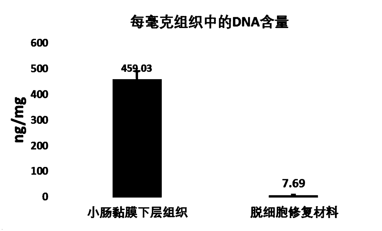
[0031] 3) Place the pre-treated small intestinal submucosa tissue obtained in step 2) in a sterile buffer solution containing 0.2-1% surfactant and 200-600 U / mL nuclease by mass percentage, shake at 37°C for 1- 4h, the decellularized small intestinal submucosa tissue was obtained;

[0032] Described su...

Embodiment 1

[0046] (1) Select the small intestine tissue of healthy pigs, remove the small intestine mucosa, muscular layer and serosa, and repeatedly wash 3 times with a sterile buffer solution containing 100ug / ml penicillin-streptomycin;

[0047] (2) Cut the submucosa tissue of the small intestine into small sections of 5 cm, cut it open from the middle along the midline, and soak it in 250 ml of sterile buffer;

[0048] (3) Place the small intestinal submucosa tissue in a sterile buffer solution containing sodium cocoyl glutamate (1%) and nuclease (200U / ml), and shake it at 37°C for 2 hours;

[0049] (4) Place the small intestinal submucosa tissue in a sterile buffer solution, shake and wash for 10 minutes, and repeat the washing 6 times.

[0050] (5) Exhibit the washed small intestinal submucosa tissue on a sterile stainless steel plate, divide it into small pieces along the natural vein texture, and keep the central matrix membrane part.

[0051] (6) Small intestinal submucosa tissu...

Embodiment 2

[0058] (1) Select 4 healthy male New Zealand white rabbits weighing 3-3.5 kg, and create a 2×2 cm conjunctival defect on the ocular surface of the right eye of each rabbit;

[0059] (2) Rehydrate the soft tissue repair material with normal saline for 1 to 2 minutes;

[0060] (3) Cut it into a patch of the same size as the ocular surface defect under an operating microscope, and suture it at the conjunctival defect.

[0061] (4) Add dexamethasone eye ointment twice a day;

[0062] (5) One week later, the sutures were removed, and the repair of conjunctival defects and the degree of inflammatory response were detected, and sodium fluorescein was added dropwise to detect the epithelial growth.

[0063] result evaluation

[0064] The photos of four New Zealand white rabbits repaired conjunctival defects with soft tissue repair materials for one week are as follows: Figure 5 As shown, among them, Figure 5 a, 5b, 5c, and 5d are parallel comparisons, and are photos of four diff...


PUM


Abstract

The invention relates to a soft tissue repair material and a method for preparing the same, and belongs to the technical field of biomedical material processing. The method includes steps of 1), removing small intestine mucous membranes, muscular layers and serous membranes on small intestine tissues of mammals and carrying out washing by the aid of sterile buffer solution with antibiotics by 3-5times to obtain washed lower-layer tissues of the small intestine mucous membranes; 2), clipping the lower-layer tissues to obtain sections, and splitting and spreading the sections along median linesto obtain pretreated lower-layer tissues of the small intestine mucous membranes; 3), placing the pretreated lower-layer tissues in sterile buffer solution with surfactants and nuclease and carryingout oscillation treatment at the temperatures of 37 DEG C for 1-4 h to obtain acellular lower-layer tissues of the small intestine mucous membranes; 4), carrying out oscillation cleaning, cutting anddrying to obtain dried central basement membranes; 5), carrying out irradiation sterilization to obtain the soft tissue repair material. The soft tissue repair material and the method have the advantage that the safe and effective soft tissue repair material with excellent bioactivity and biocompatibility can be prepared by the aid of the method.

Description

technical field [0001] The invention relates to the technical field of biomedical material processing, in particular to a soft tissue repair material and a preparation method thereof. Background technique [0002] An ideal scaffold material for tissue repair needs to provide a similar microenvironment (including the protein composition, structure, and biomechanical properties of the extracellular matrix) to the autologous tissue extracellular matrix for the regeneration of planted cells or cells in vivo. Due to the complex and delicate microstructure of natural extracellular matrix and the limitations of existing technologies, it is still a difficult problem to use artificial methods to prepare materials that can replace natural extracellular matrix. Using the method of decellularization to remove cellular components from allogeneic tissues and retain the natural extracellular matrix provides a new and effective solution to this problem. [0003] The submucosa of the small ...

Claims


Application Information

Patent Timeline
IPC IPC(8): A61L27/36A61L31/00A61L27/58A61L31/14A61K35/38A61P19/04C12N5/00
CPCA61K35/38A61L27/3629A61L27/3687A61L27/58A61L31/005A61L31/148A61L2430/34A61P19/04C12N5/0062C12N2513/00C12N2533/90
Inventor 史伟云赵龙周庆军
Owner SHANDONG EYE INST
Why Eureka
Try Eureka