
Methods and compositions for repair of cartilage using an in vivo bioreactor

a bioreactor and cartilage technology, applied in the field of cartilage repair, can solve the problems of low porosity of scaffolds, insufficient supply of nutrients to cells, and insufficient support of cell growth, so as to reduce risks, improve the functioning of scaffolds, and reduce the chance of surgical complications and re-interventions.

Active Publication Date: 2007-08-09
SPINALCYTE
View PDF12 Cites 51 Cited by
  • Summary
  • Abstract
  • Description
  • Claims
  • Application Information

AI Technical Summary

Benefits of technology

[0027]It is an exemplary object of the present invention to provide a method intended to repair a degenerated intervertebral disc, e.g. restore intervertebral disc anatomy and improve its functioning. In particular aspects of the invention, there is provided a method to repair damaged disc using a hybrid structure made of an inert containing device intended to feed and differentiate an inner living core. Therefore, the inert structure acts as a delivery system of nutrients and growth factors and as a bioreactor able to differentiate autologous dermal fibroblasts into chondrocyte-like cells. Under mechanical stress (such as intermittent hydrostatic pressure, and / or fluid shear stress), the cells will acquire the characteristics of nucleus cells in the central part and annulus cells in the periphery. Exemplary fibroblast-derived chondrocyte-like cells may be harvested from skin, such as by a biopsy, and then seeded onto three-dimensional polymer scaffold for use of the repair of the disc. This would obviate the need for invasive technique to harvest autologous chondrocytes, in particular aspects. An advantage of certain aspects of the inventive hybrid construct that combines both an inert biomaterial acting as a nutrient-delivery system and living cells easily harvested from skin, for example, is that it is capable of self-maintenance or remodeling and may restore the disc function using a minimally invasive posterior surgical approach, for example.
[0028]In certain aspects of the invention, the damaged cartilage from the joint or intervertebral space is removed and the hybrid structure is installed within the space provided by the foregoing removal. In some embodiments of the invention, the device is implanted using a minimally invasive surgical procedure. In specific embodiments, an exemplary surgical technique is employed. In general embodiments for intervertebral discs, when an intervertebral disc must be removed from between two adjacent vertebrae, e.g. in the lumbar spine, it is less invasive to surgically proceed posteriorly from the back of the patient. This minimally invasive procedure allows proceeding with the curettage of the inter-somatic space through a small aperture within the annulus (annulotomy) for removing the degenerated fragments of the disc's nucleus. Using this small annulus opening, the present invention employs a novel intervertebral repairing pack that can be slid through the aforementioned incision and then expanded into the area generated by the nucleus removal within the inter-somatic space, for example. In specific embodiments, the removal of the damaged disc and the installation of the tissue-engineered construct are done in the same posterior operation, thereby minimizing risks, chances of surgical complications and re-interventions, as well as surgery time.
[0031]In some embodiments, there is a hybrid structure for cartilage repair comprising an encapsulating device comprised of inert material and a living core comprised of chondrocyte-like cells. This encapsulating device acts as an in vivo bioreactor for cartilage engineering. It allows in vivo growth and differentiation of cartilage cells by providing growth factors and nutrients and transmitting a physiologic loading regimen.

Problems solved by technology

However, conventional fabrication techniques generally result in scaffolds with low porosities that do not adequately support cell growth.
The pores on the surface of the scaffold are often blocked, nutrients are not sufficiently supplied to the cells, and cells have difficulties in growing into the scaffold.
Despite such progress in the engineering of scaffolds with improved diffusion of nutrients, the scaffold once transplanted to the patient suffers from a limited supply of nutrients.
In degenerative disc disease, the vertebral endplates of vertebrae are not well-functioning and do not allow sufficient diffusion of nutrition to the implanted cell-scaffold.
These cells are often procured from an autologous source since the procurement of heterologous cells or cells from cadavers carries the inherent risk of transfer of pathogens.
However, these cell sourcings raise many issues.
Chondrocytes from intervertebral disc are difficult to harvest, because the autologous cells are obtained from the patient's disc and therefore it requires an invasive procedure (back surgery) to perform a biopsy.
If cells are harvested from a healthy disc, it jeopardizes the functioning of the healthy disc.
Moreover, chondrocytes are difficult to expand in culture since they de-differentiate.
MSCs also have some disadvantages, because they require a bone marrow biopsy.
While a large quantity of cells is needed for tissue engineering, it is difficult to obtain a large quantity of adult stem cells.
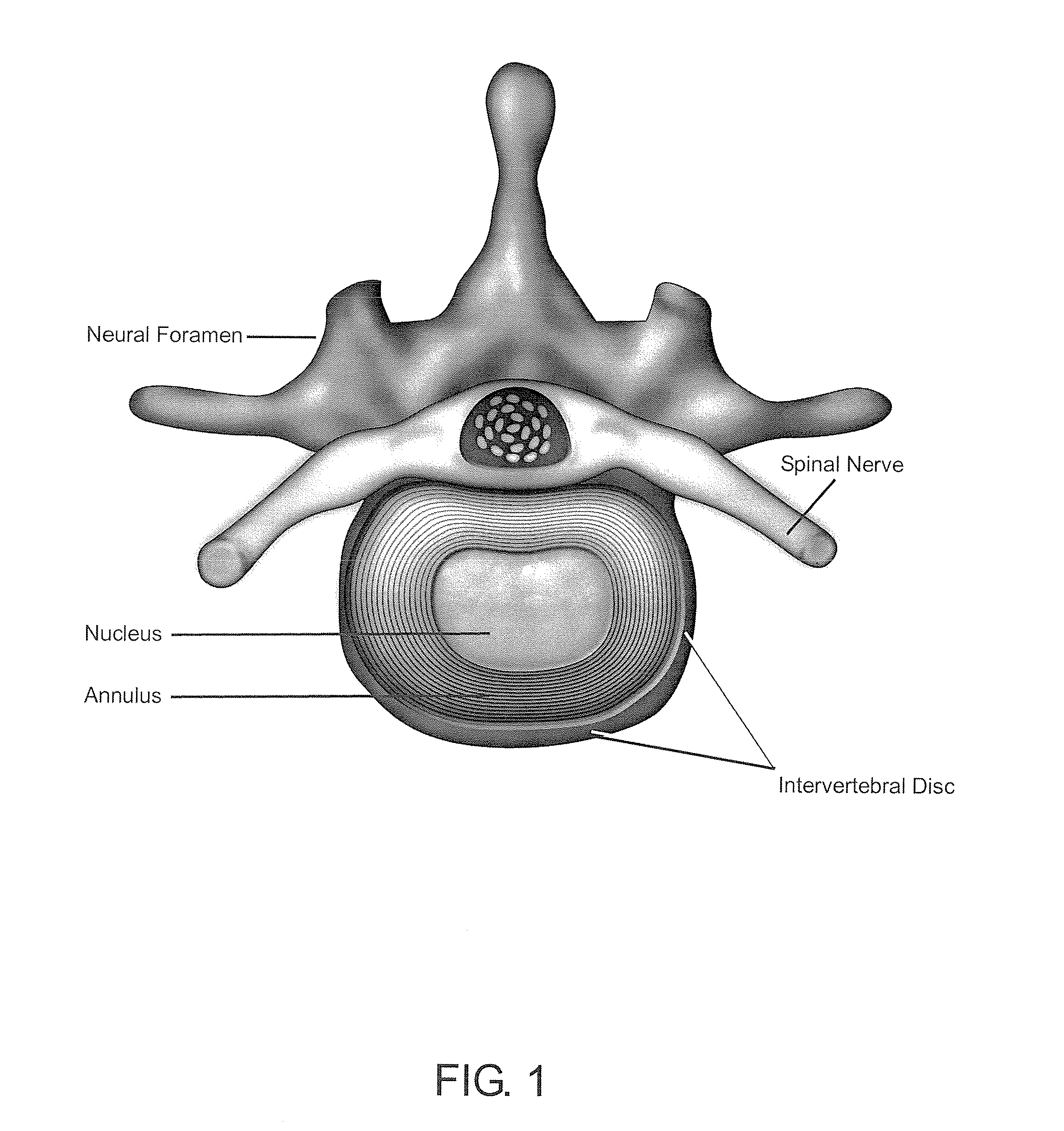
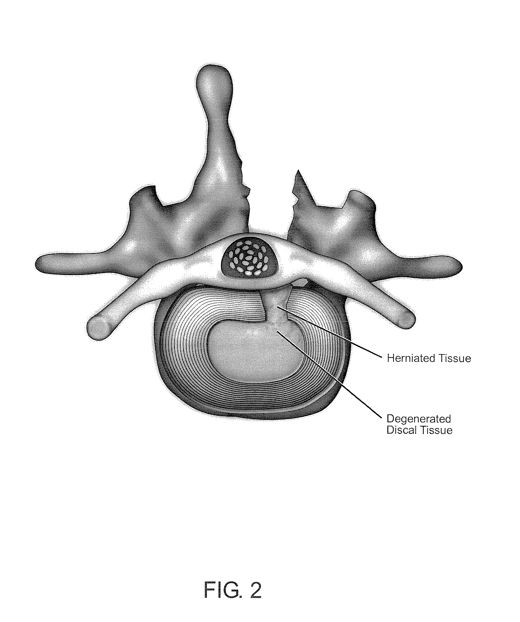
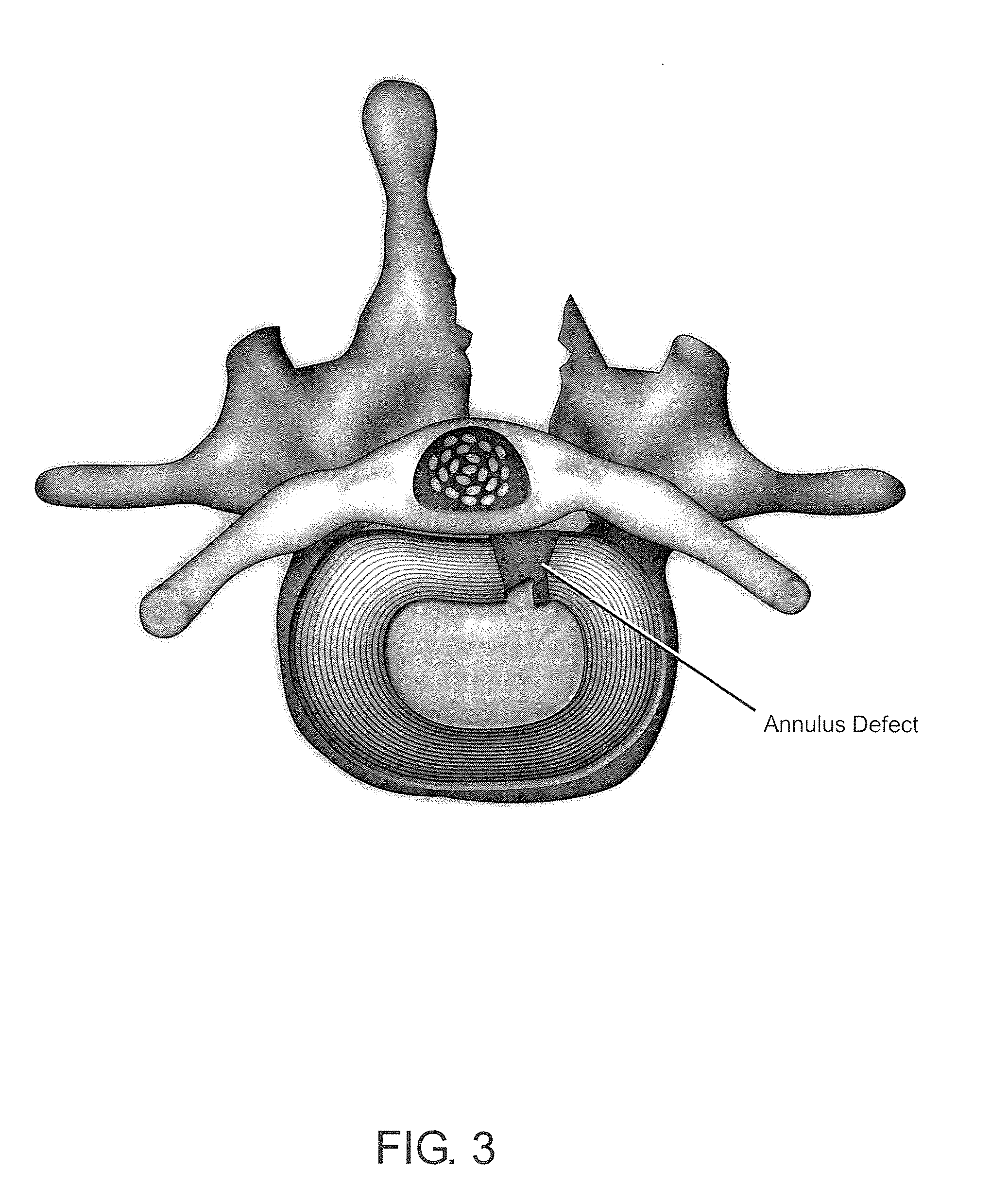
Once damaged, the degenerated disc may bulge or extrude, and therefore needs to be removed.
Although this procedure is ideal for decompressing and relieving the nervous system (root or cauda equina), it is a poor operation for the spine, because it creates a potentially disabling condition that leads to a degenerative cascade that may require an additional invasive surgical procedure, like fusion or arthroplasty, for example.
Discectomy brings a good short-term effect in relieving radicular pain, but it causes disc height reduction with neuro-foramen stenosis, instability of the treated level, poor result on back pain, and / or complications, such as spinal stenosis or facet pain, for example.
Over 200,000 spinal fusions are performed each year in the U.S. alone, but by eliminating the motion, the spinal fusion alters the biomechanical properties of the inter-vertebral disc and increases stress and strain on the discs that are adjacent to the fused disc.
In fact, both discectomy and fusion worsen the condition of the affected disc, adjacent discs, and surrounding tissues (such as facet joints), leading to further degeneration.
Today, two kinds of devices are marketed: the total disc replacement and the nuclear replacement, but both of them have major pitfalls.
Its design allows its implantation through a posterior approach, but the major limitation of such nucleus prosthesis is that it can be used only in patients in whom disc degeneration is at an early or intermediate stage, because it requires the presence of a competent natural annulus.
As a hydrogel-based device, it is fragile, and so does not resist the outstanding bio-mechanical constraints of the lumbar spine (shear forces).
As inert materials, they may lose their mechanical properties over time, and tears and breakages have been reported.
The main disadvantage of ACT for disc repair is that it requires a disc biopsy.

Method used


Image

  • Methods and compositions for repair of cartilage using an in vivo bioreactor
  • Methods and compositions for repair of cartilage using an in vivo bioreactor
  • Methods and compositions for repair of cartilage using an in vivo bioreactor

Examples


example 1

Exemplary Materials and Methods

[0153]Exemplary embodiments of materials and methods for use in the invention are described in this Example.

Cell Culture

[0154]While autologous HDFs harvested from the patient are used to construct the implant, preliminary studies are performed using neonatal foreskin fibroblasts, as there are convenient sources of cells for experimental purpose, for example. Further studies using autologous cells harvested from the patient are performed to demonstrate that the procedure works with these cells.

[0155]Neonatal foreskin HDFs are obtained from Cascade Biologics (Portland, Oreg.) and expanded in vitro with DMEM (Invitrogen, Carlsbad, Calif., USA), containing 10% FBS (Invitrogen) and antibiotics. Suspensions of HDFs are seeded in alginate or in monolayer culture as described below.

[0156]To generate alginate gel cultures, cells are suspended at high density (107 cells / ml) in 2% wt / vol medium viscosity alginate (Sigma-Aldrich, St. Louis, Mo.), and 25 mL drople...

example 2

Assessment of Cell Viability of HDFs in Alginate Beads

[0167]The viability of HDFs in alginate beads cultured in chondrogenic medium is tested by light microscopy and / or viability test, in specific aspects of the invention. Light microscopy is employed to study morphology and proliferation of HDFs. In an exemplary viability test, alginate beads are dissolved in dissolving-buffer (0.55 M Na-Citrate, 1.5 M NaCl, and 0.5 M EDTA), cells are centrifuged, and the pellet is treated with collagenase for 1 h. Cells are resuspended in DMEM, and viability is determined using a Neubauer chamber and the trypan blue exclusion method, for example.

example 3

Assessment of Chondrogenic Differentiation

[0168]In specific embodiments, HDFs are characterized by the production of collagen of type I, III and V, while chondrocytes are characterized by the production of collagen of type II, IX, XI and the production of sulfated proteoglycans.

[0169]Chondrogenic differentiation is assessed by measuring sulfated glycosaminoglycan (sGAG ) content and collagen I et II production by western blotting. The rate of collagen synthesis is measured by [3H]-proline incorporation.

Total DNA and sGAG Content

[0170]Cells in alginate beads are recovered from the alginate using 55 mM sodium citrate, 0.9% NaCl solution. Then the cells are lysed in 300 μl of 0.5% v / v Nonidet P-40 buffer (50 mM Tris-Cl, 100 mM NaCl, 5 mM MgCl2). The lysate is transferred to microcentrifuge tubes, spun, and DNA is measured in a 100 μl supernatant aliquot using the Hoescht dye method (DNA Quantification Kit, Sigma, St. Louis, Mo.) with calf thymus DNA as standard. The remaining lysis bu...


PUM

PropertyMeasurementUnit
Timeaaaaaaaaaa
Forceaaaaaaaaaa
Structureaaaaaaaaaa

Abstract

Methods and compositions for the biological repair of cartilage using a hybrid construct combining both an inert structure and living core are described. The inert structure is intended to act not only as a delivery system to feed and grow a living, core component, but also as an inducer of cell differentiation. The inert structure comprises concentric internal and external and inflatable / expandable balloon-like bio-polymers. The living core comprises the cell-matrix construct comprised of HDFs, for example, seeded in a scaffold. The method comprises surgically removing a damaged cartilage from a patient and inserting the hybrid construct into the cavity generated after the foregoing surgical intervention. The balloons of the inert structure are successively inflated within the target area, such as a joint, for example. Also disclosed herein are methods for growing and differentiating human fibroblasts into chondrocyte-like cells via mechanical strain.

Description

CROSS-REFERENCE TO RELATED APPLICATIONS[0001]The present application claims priority to U.S. Provisional Application Ser. No. 60 / 771,172, entitled Method for Repairing an Intervertebral Disc, and filed on Feb. 7, 2006 and also claims priority to U.S. Provisional Application Ser. No. 60 / 785,478, entitled Multi-Layered Multi-Compartmental Three-Dimensional Polymeric Scaffold For Intervertebral Disc Repair, and filed on Mar. 24, 2006, both of which are incorporated by reference herein in their entirety.FIELD OF THE INVENTION[0002]The present invention generally concerns at least the fields of medicine, surgery, anatomy, biology, cell biology, and / or molecular biology. In particular aspects, the present invention concerns the fields of cartilage repair, such as articular cartilage repair. More particularly, the field of the invention concerns cell-matrix encapsulation devices for growing, proliferating, and / or differentiating cells into chondrocyte-like cells under mechanical stress.BAC...

Claims


Application Information

Patent Timeline
IPC IPC(8): A61K35/12A61F2/02A61K35/32A61K35/33C12N5/077
CPCA61F2/30756A61F2/3099A61K35/33A61K35/32A61K45/06C12N2533/74C12N2521/00A61F2/32A61F2/38A61F2/3804A61F2/40A61F2/441A61F2002/2817A61F2002/286A61F2002/30062A61F2002/30586A61F2002/30677A61F2002/30762A61F2002/30764A61F2002/30766A61F2002/444A61F2002/4445A61F2002/445A61F2002/4635A61F2210/0004A61K38/18A61L27/3817A61L27/3852A61L27/3895C12N5/0655C12N2500/02C12N2500/38C12N2501/155C12N2506/1307A61K2300/00A61P19/00A61P19/04A61P31/04A61P31/10A61P31/12A61P41/00A61P43/00A61L2430/06
Inventor SEVRAIN, LIONEL C.VERDIER-SEVRAIN, SYLVIE Y.
Owner SPINALCYTE
Learn More