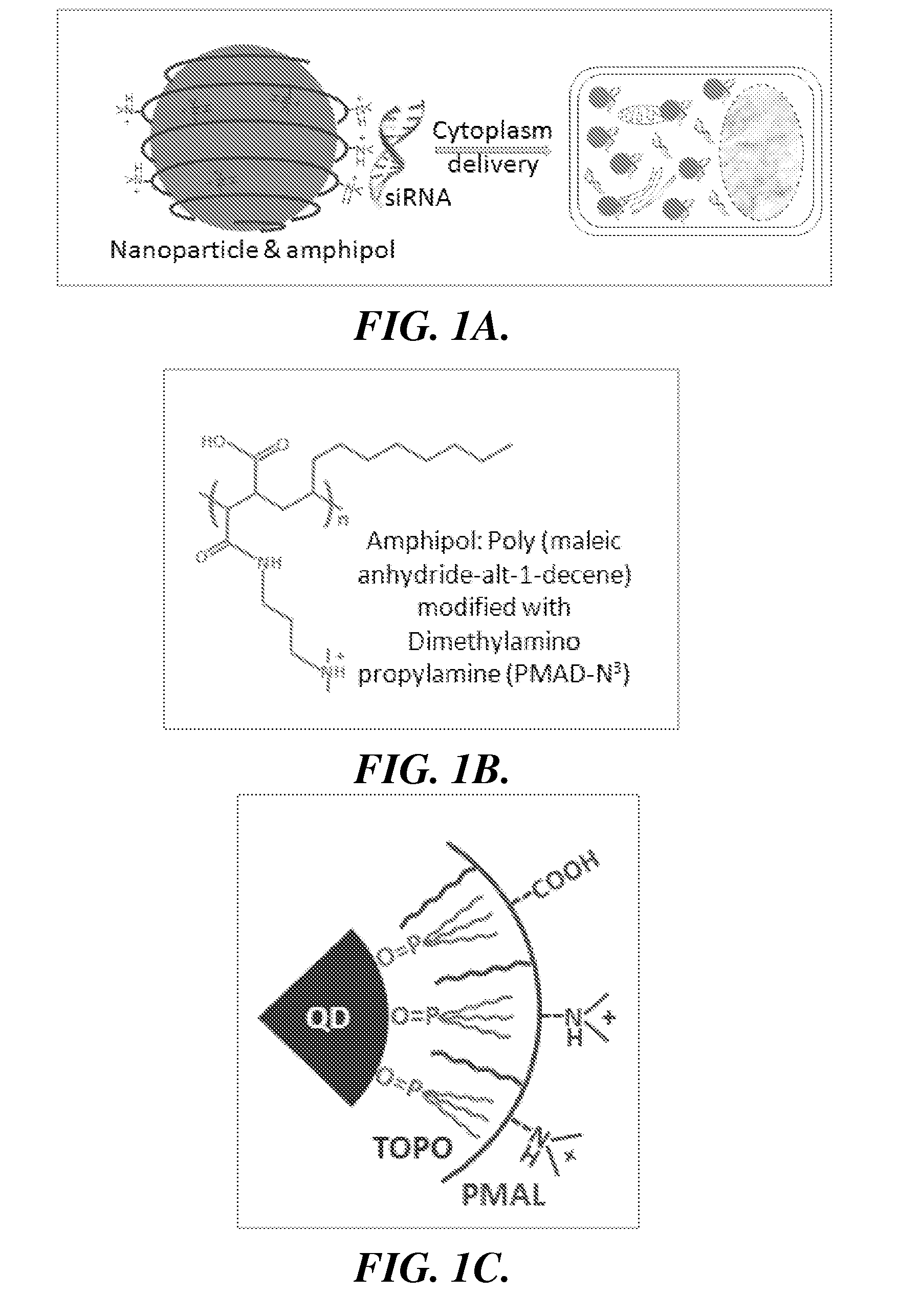
[0073]The effectiveness of the invention is described below utilizes a representative amphipol: poly(
maleic anhydride-alt-1-
decene) modified with dimethylamino
propylamine (PMAL, m.w. 18.5K) (FIG. 1B). PMAL's
hydrocarbon chains interact with (e.g., bind to) the hydrocarbons on the surface of QDs via multivalent hydrophobic interactions, leading to the formation of stable and water-soluble organic-inorganic
hybrid structures (FIG. 1C). At
neutral pH, the overall
surface charge of the
hybrid structure is highly positive, which allows immobilization of negatively charged biomolecules (e.g., siRNAs) and interaction with negatively charged
cell surface. Amphiphilic copolymers have been used for QD solubilization and
bioconjugation for
cell labeling. However, in contrast to the nanocomplexes of the invention, those polymers employ a dense layer of carboxylic acids, which prevents interaction with siRNA molecules. The clustered
tertiary amines grafted on the PMAL backbone have strong
proton absorbing capability inside acidic cellular compartments, such as endosomes, leading to osmotic swelling and
endosome rupture. In addition to the
tertiary amines, it has also been shown that the pKa of
carboxylic acid groups in polymaleic anhydrides is also around 5 to 6, resulting in a second chemical group for
proton absorption. The co-existence of
tertiary amine and
carboxylic acid groups weakens the interaction between siRNA and nanoparticles, which is facilitates siRNA release inside cells. Indeed, it has been found that when polyethyleneimine (PEI) are chemically modified to reduce electrostatic binding, the
gene delivery activity is increased by 20-60 fold. Furthermore, the zwitterionic surface of QD-PMAL provides an important feature for
in vivo applications, because zwitterionic charge reduces
serum protein adsorption onto NP surfaces, which not only slows NP uptake by the reticuloendothelial systems (RES), but also helps NP renal clearance when the particles are made smaller than 5.5 nm.
[0074]The PMAL encapsulated QDs were prepared by a molecular self-
assembly approach. QDs coated with hydrophobic ligands (tri-n-octylphosphine
oxide or TOPO) were mixed with PMAL at a
molar ratio of 1:500. Because of the strong multivalent hydrophobic interactions between TOPO and the PMAL hydrocarbons, QD and PMAL bind to each other and form highly stable complexes (at least 6 months).
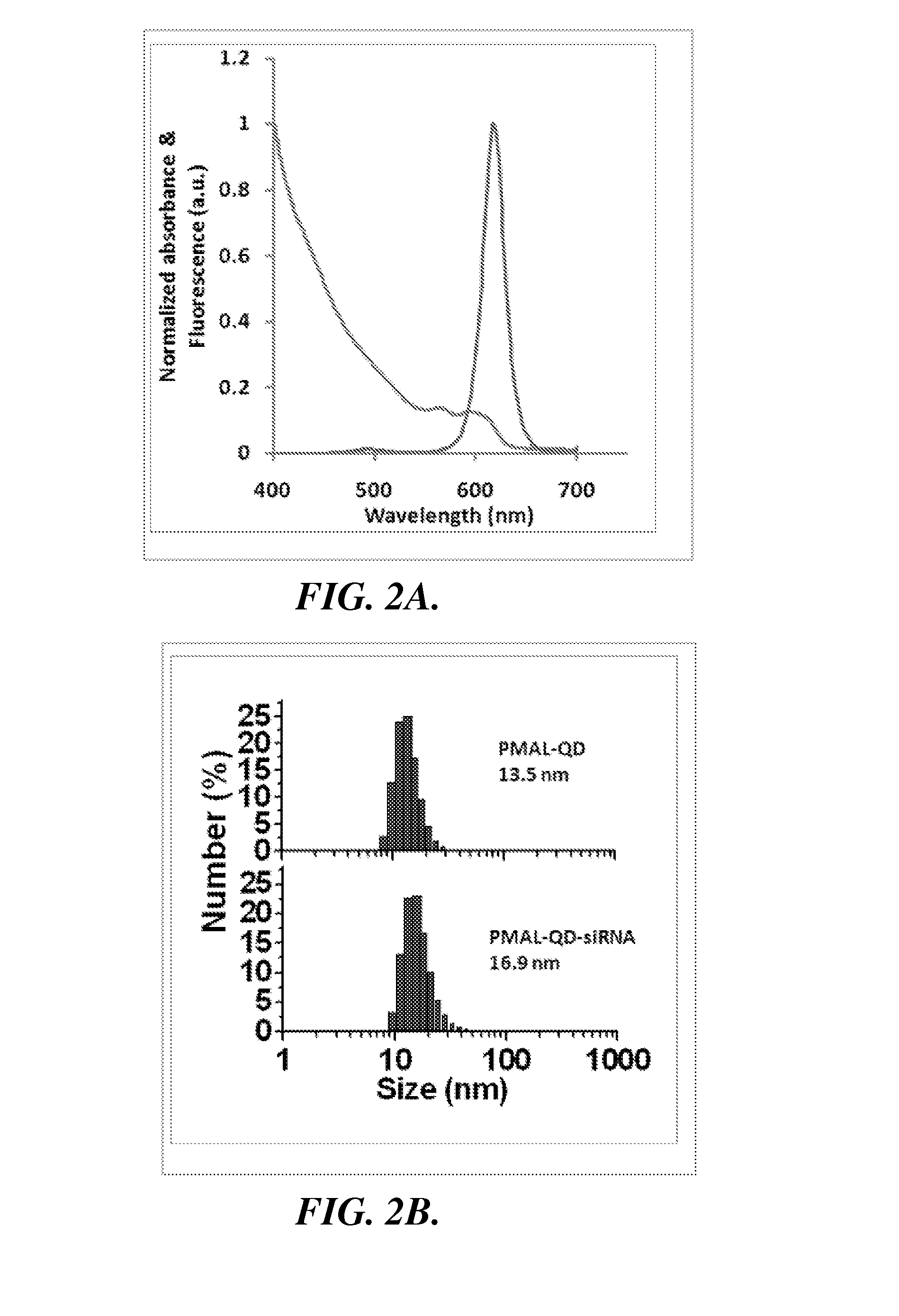
Transmission electron microscopy (TEM),
dynamic light scattering (DLS), and
spectroscopy measurements were obtained to characterize the size and optical properties of purified QD-PMAL and its siRNA complex. The PMAL encapsulated QDs have excellent optical properties and narrow size distributions, with comparable
quantum yield values as that of the original dots suspended in
chloroform (FIG. 2A).
Dynamic light scattering measurements (FIG. 2B) shows that QD-PMAL has a hydrodynamic
diameter of 12.1±1.5 nm (1.5 nm is the standard deviation of three different samples, rather than the size spread in one sample). Considering the QD core is 5.5±0.7 nm in
diameter (FIG. 2C), the larger
hydrodynamic radius in aqueous buffers is likely due to the physical size of the positively-charged PMAL
polymer, as well as its
strong interaction with the
solvent. This
surface charge is sufficient to carry small oligonucleotides and deliver them into mammalian cells. When bound to siRNA, the size of the
nanoparticle complexes further increases to 15.9±1.0 nm (1.0 nm is the standard deviation of three different samples), suggesting that QDs remain mainly single with siRNA on the surface, a result that was also confirmed by the ‘blinking’ feature of QDs under
fluorescence microscopy. The compact size of single particles is highly desirable because large particles enter cells at a much slower rate, and can be eliminated quickly by the RES
system in vivo. In contrast, previously reported gene deliveries based on silica and gold nanoparticles often form 100-200 nm aggregates likely because the size mismatch of large
plasmid DNA to small nanoparticles, and consequently many NPs are required to work together (forming clusters with the
DNA plasmid) for successful
transfection.
[0075]To investigate the number of siRNAs that can be loaded onto individual QDs, siRNA molecules were labeled with FITC dye (green) and mixed the siRNA (constant siRNA quantity at 10 pmol) with red QDs at various
molar ratios. As shown in the
gel electrophoresis data (FIG. 3A), the
fluorescence intensity of the siRNA band gradually decreases as QD concentration increases, and disappears when the siRNA / QD ratio is below 10, indicating that approximately 10 siRNA molecules can be immobilized onto the surface of individual QDs. To ensure that the result was not an artifact due to the
detection limit of
gel electrophoresis, siRNA-FITC samples of various quantity
ranging from 10-0.1 pmol were also studied under similar experiment conditions. As shown in FIG. 7, siRNA of 0.2 pmol is still detectable, suggesting that the disappearance of siRNA bands in the siRNA / QD ratio studies is indeed due to siRNA-QD binding. Focusing on the siRNA / QD ratio of 1:1, which is used in the siRNA
intracellular delivery experiment described below, two additional assays were conducted to confirm siRNA-QD association. First,
zeta potential measurements show that QD-PMAL has a
zeta potential value of 21.3 mV before siRNA binding and it reduces to 18.2 mV after siRNA binding, because negatively charged siRNA partially neutralizes the positive charge on QD surface. Second, the interaction of siRNA to QDs can also be characterized by
fluorescence quenching of FITC-labeled siRNA due to fluorescence
resonance energy transfer or FRET (FIG. 8).
[0076]The association of siRNA to QDs provides a mechanism for siRNA protection against
enzymatic degradation. This is a very important feature because RNAs in general are susceptible to
nuclease digestion. Enhanced resistance to
nuclease degradation should increase siRNA lifetime in the
cell and the subsequent interference effect on target mRNAs.
Gel electrophoresis experiments show that QD-bound siRNAs are degraded at a significantly slower rate (75% intact) compared with free siRNA (undetectable) under the same experiment conditions (FIG. 3B). Similar results have been previously observed with
plasmid DNA and short oligonucleotides on silica and gold NPs, and have been attributed to the NP steric hindrance to
nuclease activities.
[0077]To evaluate the RNAi efficiency using QD-PMAL
delivery vehicle, a model
gene silencing experiment was designed using
human breast adenocarcinoma cell line (SK—BR-3) and siRNA targeting Her-2 / neu. Her-2 / neu, a
cell surface receptor tyrosine kinase, is over-expressed in approximately 30% of breast tumors, and is an excellent
model system because it is involved in
signal transduction pathways leading to
cell growth and differentiation. FIGS. 4A-4D show that Her-2 / neu expression was suppressed to 36±2% using QD-PMAL in serum-free media. In comparison, when the two common
transfection reagents (
Lipofectamine™ and PEI) are used, the
target gene expression was reduced to 29±5% and 58±13%, respectively. When used in complete
cell culture media (contains serum), QD-PMAL reduces Her-2 expression to 35±4%, similar to the values achieved with serum-free media.
Lipofectamine™ and PEI reduce Her-2 expression to 48±7% and 62±5%. These results demonstrate that QD-PMAL is efficient in siRNA
intracellular delivery for both serum-free and complete media. In contrast, Lipofectamine™ only works well in serum-free media, and the QD-PMAL also outperforms PEI under both conditions.
[0078]The high
delivery efficiency of QD-PMAL could be explained by its structural and surface properties. First, when form complexes with siRNA, QDs remain single, and the small sizes facilitate their
diffusion and entry into cells. Second, after the nanostructures are endocytosed, both the tertiary amines and carboxylic groups on QD surface play important roles in
endosome escape. At low pH values carboxylic and amine groups are protonated, and at high pH values they will be de-protonated. Therefore, the zwitterionic surface behaves like a buffer
system that can quickly neutralize excess protons in
endosome, which also lead to a net influx of
chloride ions. The
osmotic pressure building along this
proton buffering process will eventually rupture the endosomes, a process known as the proton
sponge effect.

