
Two-way photodynamic therapy stereo colposcope for diagnosing and treating diseases of the female genital tract

a colposcope and two-way photodynamic technology, applied in the field of medical imaging, can solve the problems of poor program, high event incidence, lack of new medical devices for diagnosis and treatment,

Inactive Publication Date: 2014-01-30
DIAZ SANCHEZ JOEL GERARDO +1
View PDF14 Cites 3 Cited by
  • Summary
  • Abstract
  • Description
  • Claims
  • Application Information

AI Technical Summary

Benefits of technology

The new stereocolposcope is a device that helps detect and treat sexually transmitted diseases and neoplasms caused by human papilloma virus (HPV) by using fluorescence, photodynamic therapy, and purifying-ozone production electrotherapy. The device has various filters to analyze the fluorescence and eliminate unwanted wavelengths of light. It also has a housing with a d'Arsonval System, a vacuum glass tube, and a balanced mechanism for easy maneuverability during gynecologic procedures. The invention uses graphics and polymerization to create a cost-effective and reliable tool for detecting and treating HPV-related diseases.

Problems solved by technology

The high event incidence reflects poor programs in early disease detection, and a lack of new medical devices for diagnosis and treatment thereof.
Many attempts were then flawed due to impurities present in these photosensitizers.
Damage is due to high oxygen molecule reactions producing oxygen intermediates such as singlet oxygen, the main death cause of tumor cells.
Since early in twentieth century to early 50's, thousands of these devices were built within the United States of America, these devices being known as “violet ray machine.” These devices were out from trade in the U.S. about 1954 due to chemical and pharmaceutical industry pressure and also due to medical abuse thereon.
Infectious etiology female genital tract diseases including the lesions caused by human papillomavirus (HPV), which are known as cervical intraepithelial neoplasm (CIN) are often undiagnosed and not easily treated.
This is due to a lack of equipment and tests in doctor's office setting easing the diagnosis of said diseases causing that the gynecologist turns to laboratory tests with wide margin of error, or to the use of sophisticated and high cost equipment, for example, those of spectroscopy and optical coherence tomography requiring a long training and only available in industrialized countries but not in developing countries due to a number of factors.
This results in patients suffering of long waiting times and multiple doctor visits.
As to currently used therapy techniques for cervical cancer and especially for neoplasms (CIN), their use leads to cervical tissue loss and leaves lasting remnants for life, sometimes producing uterine or cervical synechiae which are scars in the uterus or cervix cavity where tissues adhere by fibrous membranes, causing pelvic infections, or the formed fibrous tissue may potentially cause infertility by preventing the passage of sperm or endometriosis by impeding menstrual outflow.
It may also cause the so-called “incompetent cervix”, i.e., cervical muscle fibers providing firmness thereto and used to support the baby weight during the nine pregnancy months when cut or injured, lose their strength when a woman becomes pregnant which leads to premature membrane rupture (MPR) causing immature baby deliveries and having several consequences such as newborn retinopathy which is virtually permanent blindness in the product, or even death thereof.
Currently there is significant controversy for their use in the U.S. and Europe.
Costs of both vaccines are high for both government administrations and individuals, the biggest problem being that real immunization time is not yet known in each of the viral types providing protection.

Method used


Image

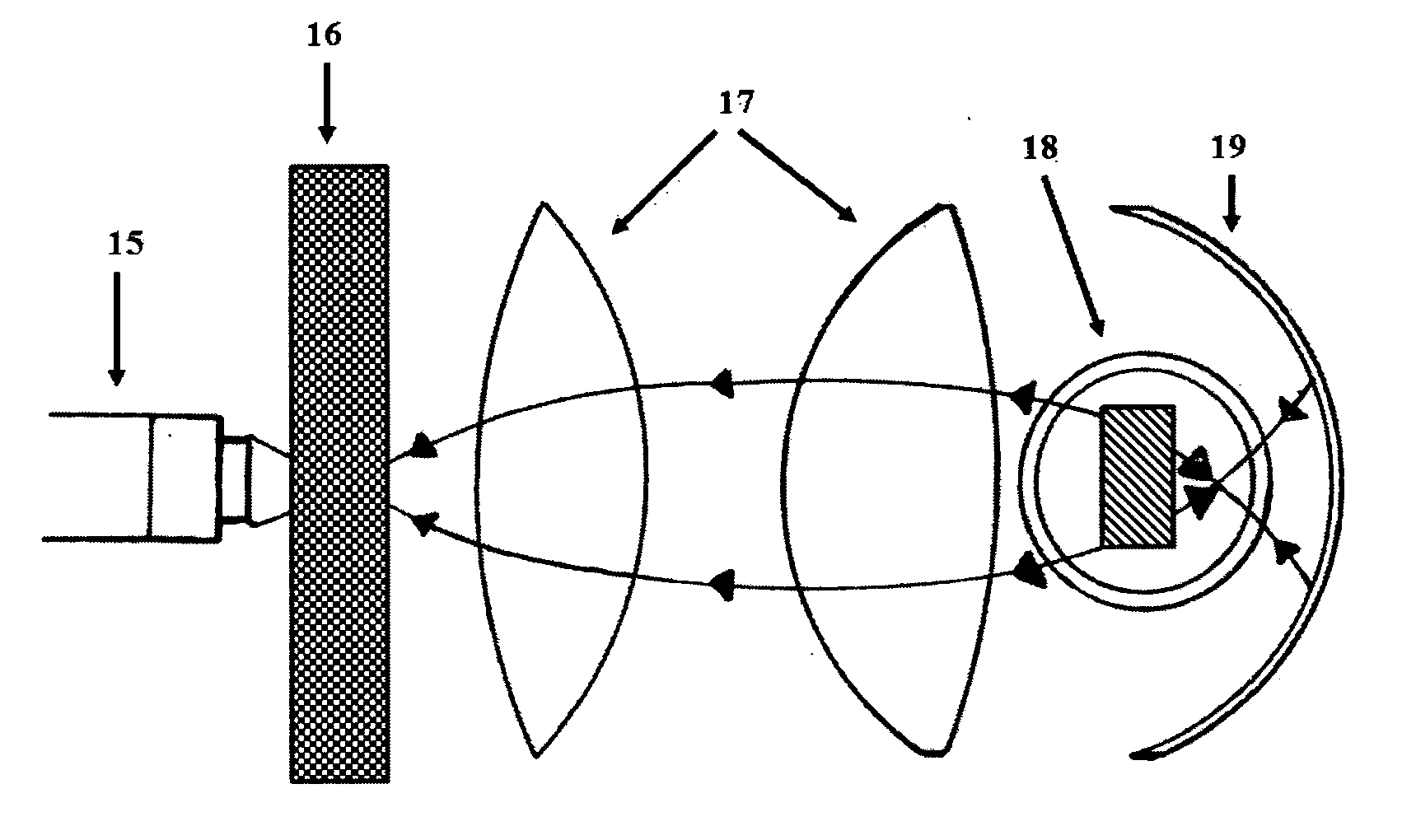
  • Two-way photodynamic therapy stereo colposcope for diagnosing and treating diseases of the female genital tract
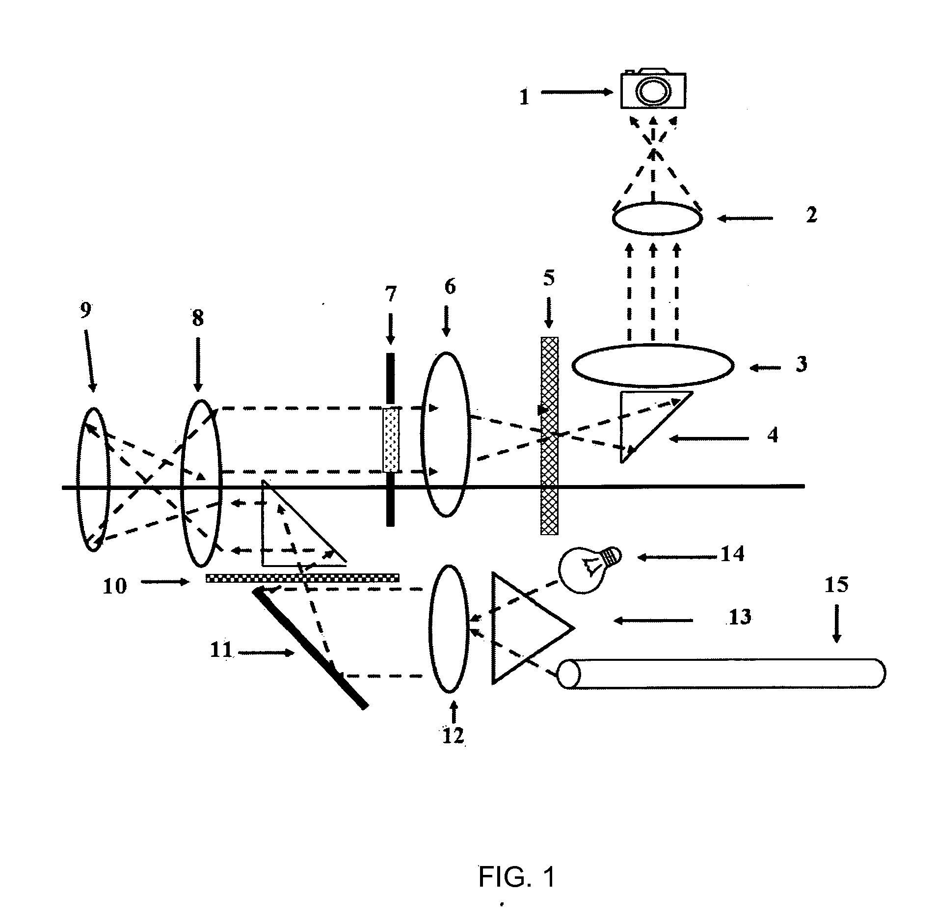
  • Two-way photodynamic therapy stereo colposcope for diagnosing and treating diseases of the female genital tract
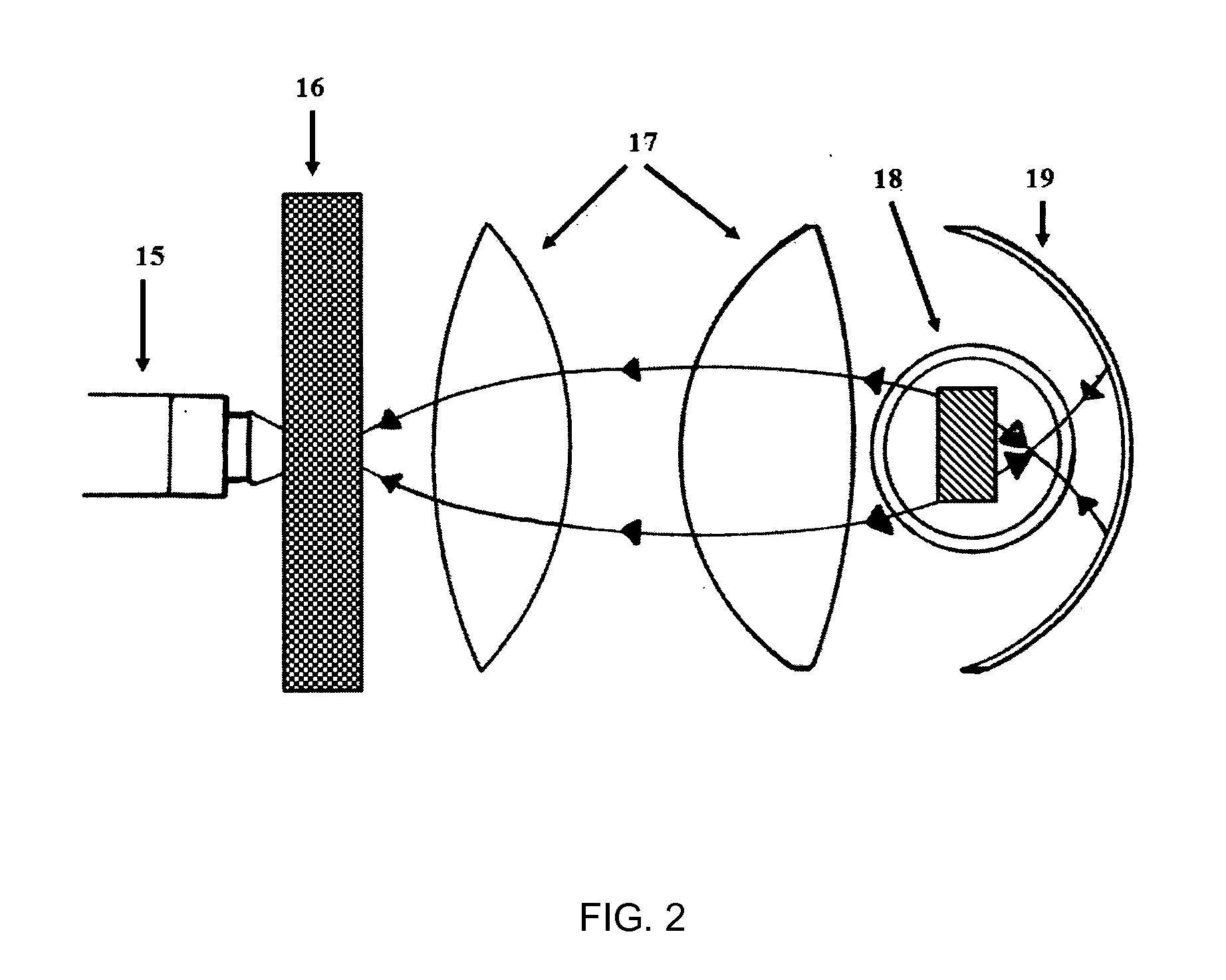
  • Two-way photodynamic therapy stereo colposcope for diagnosing and treating diseases of the female genital tract

Examples


example 1

Diagnosis for Human Papillomavirus Lesions Using a Fluorochrome: Fluorescein

Clinical Method.

[0093]The fluorescein isothiocyanate (FITC) fluorochrome was used in this case for several reasons:

[0094]This is widely used by ophthalmologists in fluoroangiographies (retina), and in local solution to see the corneal lesions. They use it with a lamp called “Actinic Lamp”, which has a cobalt filter and the emission spectrum is blue. This lamp used by them has no suppresor filters, we have chosen this fluorochrome since it has no adverse or toxic effects in humans.

Procedure:

[0095]Place the patient comfortably in lithotomy position. Study duration is 15-20 minutes.[0096]Introduce a plastic speculum to prevent unwanted reflections.[0097]Locate the cervix.[0098]Take vaginal pH (acidity or alkalinity): HPV infection often coexists with other bacterial or parasitic diseases so that the pH is alkaline.[0099]Take samples from endocervix with cervical brush.[0100]Take samples of ectocervix with Ayre ...

example 2

Photodynamic Diagnosis (PPD) for Human Papillomavirus Lesions Using a Photosensitizing Agent, 5-Aminolevulinic Acid (ALA)

Clinical Method.

[0111]Procedure:[0112]Place the patient comfortably in lithotomy position.[0113]Introduce a vaginal speculum.[0114]Locate the cervix.[0115]Clean the cervix and vaginal walls with normal saline.[0116]Apply the cream with the photosensitizer.[0117]Putting the patient at rest for 2 hours.[0118]Repeat the first three steps again.[0119]Select the frequency in the head variable linearity excitation filter to a range of 405 nanometers (blue-violet).[0120]Select the variable linearity suppressor filters at a range of 635 nanometers (red).[0121]Make direct observation or make video-photographic records with three-dimensional cameras mounted on the head.

[0122]When a patient is positive to photodynamic diagnosis (PPD), the lesion image is seen in bright red on a black background, if the patient is negative the black background is simply seen without red image...

example 3

Photodynamic Therapy (PPT) Application for Human Papillomavirus Lesions Using 5-Aminolevulinic Acid (ALA) Photosensitizing Agent

Clinical Method.

[0123]This application is performed depending on status, clinical judgment and light irradiation required power in three modes.

Procedure 1:

[0124]Place the patient comfortably in lithotomy position.[0125]Introduce a vaginal speculum.[0126]Locate the cervix.[0127]Clean the cervix and vaginal walls with normal saline.[0128]Apply the cream with the photosensitizer.[0129]Put the patient at rest for 4 hours.[0130]Repeat the first three steps back.[0131]Select the frequency in the head variable linearity excitation filter at a range of 685 nanometers (red).[0132]Depending on the lesion, irradiate it by keeping the stereocolposcope focus distance for the necessary time.

This procedure is recommended for small lesions.

Procedure 2:

[0133]Place the patient comfortably in lithotomy position.[0134]Introduce a vaginal speculum.[0135]Locate the cervix.[0136]...


PUM


Abstract

A stereo colposcope having variable linearity filter systems for both the excitation step and the suppression step, and can be used universally with any fluorescent compound or drug, as is the case of photodynamic diagnosis (PDD). The colposcope is a two-way colposcope because the treatment can be administered by an optical system or by a light-producing radio-frequency electrical current with a diathermic effect which facilitates photodynamic treatment. The colposcope produces ozone, which has an antiseptic effect when applied to the genital tract. A monitor provides for three-dimensional viewing through the use of two video cameras with the DLP (Digital Light Processing) and HDTV (High Definition Television) systems with the use of active lenses.

Description

TECHNICAL FIELD[0001]I. Medical field: a) Medical Imaging, b) Phototherapy, c) Electrotherapy[0002]II. Technology field: a) Surgical microscopes, b) Stereomicroscopes, c) Medicine-applied Optics, d) Medical Instruments and EquipmentSTATE OF THE ART[0003]Since the second half of the nineteenth century, photonics and electronics technologies have led to a plethora of scientific and technological developments for medical diagnosis and treatment, resulting in new devices with which research and a better understanding about the interaction of medical instrumentation, optics and electronics with biological tissues belonging to the field of immunohistochemistry and pathology have been conducted, targeting, as in our case, the study, diagnosis and treatment of female genital tract.[0004]As sexually transmitted diseases are currently considered emerging, there has been accordingly an increasing demand to solve women's genital tract diseases, leading to new medical device development in the l...

Claims


Application Information

Patent Timeline
Patent Type & Authority Applications(United States)
IPC IPC(8): A61B1/00A61N5/06A61N1/00A61B1/05A61B1/07A61B5/00A61B1/303A61B1/06
CPCA61B1/00087A61B1/303A61B1/00193A61N5/0603A61N5/062A61B5/0084A61B1/05A61B1/07A61B5/4836A61B5/4337A61N1/00A61B1/0676G01N21/6456G01N2021/6419G01N2021/6421A61B1/043A61B1/00186A61B1/0646A61B5/0036A61B1/00194
Inventor DIAZ SANCHEZ, JOEL GERARDOZERTUCHE ZUANI, JOSE GERARDO
Owner DIAZ SANCHEZ JOEL GERARDO
Learn More