
DNA diagnostics based on mass spectrometry

a mass spectrometry and dna technology, applied in the field of dna diagnostics based on mass spectrometry, can solve the problems of only being able to identify, genetic disease, and protein with altered or in some cases even lost biochemical activities, and achieve the effects of increasing mass resolution, mass accuracy, and partial or complete digestion of target nucleic acids

Inactive Publication Date: 2007-04-03
AGENA BIOSCI
View PDF491 Cites 223 Cited by
  • Summary
  • Abstract
  • Description
  • Claims
  • Application Information

AI Technical Summary

Benefits of technology

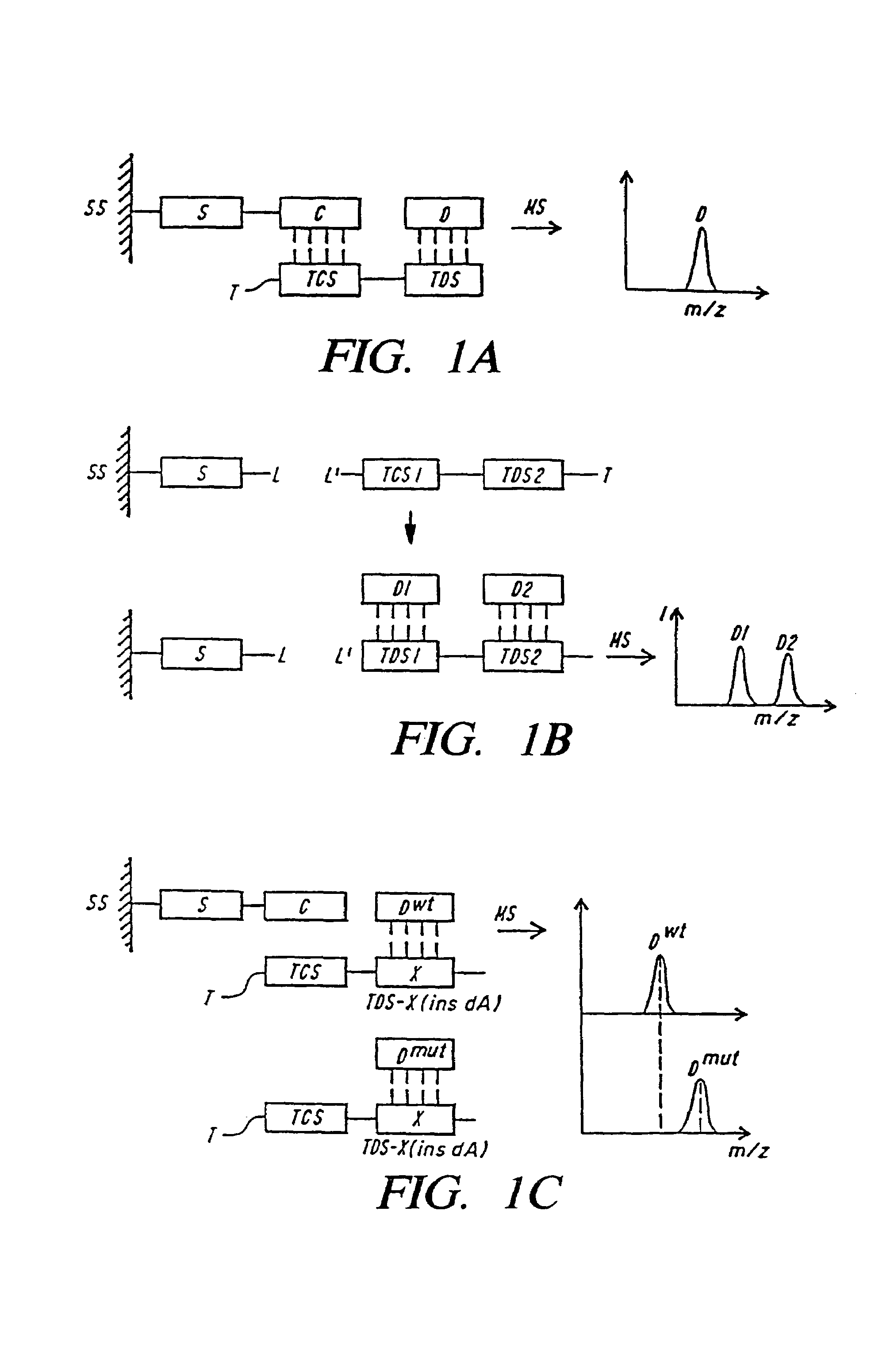
[0019]In certain embodiments the DNA is immobilized on the support via a selectively cleavable linker. Selectively cleavable linkers include, buta are not limited to photocleavable linkers, chemically cleavable linkers and an enzymatically (such as a restriction site (nucleic acid linker), a protease site) cleavable linkers. Inclusion of a selectively cleavable linker expands the capabilities of the MALDI-TOF MS analysis because it allows for all of the permutations of immobilization of DNA for MALDI-TOF MS, the DNA linkage to the support through the 3′- or 5′-end of a nucleic acid; allows the amplified DNA or the target primer to be extended by DNA synthesis; and further allows for the mass of the extended product (or degraded product via exonuclease degradation) to be of a size that is appropriate for MALDI-TOF MS analysis (i.e., the isolated or synthesized DNA can be large and a small primer or a large primer sequence can be used and a small restriction fragment of a gene or single strand thereof hybridized thereto).
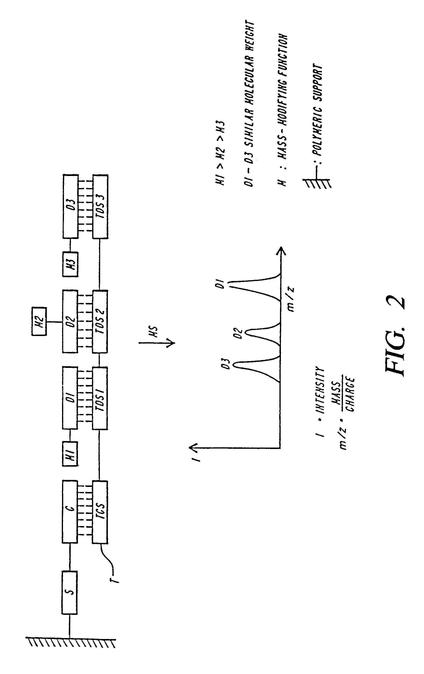
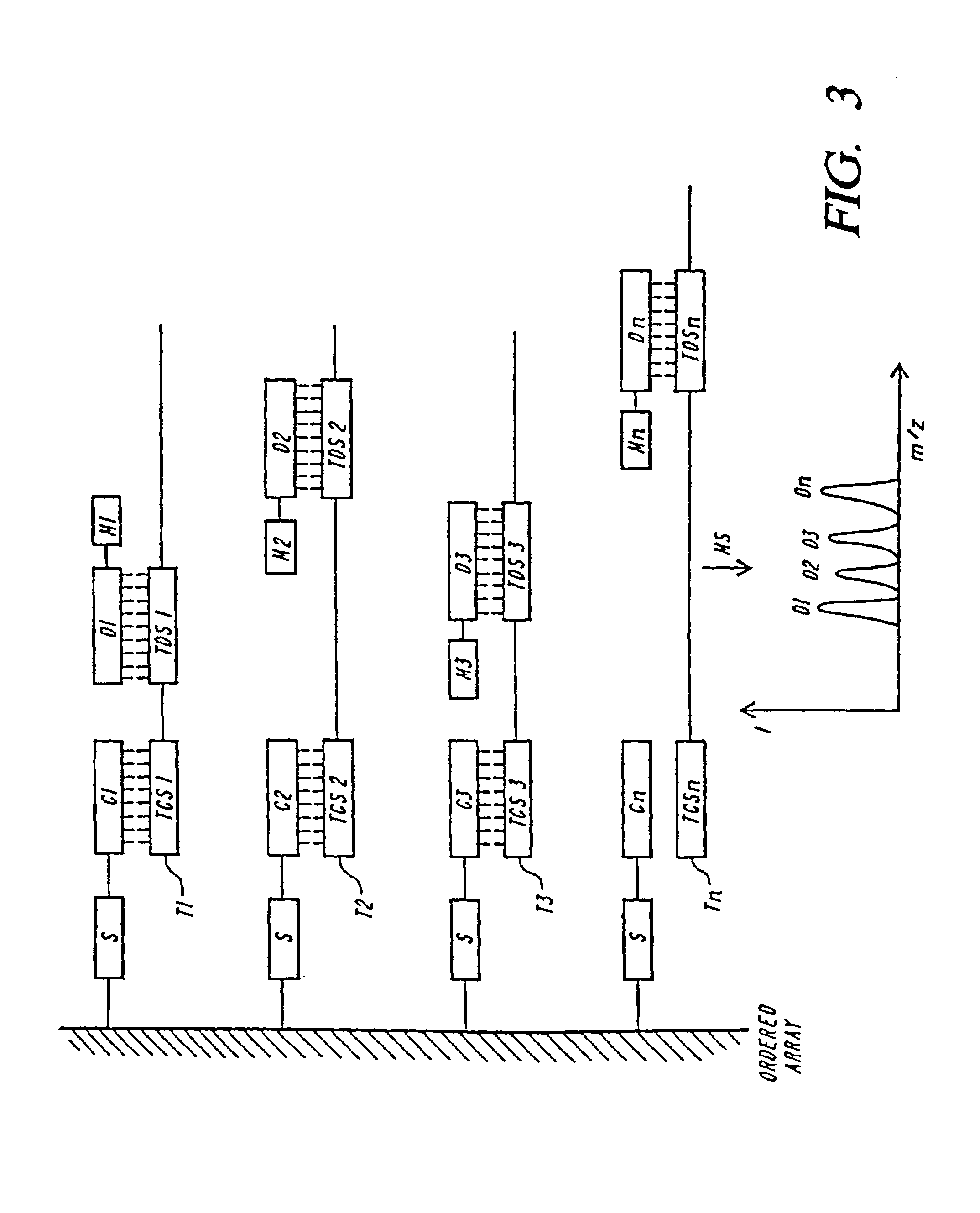
[0021]Methods for sequencing long fragments of DNA are provided. To perform such sequencing, specific base terminated fragments are generated from a target nucleic acid. The analysis of fragments rather than the full length nucleic acid shifts the mass of the ions to be determined into a lower mass range, which is generally more amenable to mass spectrometric detection. For example, the shift to smaller masses increases mass resolution, mass accuracy and, in particular, the sensitivity for detection. Hybridization events and the actual molecular weights of the fragments as determined by mass spectrometry provide sequence information (e.g., the presence and / or identity of a mutation). In a preferred embodiment, the fragments are captured on a solid support prior to hybridization and / or mass spectrometry detection. In another preferred embodiment, the fragments generated are ordered to provide the sequence of the larger nucleic acid.
[0022]One preferred method for generating base specifically terminated fragments from a nucleic acid is effected by contacting an appropriate amount of a target nucleic acid with an appropriate amount of a specific endonuclease, thereby resulting in partial or complete digestion of the target nucleic acid. Endonucleases will typically degrade a sequence into pieces of no more than about 50–70 nucleotides, even if the reaction is not run to full completion. In a preferred embodiment, the nucleic acid is a ribonucleic acid and the endonuclease is a ribonuclease (RNase) selected from among: the G-specific RNase T1, the A-specific RNase U2, the A / U specific RNase PhyM, U / C specific RNase A, C specific chicken liver RNase (RNase CL3) or crisavitin. In another preferred embodiment, the endonuclease is a restriction enzyme that cleaves at least one site contained within the target nucleic acid. Another preferred method for generating base specifically terminated fragments includes performing a combined amplification and base-specific termination reaction (e.g., using an appropriate amount of a first DNA polymerase, which has a relatively low affinity towards the chain-terminating nucleotides resulting in an exponential amplification of the target; and a polymerase with a relatively high affinity for the chain terminating nucleotide resulting in base-specific termination of the polymerization. Inclusion of a tag at the 5′ and / or 3′ end of a target nucleic acid can facilitates the ordering of fragments.
[0070]j) detecting the presence of the extended target nucleic acid, whereby the presence of a DNA fragment of a mass different from wild-type is indicative of a mutation at the target codon(s).This method eliminates one specific reagent for mutation detection compared other methods of MS mutational analyses, thereby simplifying the process and rendering it amenable to automation. Also, the specific extended product that is analyzed is cleaved from the primer and is therefore shorter compared to the other methods. In addition, the annealing efficiency is higher compared to annealing of an added primer and should therefore generate more product. The process is compatible with multiplexing and various detection schemes (e.g., single base extension, oligo base extension and sequencing). For example, the extension of the loop-primer can be used for generation of short diagnostic sequencing ladders within highly polymorphic regions to perform, for example, HLA typing or resistance as well as species typing.
[0072]Improved methods of sequencing DNA using MS are provided. In these methods thermocycling for amplification is used prior to MS analysis, thereby increasing the signal.

Problems solved by technology

Changes in a DNA sequence are known as mutations and can result in proteins with altered or in some cases even lost biochemical activities; this in turn can cause genetic disease.
Identification, however, can only be accomplished if the nucleic acid fragment is labeled with a sensitive reporter function (e.g., radioactive (32P, 35S), fluorescent or chemiluminescent).
Radioactive labels can be hazardous and the signals they produce decay over time.
Non-isotopic labels (e.g., fluorescent) suffer from a lack of sensitivity and fading of the signal when high intensity lasers are being used.
Additionally, performing labeling, electrophoresis and subsequent detection are laborious, time-consuming and error-prone procedures.
Electrophoresis is particularly error-prone, since the size or the molecular weight of the nucleic acid cannot be directly correlated to the mobility in the gel matrix.
It is known that sequence specific effects, secondary structure and interactions with the gel matrix are causing artifacts.
Nucleic acids, however, are very polar biopolymers that are very difficult to volatilize.
Consequently, mass spectrometric detection has been limited to low molecular weight synthetic oligonucleotides for confirming an already known oligonucleotide sequence by determining the mass of the parent molecular ion, or alternatively, confirming a known sequence through the generation of secondary ions (fragment ions) via CID in an MS / MS configuration using, in particular, for the ionization and volatilization, the method of fast atomic bombardment (FAB mass spectrometry) or plasma desorption (PD mass spectrometry).

Method used


Image

  • DNA diagnostics based on mass spectrometry
  • DNA diagnostics based on mass spectrometry
  • DNA diagnostics based on mass spectrometry

Examples


example 1

MALDI-TOF Desorption of Oligonucleotides Directly on Solid Supports

[0315]1 g CPG (Controlled Pore Glass) was functionalized with 3-(triethoxysilyl)-epoxypropan to form OH-groups on the polymer surface. A standard oligonucleotide synthesis with 13 mg of the OH-CPG on a DNA synthesizer (Milligen, Model 7500) employing β-cyanoethyl-phosphoamidites (Köster et al. (1994) Nucleic Acids Res. 12:4539) and TAC N-protecting groups (Köster et al. (1981) Tetrahedron 37:362) was performed to synthesize a 3′-T5-50 mer oligonucleotide sequence in which 50 nucleotides are complementary to a “hypothetical” 50 mer sequence. T5 serves as a spacer. Deprotection with saturated ammonia in methanol at room temperature for 2 hours furnished according to the determination of the DMT group CPG which contained about 10 umol 55 mer / g CPG. This 55 mer served as a template for hybridizations with a 26-mer (with 5′-DMT group) and a 40-mer (without DMT group). The reaction volume is 100 μl and contains about 1 nmo...

example 2

Electrospray (ES) Desorption and Differentiation of an 18-mer and 19-mer

[0316]DNA fragments at a concentration of 50 pmole / ul in 2-propanol / 10 mM ammoniumcarbonate (1 / 9, v / v) were analyzed simultaneously by an electrospray mass spectrometer.

[0317]The successful desorption and differentiation of an 18-mer and 19-mer by electrospray mass spectrometry is shown in FIG. 12.

example 3

Detection of The Cystic Fibrosis Mutation ΔF508, by Single Step Dideoxy Extension and Analysis by MALDI-TOF Mass Spectrometry (Competitive Oligonucleotide Simple Base Extension=COSBE)

[0318]The principle of the COSBE method is shown in FIG. 13, N being the normal and M the mutation detection primer, respectively.

Materials and Methods

[0319]PCR Amplification and Strand Immobilization. Amplification was carried out with exon 10 specific primers using standard PCR conditions (30 cycles: 1′@95° C., 1′@55° C., 2′@72° C.); the reverse primer was 5′ labelled with biotin and column purified (Oligopurification Cartridge, Cruachem). After amplification the amplified products were purified by column separation (Qiagen Quickspin) and immobilized on streptavidin coated magnetic beads (Dynabeads, Dynal, Norway) according to their standard protocol; DNA was denatured using 0.1 M NaOH and washed with 0.1M NaOH, 1×B+W buffer and TE buffer to remove the non-biotinylated sense strand.

[0320]COSBE Conditi...


PUM

PropertyMeasurementUnit
mass spectrometryaaaaaaaaaa
affinityaaaaaaaaaa
thermostableaaaaaaaaaa

Abstract

Fast and highly accurate mass spectrometry-based processes for detecting a particular nucleic acid sequence in a biological sample are provided. Depending on the sequence to be detected, the processes can be used, for example, to diagnose a genetic disease or chromosomal abnormality; a predisposition to a disease or condition, infection by a pathogenic organism, or for determining identity or heredity.

Description

RELATED APPLICATIONS[0001]This application is a divisional of U.S. application Ser. No. 09 / 297,576, filed Jun. 28, 1999, abandoned, to Köster et al., entitled “DNA DIAGNOSTICS BASED ON MASS SPECTROMETRY”, which is the U.S. National Stage of PCT / US97 / 20444, filed Nov. 6, 1997. U.S. application Ser. No. 09 / 297,576 is a continuation-in-part of U.S. application Ser. No. 08 / 744,481, filed Nov. 6, 1996, U.S. Pat. No. 6,428,955, to Köster, entitled “DNA DIAGNOSTICS BASED ON MASS SPECTROMETRY”. This application is also a continuation-in-part of U.S. application Ser. No. 08 / 744,481, filed Nov. 6, 1996, to Köster, entitled “DNA DIAGNOSTICS BASED ON MASS SPECTROMETRY”. This application is also a continuation-in-part of U.S. application Ser. No. 08 / 744,590, filed Nov. 6, 1996, U.S. Pat. No. 6,074,823, Ser. No. 08 / 746,036, filed Nov. 6, 1996, U.S. Pat. No. 5,900,481, Ser. No. 08 / 746,055, filed Nov. 6, 1996, abandoned, Ser. No. 08 / 786,988, filed Jan. 23, 1997, Ser. No. 08 / 787,639, filed Jan. 23, ...

Claims


Application Information

Patent Timeline
Patent Type & Authority Patents(United States)
IPC IPC(8): C12Q1/68C12P19/34B01J19/00B01L3/02C07B61/00C07F9/24C07H21/00C40B40/06C40B40/10C40B60/14G01N35/00G01N35/10
CPCB01J19/0046B01L3/0268C07F9/2408C07F9/2429C07H21/00C12Q1/6837C12Q1/6858C12Q1/6865C12Q1/6872G01N35/1067B01J2219/00315B01J2219/00317B01J2219/00387B01J2219/00468B01J2219/00497B01J2219/005B01J2219/00504B01J2219/00511B01J2219/0052B01J2219/00527B01J2219/00585B01J2219/00596B01J2219/00605B01J2219/0061B01J2219/00612B01J2219/00626B01J2219/0063B01J2219/00637B01J2219/00641B01J2219/00648B01J2219/00659B01J2219/00707B01J2219/0072B01J2219/00722B01J2219/00725C07B2200/11C40B40/06C40B40/10C40B60/14G01N35/1074G01N2035/00237G01N2035/1037G01N2035/1069H01J49/00C12Q2565/507C12Q2565/537C12Q2523/10C12Q2565/627C12Q2525/197C12Q2531/113C12Q2565/518C12Q2535/101
Inventor KOSTER, HUBERTBRAUN, ANDREASVAN DEN BOOM, DIRKJURINKE, CHRISTIANRUPPERT, ANDREASHILLENKAMP, FRANZLITTLE, DANIEL P.
Owner AGENA BIOSCI
Learn More