Gels for encapsulation of biological materials

a technology of biological materials and gels, applied in the field of gels for encapsulation of biological materials, can solve the problems of loss of biological activity, cell death or functional impairment, and often exposed cell processing conditions

Inactive Publication Date: 2008-08-19
BOARD OF RGT THE UNIV OF TEXAS SYST
View PDF18 Cites 22 Cited by
  • Summary
  • Abstract
  • Description
  • Claims
  • Application Information

AI Technical Summary

Benefits of technology

[0027]Ultrathin membranes can be formed by the methods described herein. These ultrathin membranes allow for optimal diffusion of nutrient and bioregulator molecules across the membrane, and great flexibility in the shape of the membrane. Such thin membranes produce encapsulated material with optimal economy of volume. Biological material thus coated can be packed into a relatively small space without interference from bulky membranes.
[0028]The thickness and pore size of membranes formed can be varied. This variability allows for “perm-selectivity”—membranes can be adjusted to the desired degree of porosity, allowing only preferred molecules to permeate the membrane, while acting as a barrier against larger undesired molecules. Thus, the membranes are immunoprotective in that they prevent the transfer of antibodies or cells of the immune system.
[0029]When the encapsulated biological material is cellular in nature, the absence of small monomers in the polymerization solution prevents the diffusion of toxic molecules into the cell. In this manner the present invention provides a polymerization system which is more biocompatible than any available in the prior art.
[0031]According to the present invention, membrane formation occurs under physiological conditions. Thus, no damage is done to the enclosed biological material due to harsh pH, temperature, or ionic conditions.
[0032]Because the membrane adheres to the biological material, the membrane can be used as an adhesive to fasten more than one biological substance together. The macromers are polymerized in the presence of these substances which are in close proximity. The membrane forms in the interstices, effectively gluing the substances together.

Problems solved by technology

During encapsulation using prior methods, cells are often exposed to processing conditions, which are potentially cytotoxic.
These conditions include heat, organic solvents and non-physiological pH, which can kill or functionally impair cells.
Proteins are often exposed to conditions that are potentially denaturing and can result in loss of biological activity.
Further, even if cells survive processing conditions, the stringent requirements of encapsulating polymers for biocompatibility, chemical stability, immunoprotection and resistance to cellular overgrowth, restrict the applicability of prior art methods.
For example, the encapsulating method based on ionic crosslinking of alginate (a polyanion) with polylysine or polyornithine (polycation) (Goosen, et al., 1985) offers relatively mild encapsulating conditions, but the long-term mechanical and chemical stability of such ionically crosslinked polymers remains doubtful.
Moreover, these polymers when implanted in vivo, are susceptible to cellular overgrowth (McMahon, et al., 1990), which restricts the permeability of the microcapsule to nutrients, metabolites, and transport proteins from the surroundings.
This has been seen to possibly lead to starvation and death of encapsulated islets of Langerhans cells (O'Shea et al., 1986).
These biomaterials are considered biocompatible if they produce a minimal or no adverse response in the body.
Most synthetic materials, however, do not elicit such a reaction.
They can thus interact with implanted material through any of these various regions, resulting in cellular proliferation at the implant surface.
However, these gels have undesirable mechanical properties.
Agarose forms a weak gel, and the polyacrylates must be precipitated from organic solvents, thus increasing the potential for cytotoxicity.
However, the polymerization process, if allowed to proceed rapidly to completion, generates local heat and requires the presence of toxic cross-linkers.
This usually results in mechanically weak gels whose immunoprotective ability has not been established.
However, implantation for periods up to a week has resulted in severe fibrous overgrowth on these microcapsules (McMahon, et al.
The free volume exclusion effect makes the approach of a macromolecule (viz., a protein) close to a surface which has grafted PEO chains sterically unfavorable (Miyama, et al., 1988; Nagoaka, et al.
The use of graft copolymers, however, still does not guarantee that the surface “seen” by a macromolecule consists entirely of PEO.
However, use of an electron bean precludes the presence of any living tissue due to the sterilizing effect of this radiation.
Also, the networks produced are difficult to characterize due to the non-specific cross-linking induced by the electron beam.
However, yeast cells are widely known to be much hardier, resistant to adverse environments and elevated temperatures, and more difficult to kill when compared to mammalian cells and human tissues.
None of these references, however, discuss the exposure of sensitive eukaryotic tissue, organisms, or sensitive molecules to the chemical conditions used during polymerization because their polymerization conditions are incompatible with sensitive materials.
In the original presentation of the technique (Fukui et al., 1976), the authors state in the final sentence that the technique would be appropriate for microbial cells, but provide no indication of usefulness for more sensitive cells.
This is an important issue, since bioincompatibility in biomedical applications leads to xenograft failure in therapeutically transplanted cells for the evaluation of drug efficacy (O'Shea at al., 1986) and to xenograft failure in diagnostically transplanted cells (McMahon et al., 1990).
Similarly, bioincompatibility would lead to the failure of encapsulated enzymes (for example, therapeutic enzymes encapsulated and circulating or implanted in a blood-rich tissue).
No biological properties of these materials have boon reported, however.
Thus, the prior art contains no description of methods to form biocompatible PEO networks on three-dimensional living tissue surfaces without damaging encapsulated tissue.
First, because the cylindrical gel is broken along random planes, shearing will often occur through pockets of cells, leaving some cells exposed to the host immune system.

Method used


Image

  • Gels for encapsulation of biological materials
  • Gels for encapsulation of biological materials
  • Gels for encapsulation of biological materials

Examples


example 1

Synthesis of PRO 6 kD Diacrylate

[0102]PEG acrylates of molecular weights 400 Da and 1,000 Da ware commercially available from Sartomer and Dajac Inc., respectively. PEG 6 kD (20 g) was dissolved in 200 mL dichloromethane in a 250 mL round bottom flask. The flask was cooled to 0° C. and 1.44 mL of triethyl amino and 1.3 mL of acryloyl chloride were added with constant stirring under a dry nitrogen atmosphere. The reaction mixture was then brought to room temperature and stirred for 12 hr under a nitrogen atmosphere. It was then filtered, and the filtrate was precipitated by adding to a large excess of hexane. The crude monomer was purified by dissolving in dichloromethane and precipitating in hexane. Yield 69%.

example 2

Synthesis of PEG 18.4 kD Tetraacrylate

[0103]A tetrafunctional water soluble PEG (30 g; m.w. 18.5 kD) having the following structure was purchased from Polysciences, Inc.:

[0104]

where F1═CONH, COO or NHCOO[0105]X═H, CH3, C2H5, C6H5, Cl, Br, OH or CH2COOH[0106]F2═COO, CONH, O or C6H4, and[0107]R═CH2 or -alkyl-.

[0108]The PEG was dried by dissolving in benzene and distilling off the water-benzene azeotrope. PEG 18.5 kD (59 g) was dissolved in 300 mL of benzene in a 500 mL flask. To this, 3.6 mL of triethylamine and 2.2 mL of acryloyl chloride were added under nitrogen atmosphere and the reaction mixture was refluxed for 2 hours. It was then cooled and stirred overnight. The triethyl amine hydrochloride was separated by filtration and the copolymer was recovered from filtrate by precipitating in a largo excess of hexane. The polymer was further purified by dissolving in methylene chloride and reprecipitating in hexane. The polymer was dried at 50 degrees C. under vacuum for 1 day. Yield 6...

example 3

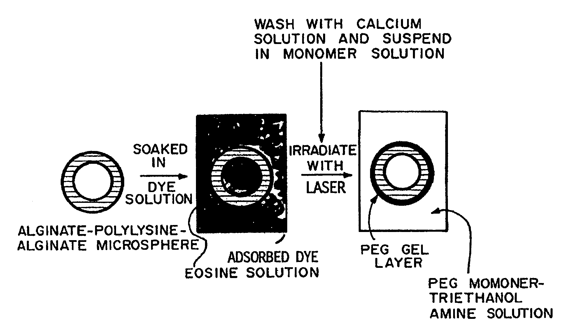
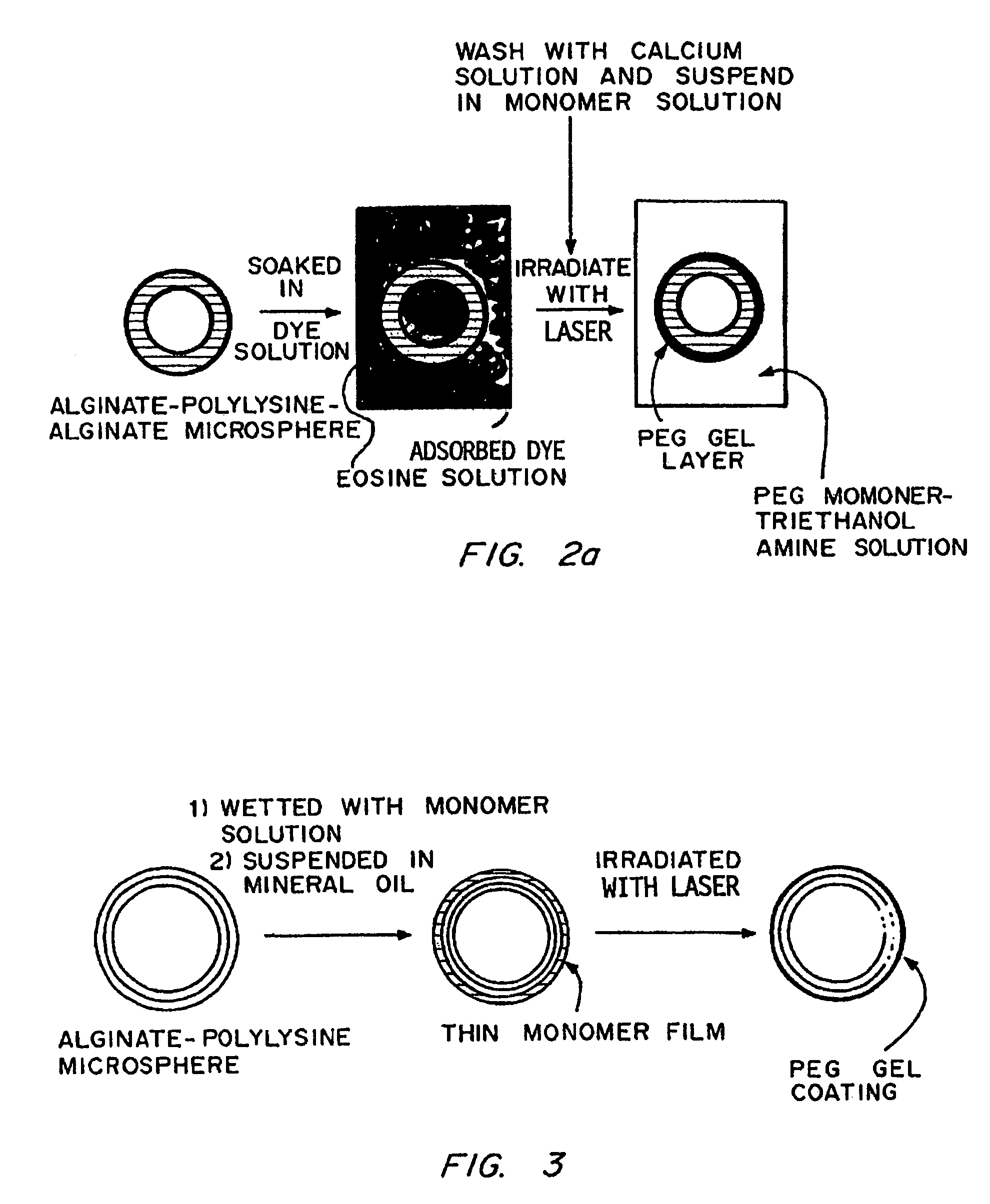
Coating of Islet-Containing Alginate-PLL Microspheres by Surface Dye Adsorption

[0109]The microcapsule interfacial polymerization method was used to form membrane around alginate-PLL microcapsules containing islets. Alginate-PLL coacervated microspheres, containing one or two human pancreatic islets each, were suspended in a 1.1% CaCl2-solution and aspirated free of excess solution to obtain a dense plug of microspheres. A solution of ethyl eosin (0.04% w / v) was prepared in a 1.1% CaCl2 solution. This solution was filter-sterilized by passage through a 0.45 μm filter. The plug of microspheres was suspended in 10 mL of the eosin solution for 2 min to allow uptake of the dye. The microspheres were then washed four times with fresh 1.1% CaCl2 to remove excess dye. A solution of PEG 18.5 tetraacrylate (2 mL; 23% w / v) containing 100 μL of a 3.5% w / v solution of triethanolamine in HEPES buffered saline was added to 0.5 mL of those microspheres. The microspheres were exposed to argon ion la...


PUM

PropertyMeasurementUnit
wavelengthaaaaaaaaaa
pore sizesaaaaaaaaaa
molecular weightaaaaaaaaaa

Abstract

This invention provides novel methods for the formation of biocompatible membranes around biological materials using photopolymerization of water soluble molecules. The membranes can be used as a covering to encapsulate biological materials or biomedical devices, as a “glue” to cause more than one biological substance to adhere together, or as carriers for biologically active species. Several methods for forming these membranes are provided. Each of these methods utilizes a polymerization system containing water-soluble macromers, species, which are at once polymers and macromolecules capable of further polymerization. The macromers are polymerized using a photoinitiator (such as a dye), optionally a cocatalyst, optionally an accelerator, and radiation in the form of visible or long wavelength UV light. The reaction occurs either by suspension polymerization or by interfacial polymerization. The polymer membrane can be formed directly on the surface of the biological material, or it can be formed on material, which is already encapsulated.

Description

[0001]This application is a divisional of U.S. application Ser. No. 10 / 761,180, filed Jan. 20, 2004 now abandoned which is a continuation of U.S. application Ser. No. 09 / 811,901, filed Mar. 19, 2001, now U.S. Pat. No. 6,911,227, which is a continuation of U.S. application Ser. No. 08 / 783,387, filed Jan. 13, 1997, now U.S. Pat. No. 6,258,870, which is a divisional of U.S. application Ser. No. 08 / 484,160, filed Jun. 7, 1995, now abandoned, which is a divisional of U.S. application Ser. No. 07 / 958,870, filed Oct. 7, 1992, now U.S. Pat. No. 5,529,914, which is a continuation-in-part of U.S. application Ser. No. 07 / 870,540, filed Apr. 20, 1992, now abandoned, which is a continuation-in-part of U.S. application Ser. No. 07 / 843,485, filed Feb. 28, 1992, now abandoned, which is a continuation-in-part of U.S. application Ser. No. 07 / 598,880, filed Oct. 15, 1990, now abandoned. All of the above-identified patents and applications are expressly incorporated herein by reference.BACKGROUND[0002]...

Claims


Application Information

Patent Timeline
Patent Type & Authority Patents(United States)
IPC IPC(8): C08J7/04C08F2/50A61K9/50A61K9/16A61K35/12A61K35/30A61K35/39A61K38/42A61K38/44A61K47/48A61L24/00A61L24/04A61L26/00A61L27/34A61L27/38A61L27/52A61L29/08A61L31/10A61L31/14C08F290/00C08F290/02C08F290/06C08F291/00C09D153/00C12N5/00C12N5/071C12N11/04
CPCA61K9/1635A61K35/39A61K9/5031A61K9/5073A61K38/42A61K38/44A61K47/48315A61K47/48869A61L24/0031A61L24/0042A61L24/046A61L26/0019A61L27/34A61L27/38A61L27/52A61L29/085A61L31/10A61L31/145A61L31/148C08F290/00C08F290/02C08F290/06C08F290/062C08F290/14C08F291/00C09D153/00C12N5/0012C12N5/0677C12N11/04A61K9/1647A61K35/30C08L71/02A61K2035/128C12N2533/30C12N2533/32C12N2533/40C12N2533/74A61K47/645A61K47/6925
Inventor HUBBELL, JEFFREY A.PATHAK, CHANDRASHEKHAR P.SAWHNEY, AMARPREET S.DESAI, NEIL P.HOSSAINY, SYED F. A.
Owner BOARD OF RGT THE UNIV OF TEXAS SYST
Why Eureka
Try Eureka