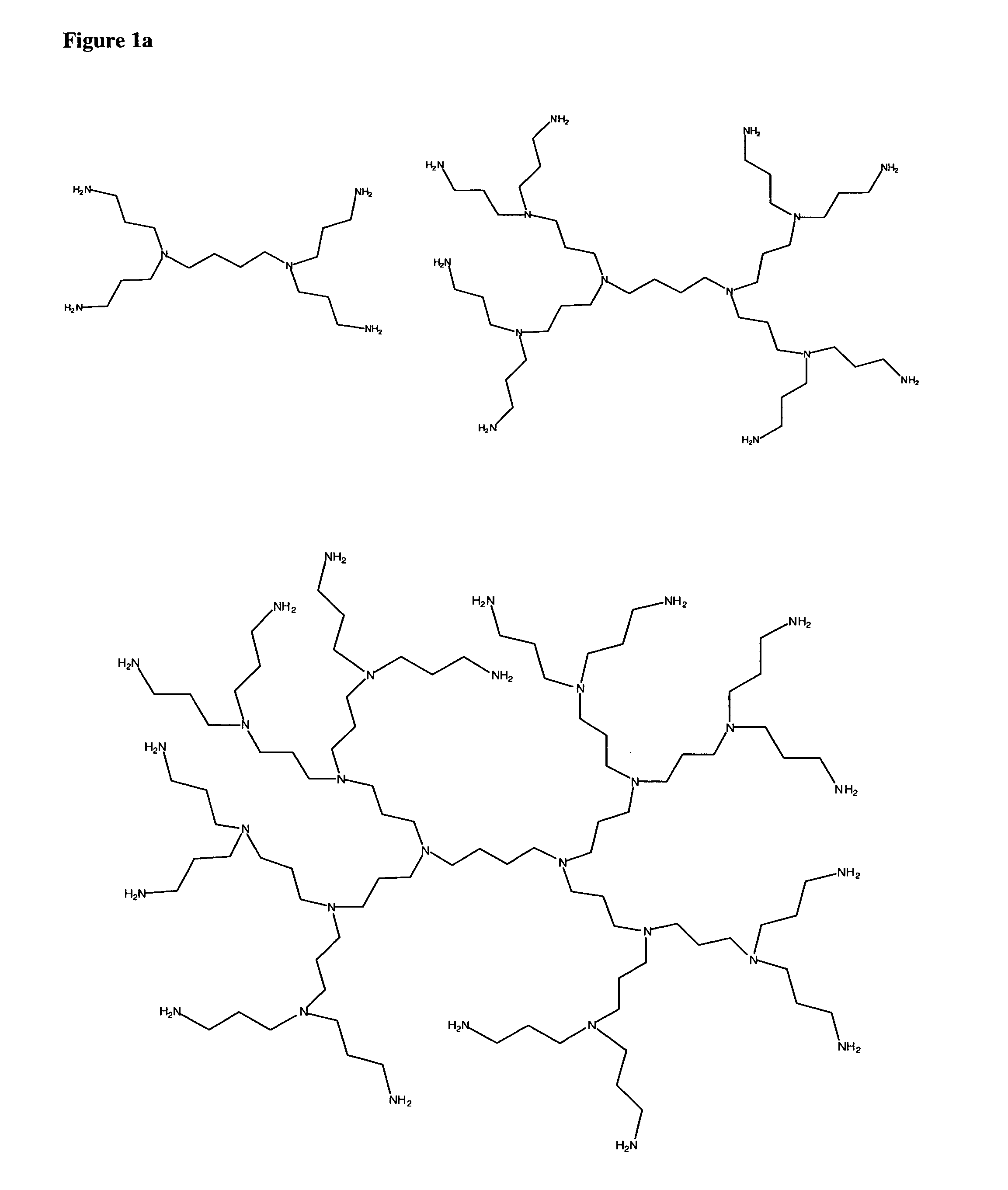
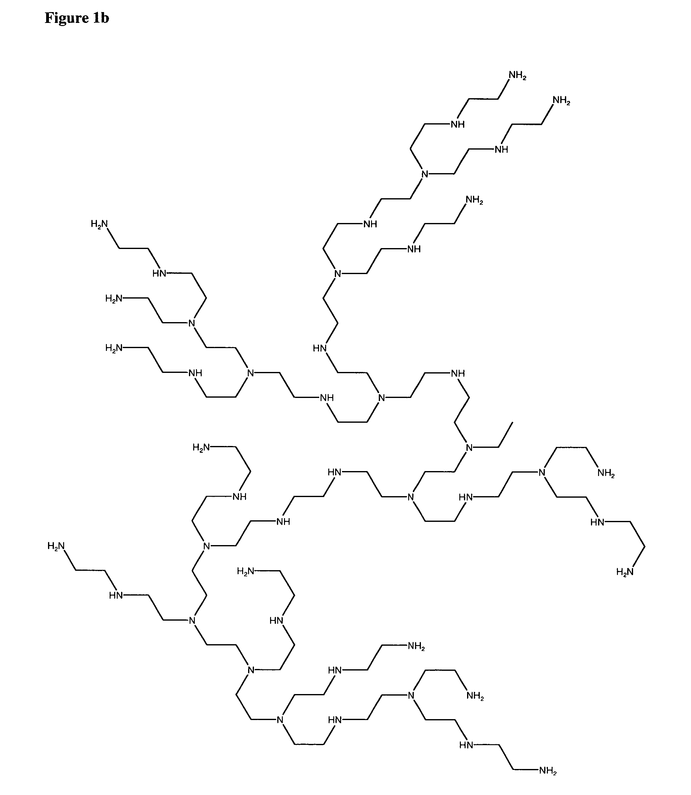
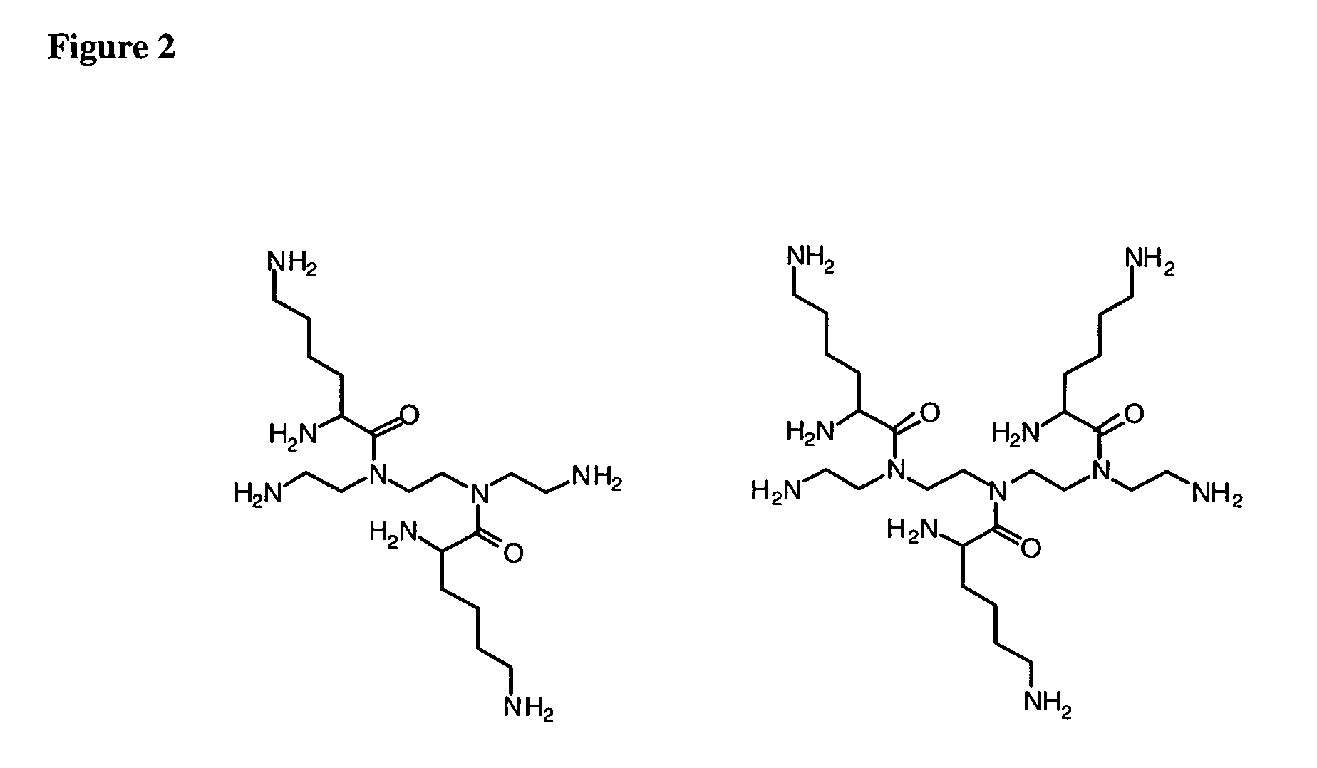
However, larger lacerations often require sutures or a glue to help seal the wound.
Liver lacerations are difficult to repair owing to the nature of liver tissue.
Liver tissue has very weak cohesive strength, and, consequently, sutures and staples are not satisfactory because they may pull through the liver tissue.
The lack of satisfactory wound treatment methods for liver lacerations combined with the fact that it is difficult to reach the veins that feed the liver renders liver lacerations particularly serious.
In fact, severe lacerations of the liver often result in the patient's death due to bleeding.
Risks associated with lung surgery include wound infection; post-surgical internal bleeding; air leaks through the lung wall; pain or numbness at the incision site; and inflammation of the lungs (pneumonia).
Further, air leakage is frequently observed after thoracic procedures, such as pulmonary resection and decortication.
Unfortunately, corneal perforations often lead to loss of vision and a decrease in an individual's quality of life.
However, the surgical procedures are difficult given the delicate composition of the cornea and the severity of the wound which increase the likelihood for leakage and severe astigmatism after surgery.
However, these “super glues” present major inconveniences.
Their monomers, in particular those with short alkyl chains, can be toxic, in part due to their ability to produce formaldehyde in situ.
They also polymerize too quickly leading to applications that might be difficult and, once polymerized, the surface of the glue is rough and hard which leads to patient discomfort and a need to wear contact lens.
The use of sutures has limitations and drawbacks.
First, suture placement itself inflicts trauma to corneal tissues, especially when multiple passes are needed.
Thirdly, corneal suturing often yields uneven healing and resultant regular and irregular astigmatism.
Postoperatively, sutures are also prone to becoming loose and / or broken and require additional attention for prompt removal.
Finally, effective suturing necessitates an acquired technical skill that can vary widely from surgeon to surgeon and can also involve prolonged operative time.
These tests for fluid flow, however, make several assumptions, including that the eye will remain well pressurized during the early postoperative period, that the hydrated wound will not be rapidly deturgesced by the corneal endothelium, and that the absence of aqueous outflow from the wound correlates with the inability of surface fluid from the tear film to flow into the wound, possibly contaminating the aqueous humor and predisposing to infection.
This separation resulted in a wedge-shaped gaping in the internal aspect of the incision.
These studies demonstrated that a transient reduction of intraocular pressure might result in poor wound apposition in clear corneal incisions, with the potential for fluid flow across the cornea and into the anterior chamber, with the attendant risk of endophthalmitis.
Some studies, however, reveal an increased incidence of postoperative endophthalmitis after clear corneal cataract incisions and a recent, retrospective, case-controlled study, reported that clear corneal incisions were a statistically significant risk factor for acute post-cataract surgery endophthalmitis when compared with scleral tunnel incisions.
Post-cataract endophthalmitis remains a potentially blinding complication of a sight-restoring procedure.
However, with trauma, this flap can become dislocated prior to healing, resulting in flap striae (folds) and severe visual loss.
The use of sutures has limitations and drawbacks as discussed above.
These visually debilitating flap complications are seen not uncommonly following the popular procedure LASIK, and are currently treated by flap repositioning and suturing (which require considerable operative time and technical skill).
These intraocular lens implants are held in place by the posterior capsule are not able to provide ocular accommodation.
Techniques commonly used for the treatment of retinal holes, such as cryotherapy, diathermy and photocoagulation, are unsuccessful in the case of complicated retinal detachment, mainly because of the delay in the application and the weak strength of the chorioretinal adhesion.
As noted previously with regard to corneal perforation treatment, the extremely rapid polymerization of cyanoacrylate glues (for example, risk of adhesion of the injector to the retina), the difficulty to use them in aqueous conditions and the toxicity are inconveniences and risks associated with this method.
Risks of retinal tear at the edge of the treated hole can also be observed because of the hardness of cyanoacrylate once polymerized.
Leaking filtering blebs after glaucoma surgery are difficult to manage and can lead to serious, vision-threatening complications.
A filtering bleb can also lead to the loss of bleb function and to the severe complications of endophthalmaitis.
The incidence of bleb leaks increases with the use of antimetabolites.
Bleb leaks in eyes treated with antimetabolities may be difficult to heal because of thin avascular tissue and because of abnormal fibrovascular response.
In a thin-walled or avascular bleb, a suture may not be advisable because it could tear the tissue and cause a larger leak.
However, this procedure can actually worsen the problem if the cyanoacrylate tears from the bleb and causes a larger wound.
One problem with techniques employing sutures or staples is that leakage may occur around the sutures or staples.
Compositions and procedures for proper sealing the consequences of a failed anastomosis are severe and frequently life-threatening.
Failures are caused by myriad factors, including poor surgical technique (e.g., sutures that were not inserted correctly; knots that were tied too tightly rendering the ends ischaemic).
As a result, articular cartilage has limited capacity for self-repair following injury or aging.
Degeneration of cartilage in the meniscus, interverebral disks, or joints can lead to severe and debilitating pain in patients.
Injuries to these tissues are often retained for many years and may eventually lead to more severe secondary damage.
Despite the large number of patients suffering from cartilage degeneration, the only widely-available treatment options for cartilage degeneration are chronic administration of anti-inflammatory agents, total joint replacement, osteotomy, or allograft transplantation, each of which leads to mixed long-term results.
This is a meticulous surgery and suffers from the limitation that pinholes produced by surgical needles can cause leakage.
Moreover, intraoperative dehydration can shrink the dura creating a difficult closure since it is difficult to approximate the edges with sutures.
In older patients, the dura is often more susceptiable to tearing when stretched and / or sutured because the dura can be thin and fragile.
Adhesives such as fibrin have been explored for repair of dura tissue, but have had limited success.
However, one complication of this procedure is that the tissue at the injection site can become infected or susceptible to poor healing.
In fact, AMD is the leading cause of visual impairment in the elderly population.
These new blood vessels tend to be very fragile and often leak blood and fluid.
Damage to the macula occurs rapidly and loss of central vision can occur quickly.
Yet, the vessels do not form properly and leaking results.
This leakage causes scarring in the macula and eventual loss of central vision.
To date poly alkyleneamines (PAIs) have been used primarily as gene transfection agents with limited success.
In general, large PAIs (25,000 molecular weight and higher) are more efficient at forming complexes and condensing with polynucleic acids, but their associated toxicity has also been reported to increase with increasing molecular weight.