Three-dimensional bioresorbable scaffolds for tissue engineering applications

a bioresorbable scaffold and tissue engineering technology, applied in the field of medical equipment and methods, can solve the problems of unsatisfactory two-dimensional scaffolds, increased bone grafting, and less satisfactory when generating functional tissues, so as to improve biocompatibility and hard tissue integration, avoid unfavorable environment, and increase initial flash spread of serum proteins

Inactive Publication Date: 2011-12-06
OSTEOPORE INT PTE
View PDF18 Cites 53 Cited by
  • Summary
  • Abstract
  • Description
  • Claims
  • Application Information

AI Technical Summary

Benefits of technology

[0023]The 3D polymer matrix has degradation and resorption kinetics of 6 to 12 months and the capability to maintain a given space under biomechanical stress / loading for 6 months. Incorporation of a bioresorbable ceramic in the bioresorbable, synthetic and natural polymer produces a hybrid / composite material support triggering the desired degradation and resorption kinetics. Such a composite material improves the biocompatibility and hard tissue integration: the HA / TCP particles, which are embedded into the synthetic polymer matrix, allow for increased initial flash spread of serum proteins compared to the more hydrophobic polymer surface. Furthermore, the basic resorption products of the HA / TCP help buffer the acidic resorption by-products of the aliphatic polyester and thereby help to avoid the formation of an unfavorable environment for the hard tissue cells due to a decreased pH.

Problems solved by technology

Conventional two-dimensional scaffolds are satisfactory for multiplying cells, but are less satisfactory when it comes to generating functional tissues.
Today, bone grafting is increasing and the failure rate is unacceptably high.
The failure rate of autografts is at the lower end of this range, but the need for a second (i.e., donor) site of surgery, limited supply, inadequate size and shape, and the morbidity associated with the donor site are all major concerns.
Furthermore, the new bone volume maintenance can be problematic due to unpredictable bone resorption.
Furthermore, not only is the operating time required for harvesting autografts expensive, but often the donor tissue is scarce, and there can be significant donor site morbidity associated with infection, pain, and hematoma.
Allografting introduces the risk of disease and / or infection; it may cause a lessening or complete loss of the bone inductive factors.
Distraction osteogenesis techniques are often laborious and lengthy processes that are reserved for the most motivated patients.
However, the cartilage repair and regeneration response is limited in terms of form and function.
While many surgical techniques and drug treatments have been proposed over the past 10 years, none have successfully regenerated long-lasting cartilage tissue to replace damaged or diseased cartilage from a clinical point of view.
In a highly porous configuration, however, their mechanical properties may be limited.
Unfortunately, autografted bone is limited in amount and desired morphology.
In addition, the use of the patient's own bone is associated with donor-site morbidity and graft resorption.
However, all metals, most ceramics, and many polymers are not designed to degrade and resorb, and the potential for replacement by host tissue does not exist.
These undesirable mechanical interactions can result in seroma formation and soft tissue inflammation.
Clinically, foreign body reactions are observed and long-term problems include a minimal potential for new bone growth and poor remodeling adjacent to the reconstructed area.
Stress shielding of nearby bone from the alloplastic transplant, due to the mismatch of the mechanical properties of the synthetic implant and host bone, can lead to local tissue atrophy and necrosis.
Due to in vivo polymerization of PMMA, a significant exothermic reaction takes place with temperatures reaching 81° C. The reaction can result in thermal injury to, and necrosis of, adjacent host bone, dura, and other soft tissues.
However, there are numerous drawbacks to using such scaffolds for tissue engineering applications.
For one thing, the pores are not fully interconnected due to the formation of skin-layers during solvent evaporation.
And the pore size varies, as it is difficult to ensure that the porogens are well-dispersed and not agglomerated to form bigger particles.
Also, the scaffolds cannot be made with thick sections inasmuch as deeply embedded porogens become too distant from the surface and residual porogens may be left in the final structure.
In addition, the aforementioned conventional scaffold fabrication techniques do not allow the fabrication of a 3-D scaffold with a varying multiple layer design.
The application of rapid prototyping (RP) technologies in medicine has, until recently, been largely restricted to the surgical planning and simulation for reconstructive surgery, e.g., to the fabrication of prosthesis models such as cranial titanium plates to repair skull defects.
However, the removal of entrapped powder is typically quite difficult.
Unfortunately, aliphatic polyesters can generally only be dissolved in highly toxic solvents such as chloroform and methylene chloride.
Other RP technologies, such as stereolithography (SLA) and Selective Laser Sintering (SLS), pose significant material (non-bioresorbable) constraints for the manufacture of tissue engineering scaffolds.
Ballistic Particular Manufacturing (BPM) is also limited by the lack of suitable biodegradable materials.

Method used


Image

  • Three-dimensional bioresorbable scaffolds for tissue engineering applications
  • Three-dimensional bioresorbable scaffolds for tissue engineering applications
  • Three-dimensional bioresorbable scaffolds for tissue engineering applications

Examples


example 1

(i) Scaffold Fabrication

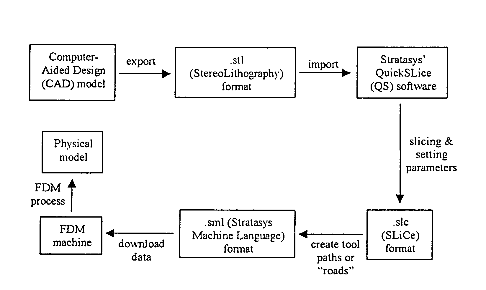
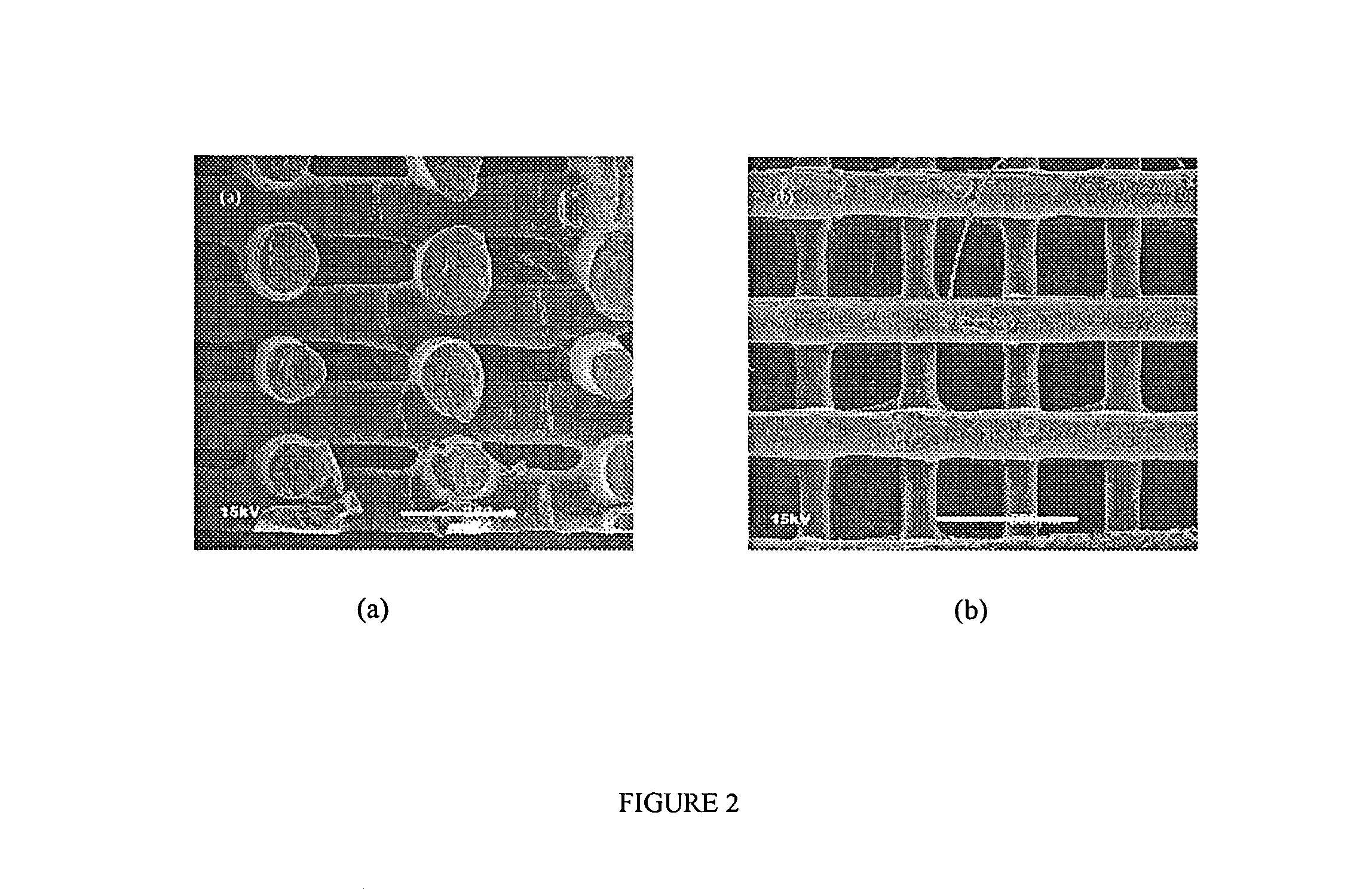
[0094]Scaffold specimens were fabricated using PCL and PCL / HA filaments as described above with a FDM 3D Modeler rapid prototyping system from Stratasys Inc. (Eden Prairie, Minn.). Blocks of 32.0 (length)×25.5 (width)×13.5 mm (height) with a 61% porosity were created directly in the Stratasys' QuickSlice (QS) software. The head speed, fill gap, and raster angle for every layer were programmed through the QS software and saved as an .SLC, the (Slice) file format. Lay-down patterns of 0 / 60 / 120° and 0 / 72 / 144 / 36 / 108° were used to give a honeycomb-like pattern of triangular and polygonal pores, respectively.

[0095]The 2D slice data were converted into QS's .SML (Stratasys Machine Language) file format that automatically generated the build paths based on the input parameters for each slice layer. The .SML data was sent to the FDM machine to fabricate the scaffold specimens using a T16 tip. The liquefier temperature was set at 120° C. and the envelope temperature re...

example 2

(i) Cell Culture Experiments

[0102]One day prior to cell seeding, the 3D scaffolds were sterilized in 70% ethanol overnight. The ethanol was removed by centrifuging three times in changes of phosphate buffered saline (PBS) for 15 minutes with 1000 rpm.

(ii) Fibroblast Cultures And Seeding

[0103]Human fibroblasts were harvested from the Anterior Cruciate Ligament (ACL) of a 34 year old male patient. The fibroblasts were isolated via enzyme digestion by incubating at 37° C., 5% CO2 for 12 hrs with a collagenase-trypsin solution (Sigma, StLouis, Mo.). After the fourth passage, cultures were trypsinized and cell viability was examined via trypan blue (Sigma, St Louis, Mo.) exclusion. The cell pellets obtained after centrifuging the trypsinized cell suspensions were resuspended in 300 μl of DMEM and 450,000 cells in 30 μl were carefully seeded onto 16 scaffolds with a micropipette. The seeded scaffolds were left untouched for 2 hours to allow for protein secretion and cell attachment. A 2 m...

example 3

(i) Seeding and Culture of Chondrocytes

[0112]A study was performed to evaluate three scaffold / cell constructs with different physical and chemical properties, in their potential for tissue engineering elastic cartilage. Group I consisted of PCL scaffolds; Group II, custom-made non-woven PGA fibers; and Group III, collagen sponges. Chondrocytes were isolated from an ear cartilage biopsy of 2 year old male piglets and seeded in combination with a collagen based hydrogel and embedded in a fibrin glue capsule to protect the transplants from an immunological reaction from the host tissue. The specimens were then cultured for 1 week. The scaffold / cell constructs and controls were placed subcutaneously on the paravertebral fascia for 4½ month.

[0113]From the analysis of the histological, immunohistochemical, mechanical, and physico-chemical results of the tissue inside the different scaffolds, it was concluded that a PCL matrix supports cartilage formation whereas a textile construct made o...


PUM

PropertyMeasurementUnit
temperaturesaaaaaaaaaa
decomposition temperatureaaaaaaaaaa
Tmaaaaaaaaaa

Abstract

The invention relates to the use of Fused Deposition Modeling to construct three-dimensional (3D) bioresorbable scaffolds from bioresorbable polymers such as polycaprolactone (PCL), or from composites of bioresorbable polymers and ceramics, such as polycaprolactone / hydroxyapatite (PCL / HA). Incorporation of a bioresorbable ceramic to produce a hybrid / composite material support provides the desired degradation and resorption kinetics. Such a composite material improves the biocompatibility and hard tissue integration and allows for increased initial flash spread of serum proteins. The basic resorption products of the composite also avoids the formation of an unfavorable environment for hard tissue cells due to a decreased pH. The scaffolds have applications in tissue engineering, e.g., in tissue engineering bone and cartilage.

Description

REFERENCE TO RELATED PATENT APPLICATIONS[0001]This application is a divisional of U.S. patent application Ser. No. 09 / 957,407, filed Sep. 20, 2001 by Swee Hin Teoh et al., now U.S. Pat. No. 6,730,252, issued May 4, 2004, for THREE-DIMENSIONAL BIORESORBABLE SCAFFOLDS FOR TISSUE ENGINEERING APPLICATIONS, which in turn claims the benefit of U.S. Provisional Patent Application Ser. No. 60 / 233,874, filed Sep. 20, 2000 by Swee Hin Teoh et al. for THREE-DIMENSIONAL BIORESORBABLE SCAFFOLDS FOR TISSUE ENGINEERING APPLICATIONS. The above-identified patent applications are hereby incorporated herein by reference.FIELD OF THE INVENTION[0002]This invention relates to medical apparatus and methods in general, and more particularly to apparatus and methods for tissue engineering.BACKGROUND OF THE INVENTION[0003]Tissue engineering is a truly multidisciplinary field, which applies the principles of engineering, life science, and basic science to the development of viable substitutes that restore, ma...

Claims


Application Information

Patent Timeline
Patent Type & Authority Patents(United States)
IPC IPC(8): B29C41/02A61L27/18A61L27/46A61L27/58D01F6/62
CPCA61L27/18A61L27/46A61L27/58D01F6/625C08L67/04A61F2/30756B33Y80/00B33Y70/00B33Y10/00B33Y70/10
Inventor TEOH, SWEE HINHUTMACHER, DIETMAR WERNERTAN, KIM CHENGTAM, KOCK FYEZEIN, IWAN
Owner OSTEOPORE INT PTE
Why Eureka
Try Eureka