When diabetes is present, either the body produces less or no
insulin, and / or does not properly
use insulin.
When
insulin is not produced or used correctly by the body, glucose remains in the bloodstream instead of being shuttled into cells for energy production, resulting in high blood glucose, or high “
blood sugar” levels.
If left untreated, diabetes can lead to death, and even diabetics undergoing doctor-supervised treatment suffer an increased death rate compared to the average
population.
Diabetics also face risk of multiple complications during their lifetime arising from the
disease.
These complications often stem from the disturbance of the body's
metabolism caused by the prolonged high
blood sugar.
This then leads to damage to or
stenosis of the blood vessels, ultimately resulting in a condition termed diabetic
microangiopathy, or literally,
disease of the capillaries related to diabetes.
Longstanding microvascular disease that is widespread may decrease the total capacity of
blood circulation within the body, which both directly and indirectly through
kidney damage contributes to the high
blood pressure condition referenced above.
The most dangerous effect of microvascular disease, is occurrence of
ischemia (decreased
blood supply).
This condition can progress with inadequate supply of
oxygen and nutrients, eventually producing devitalization and change of texture and color of the foot, often starting with a
toe or portion of the
forefoot, which can then spread to the rest of the limb.
Diabetic patients also have
increased risk of complications associated with their lower extremities, especially the feet, due to
nervous system disease, as described above, that can lead to a partial or complete loss of feeling.
A healthy person that starts to feel pain when subjected to continuous
local pressure may shift their body or make other suitable alterations to automatically lessen the discomfort; however, patients having a sensory loss are deprived of this protection and are therefore common victims of
pressure sores and
open wounds that can become ulcerated.
They also tend to balance themselves differently which can cause progressive alteration in the bony structure of the foot.
Diabetic foot lesions are an underlying cause of hospitalization, disability, morbidity, and mortality, especially among
elderly people.
Diabetes is a chronic, life-threatening disease for which there is no known cure.
This rupture can lead to increased pressure on the
dermis, resulting in
tissue ischemia and eventual death, ultimately manifest in the form of an ulcer.6
Neuropathy results in a loss of protective
sensation in the foot, exposing patients to undue, sudden or repetitive stress.
It can lead to
atrophy of the small intrinsic muscles, collapse of the arch, and loss of stability in the metatarsal-phalangeal joints.
Neuropathy leads to lack of awareness of damage to the foot as it may be occurring, physical defects and deformities9 which lead to greater physical stresses on the foot.
Hyaline
basement membrane thickening and capillary leakage may impair
diffusion of nutrients.
Musculoskeletal abnormalities (altered foot
mechanics, limited
joint mobility, bony deformities) can lead to harmful changes in
biomechanics and
gait, increasing the pressures associated with various regions of the foot.
Alteration or
atrophy of fat pads in the foot from increased pressure can lead to
skin loss or
callus, both of which increase the risk of ulceration by two orders of magnitude.
Clearly, however,
foot ulcers can occur in non-diabetics, especially ischemic ulcers seen in patients with
peripheral vascular disease and associated with atherosclerosis, hypertension and a history of smoking.
Foot
pathology associated with
vascular disease is a major source of morbidity among diabetics and a leading cause of hospitalization.
Current solutions are ineffective or incomplete.
Diabetic feet are at risk for a wide range of pathologies including infection, ulceration,
deep tissue destruction, and / or metabolic complications.
Noninvasive techniques now employed in screening for vascular related
foot disease have not proven useful in predicting or preventing disease.
There is currently no method to assess accurately, rapidly, and noninvasively the predisposition to serious foot complications, to define the real extent of disease or to track the
efficacy of therapeutics over time.
Hyaline
basement membrane thickening and capillary leakage may impair
diffusion of nutrients.
When comparing the
microcirculation of the
forearm and foot in diabetic patients with and without neuropathy, the
endothelium-dependent and
endothelium-independent cutaneous vasodilatation is lower in the foot.12 On a histologic level, it is well known that diabetes causes a thickening of the endothelial
basement membrane which in turn may lead to impaired function of the endothelial
cell.
The loss of vasodilatation is then thought to lead to early nerve dysfunction through
ischemia and
nutrient deprivation.15 As neuropathy worsens, the nociceptive C fibers are impaired leading to a loss of the ability to
mount a hyperemic response to
inflammation.16 This places the foot at risk in terms of infection and the ability to heal minor wounds.
Such approaches as this
pose an improvement over the cumbersome, expensive footwear noted above, but this method still suffers from drawbacks, such as ease of use,
mass availability, and expense.
Further, such methods are only useful for analyzing the bottom or sole of the foot and fails to account for pressure points or locations of
shear stress on other parts of the foot.
These other methods also do not take into account generalized (systemic), regional or local influences which may decrease
perfusion or
oxygenation to a given region of the foot.
The effectiveness of these systems to reducing foot ulcerations is still unanswered beyond anecdotal evidence, with groups squaring off between measuring pressure or contour as the important endpoint.
Ischemic ulcers arise from a lack of perfusion to the tissues adequate to meet the demands of maintaining tissue integrity or of healing a minor injury.
By reducing flow to the foot,
peripheral arterial disease can impede healing; reducing the supply of oxygen and nutrients that tissue requires to maintain the repair process and the viability of the dermal barrier, and significantly amplify the problems associated with diabetic microvascular and neuropathic disease.
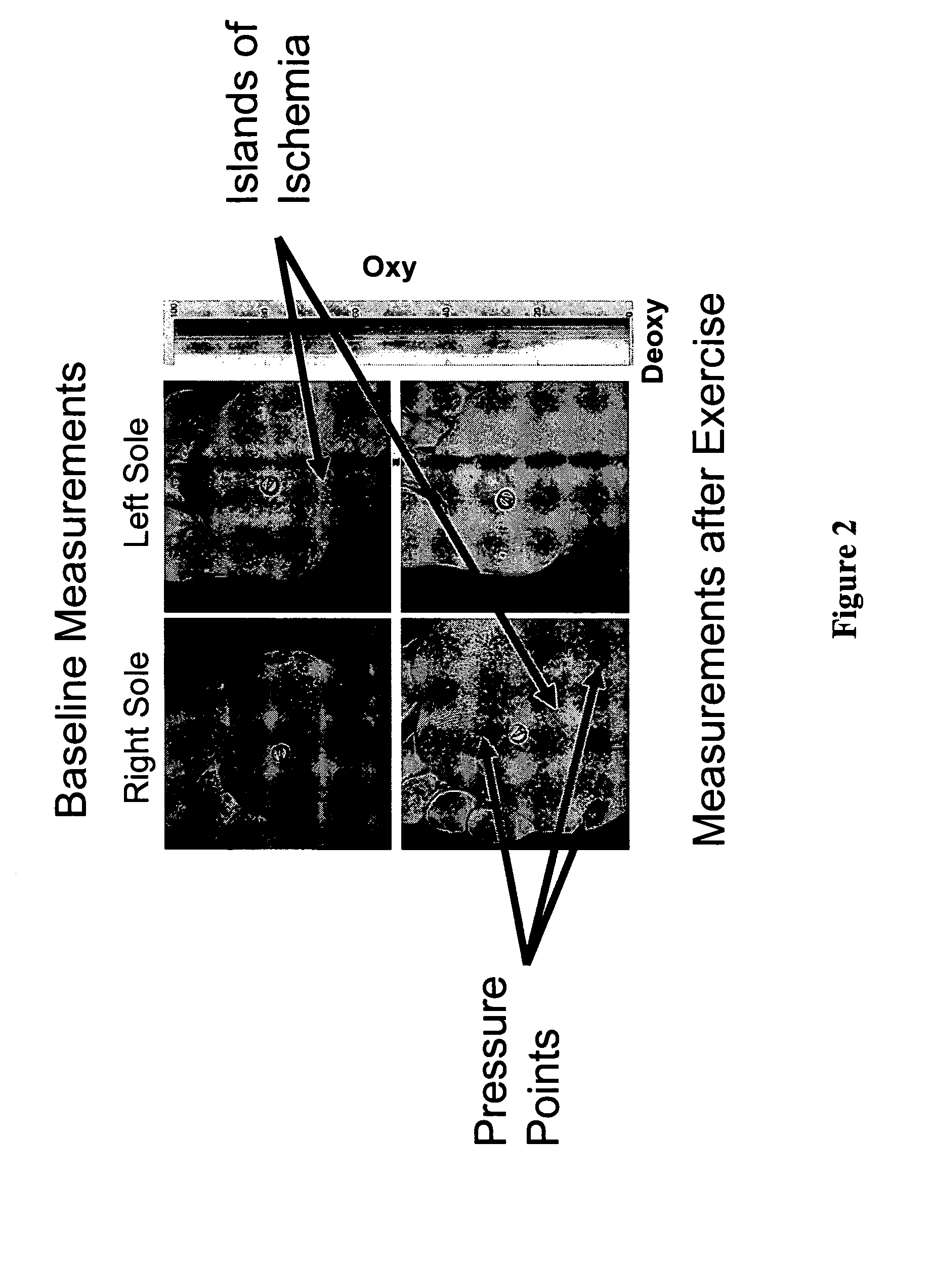
This suggests that despite adequate inflow to the extremity with
peripheral bypass, “islands of
ischemia” exist where inadequate perfusion occurs, thus making the area more susceptible to ulcer formation and inability to heal an ulcer.
If this is not possible, careful use of compression may be undertaken to help decrease the
venous pressure without compromising
arterial flow, but this can be difficult to accomplish.
Such ulcers occur in debilitated, hospitalized, paralyzed, malnourished patient groups and in other situations in which pressure is placed on a region of tissue that in some way compromises its viability.
There are also other situations in which abnormalities of
skin, vasculature or collagen lead to tissue
fragility.
Limb amputation is a significant problem due to a variety of causes including trauma, diabetic disease and atherosclerosis.
The amputee is not only challenged by having the
underlying disease or cause of
amputation to deal with but also having to learn to use the artificial limb and be beleaguered by the attendant complications that may arise from poor prosthetic fit.
This may include recurrent
residual limb breakdown predisposing the patient to pain, stump or tissue ulceration or breakdown,
osteomyelitis, and
sepsis as well as abnormal
gait which can occur with improper fit with a secondary result in safety concern, an increase in the
energy cost of ambulation and the predisposition to developing
osteoarthritis.
However, this maxim only holds true for pure compounds.
It is not nearly enough to take a spectrum from a healthy piece of tissue and a diseased piece of tissue, compare them, and make valid claims regarding their disease state.
The interpretation of in-vivo reflectance data is further complicated in that most physical situations which modify tissue
absorbance also affect tissue scattering.
No other method however provides information towards the
spatial distribution or heterogeneity of the data.
A drawback of
single point DR is that it provides no spatial information of
tissue oxygenation and for complex systems it is clearly desirable to collect spatial information to monitor local variations, as different regions within the tissue may experience vastly different levels of
blood flow, perfusion, and
oxygen extraction.
Foot or other tissue that is poorly perfused or metabolically unstable is more susceptible to the effects of pressure on the region.
Non-invasive clinical assessment of these patients is limited.

