However, the synaptic connections involved in neural circuits are continuously altered throughout the life of the individual, due to
synaptic plasticity and
cell death.
This inability to produce new
nerve cells in most mammals (and especially primates) may be advantageous for long-
term memory retention; however, it is a distinct
disadvantage when the need to replace lost neuronal cells arises due to injury or
disease.
However, this correlation may not always hold as stem cells are thought to be present in tissues (e.g., liver [Travis, Science, 259:1829, 1993]) that do not have a
high turnover of cells.
In addition to neurodegenerative diseases, acute brain injuries often result in the loss of neural cells, the inappropriate functioning of the affected
brain region, and subsequent behavior abnormalities.
This may be due to inappropriate firing of neurons, or the abnormal synthesis, release, and
processing of neurotransmitters.
Demyelination of central and
peripheral neurons occurs in a number of pathologies and leads to improper
signal conduction within the nervous systems.
Unfortunately, this type of treatment has been fraught with many complications including the limited ability to transport drugs across the blood-brain barrier and the
drug-tolerance which is acquired by patients to whom these drugs are administered long-term.
In addition, there are a number of side effects associated with
levodopa such as increased and uncontrollable movement.
However, there are limitations to this technique as well.
There is an additional drawback that O-2A cells isolated by the available procedures are capable of only a limited number of divisions [Raff Science 243:1450-1455 (1989)].
However, adrenal cells do not obtain a normal neural
phenotype, and are therefore probably of limited use for transplants where synaptic connections must be formed.
However, the potential for these cells to induce adverse immune responses, the use of retroviruses to immortalize cells, the potential for the reversion of these cells to an amitotic state, and the lack of response of these cells to
normal growth-inhibiting signals make
cell lines less than optimal for widespread use.
However, there still exists a risk of inducing an
immune reaction using currently available
cell lines.
In addition, these cells may also not achieve normal neuronal connections with the
host tissue.
However, it is likely that the expression of
membrane bound factors and the release of soluble molecules such as growth factors and
proteases will alter the normal behavior of the surrounding tissue.
Another concern that arises when fibroblasts are implanted into the CNS is the possibility that the implanted cells may lead to
tumor formation because the intrinsic inhibition of
fibroblast division is poorly controlled.
Fibroblasts are intrinsically limited in their ability to extend neuronal-like processes and form synapses with
host tissue.
Hence, although the genetic modification and implantation of fibroblasts into the CNS represents an improvement over the
current technology for the delivery of certain molecules to the CNS, the inability of fibroblasts to integrate and function as CNS tissue, their potential negative effects on CNS cells, and their limited intrinsic control of proliferation limits their practical usage for implantation for the treatment of acute or chronic
CNS injury or
disease.
While the studies noted above are encouraging, the use of large quantities of aborted
fetal tissue for the treatment of
disease raises ethical considerations and political obstacles.
In addition, there are serious doubts as to whether an adequate and constant supply of
fetal tissue would be available for
transplantation.
Moreover, the tissue may already be infected with a
bacteria or
virus, thus requiring expensive diagnostic testing for each
fetus used.
However, even diagnostic testing might not uncover all infected tissue.
For example, the diagnosis of HIV-free tissue is not guaranteed because antibodies to the
virus are generally not present until several weeks after infection.
While currently available
transplantation approaches represent a significant improvement over other available treatments for neurological disorders, they suffer from significant drawbacks.
The inability in the prior art of the transplant to fully integrate into the
host tissue, and the lack of availability of cells in unlimited amounts from a reliable source for
grafting are, perhaps, the greatest limitations of neurotransplantation.
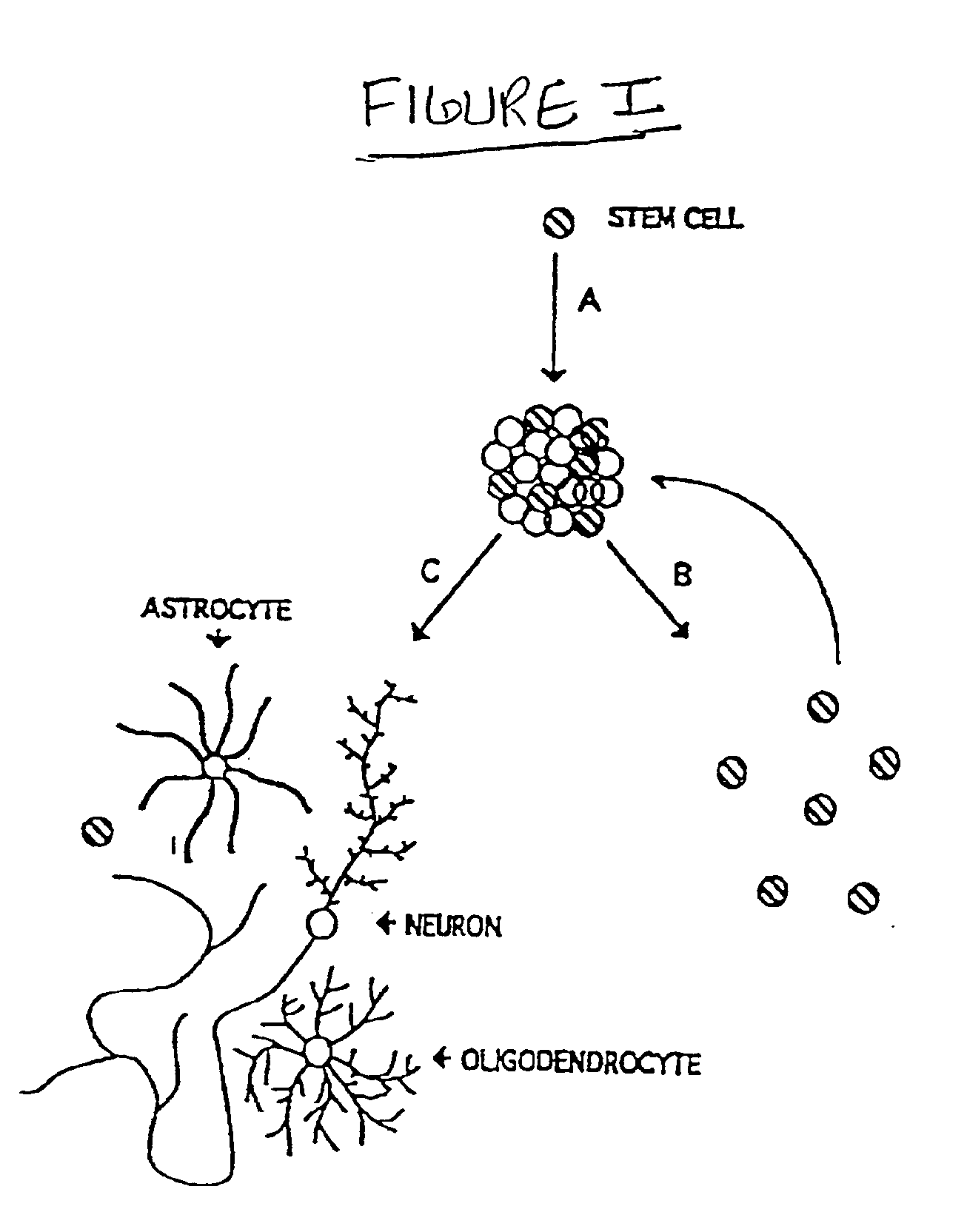
Since adult neural tissue undergoes minimal division, it does not readily meet these criteria.
While astrocytes retain the ability to divide and are probably amenable to infection with foreign genes, their ability to form synapses with neuronal cells is limited and consequently so is their extrinsic regulation of the expression and release of the foreign
gene product
Oligodendrocytes suffer from some of the same problems.
However, due to the limited proliferative ability of
oligodendrocyte progenitors, the infection and harvesting of these cells does not represent a practical source.
However, differentiated neurons do not divide and
transfection with foreign genes by chemical and physical means is not efficient, nor are they stable for long periods of time.
The infection of primary neuronal precursors with retroviral vectors
in vitro is not practical either because neuroblasts are intrinsically controlled to undergo a limited number of divisions making the selection of a large number of neurons, that incorporate and express the foreign gene, nearly impossible.
Due to the complexity of the mammalian CNS, the study of CNS developmental pathways, as well as alterations that occur in adult mammalian CNS due to dysfunction, has been difficult.
While primary CNS cultures have many advantages, they suffer from two primary drawbacks.
First, due to the limited proliferative ability of primary neural cells, new cultures must be generated from several different animals.
While great care is usually taken to obtain tissue at identical states of development and from identical brain regions, it is virtually impossible to generate primary cultures that are identical.
A second
disadvantage of primary cultures is that the tissue must be obtained from fetuses or early post-natal animals.
Due to the limited supply and ethical concerns, the culturing of primary cells from primates (both human and non-human) is not practical.
Due to the limited proliferative ability of primary neural cells, the generation of a large number of homogenous cells for studies of
neural function, dysfunction, and
drug design / screening has previously not been achieved.
While these types of cell lines are able to generate a large number of cells for screening the effects of exogenous agents on
cell survival or function, the limited number of these types of lines, the limited number of phenotypes that they are able to generate and the unknown nature of their immortalization (which may effect the function of the cells in an undefined manner) makes these types of cell lines less than ideal for
in vitro models of
neural function and discovery of novel therapeutics.
First, the addition of an
oncogene that alters the proliferative status of a cell may affect other properties of the cell (oncogenes may play other roles in cells besides regulating the
cell cycle).
Another drawback to using intentionally immortalized cells results from the fact that the
nervous system is composed of billions of cells and possibly thousands of different cell types, each with unique patterns of
gene expression and responsiveness to their environment.
While a large supply of one
neural cell type can be generated, this approach does not take into account cellular interactions between different cell types.
In addition, while it is possible to immortalize cells from a given
brain region, immortalization of a desired cell is not possible due to the lack of control over which cells will be altered by the
oncogene.
Hence, while custom designed cell lines offer a few advantages over spontaneously occurring tumors, they suffer from several drawbacks and are less than ideal for understanding CNS function and dysfunction.

