Photopolymerizable biodegradable hydrogels as tissue contacting materials and controlled-release carriers

a biodegradable, photopolymerizable technology, applied in the direction of cell encapsulation, powder delivery, granular delivery, etc., can solve the problems of poor biocompatibility, frequent formation of post-surgical adhesions involving organs of the peritoneal cavity and the peritoneal wall, and insufficient use in the united states. , to achieve the effect of excellent biocompatibility, high hydrophilicity and water solubility, and rapid poly

Inactive Publication Date: 2001-10-23
BOARD OF REGENTS
View PDF136 Cites 163 Cited by
  • Summary
  • Abstract
  • Description
  • Claims
  • Application Information

AI Technical Summary

Benefits of technology

Examples of these macromers are PEG-oligoglycolyl-acrylates. The choice of appropriate end caps permits rapid polymerization and gelation; acrylates were selected because they can be polymerized using several initiating systems, e.g., an eosin dye, by brief exposure to ultraviolet or visible light. The poly(ethyleneglycol) or PEG central structural unit (core) was selected on the basis of its high hydrophilicity and water solubility, accompanied by excellent biocompatibility. A short oligo or poly(.alpha.-hydroxy acid), such as polyglycolic acid, was selected as a preferred chain extension because it rapidly degrades by hydrolysis of the ester linkage into glycolic acid, a harmless metabolite. Although highly crystalline polyglycolic acid is insoluble in water and most common organic solvents, the entire macromer is water-soluble and can be rapidly gelled into a biodegradable network while in contact with aqueous tissue fluids. Such networks can be used to entrap and homogeneously disperse water-soluble drugs and enzymes and to deliver them at a controlled rate. Further, they may be used to entrap particulate suspensions of water-insoluble drugs. Other preferred chain extensions are polylactic acid, polycaprolactone, polyorthoesters, and polyanhydrides. Polypeptides may also be used. Such "polymeric" blocks should be understood to include timeric, trimeric, and oligomeric blocks.
These materials are particularly useful for controlled drug delivery, especially of hydrophilic materials, since the water soluble regions of the polymer enable access of water to the materials entrapped within the polymer. Moreover, it is possible to polymerize the macromer containing the material to be entrapped without exposing the material to organic solvents. Release may occur by diffusion of the material from the polymer prior to degradation and / or by diffusion of the material from the polymer as it degrades, depending upon the characteristic pore sizes within the polymer, which is controlled by the molecular weight between crosslinks and the crosslink density. Deactivation of the entrapped material is reduced due to the immobilizing and protective effect of the gel and catastrophic burst effects associated with other controlled-release systems are avoided. When the entrapped material is an enzyme, the enzyme can be exposed to substrate while the enzyme is entrapped, provided the gel proportions are chosen to allow the substrate to permeate the gel. Degradation of the polymer facilitates eventual controlled release of free macromolecules in vivo by gradual hydrolysis of the terminal ester linkages.
An advantage of these macromers are that they can be polymerized rapidly in an aqueous surrounding. Precisely conforming, semi-permeable, biodegradable films or membranes can thus be formed on tissue in situ to serve as biodegradable barriers, as carriers for living cells or other biologically active materials, and as surgical adhesives. In a particularly preferred embodiment, the macromers are applied to tissue having bound thereto an initiator, and polymerized to form ultrathin coatings. This is especially useful in forming coatings on the inside of tissue lumens such as blood vessels where there is a concern regarding restenosis, and in forming tissue barriers during surgery which thereby prevent adhesions from forming.
Examples demonstrate the use of these macromers and polymers for the prevention of postoperative surgical adhesions in rat cecum and rabbit uterine horn models. The polymer shows excellent biocompatibility, as seen by a minimal fibrous overgrowth on implanted samples. Hydrogels for the models were gelled in situ from water-soluble precursors by brief exposure to long wavelength ultraviolet (LWUV) light, resulting in formation of an interpenetrating network of the hydrogel with the protein and glycosaminoglycan components of the tissue. The degradable hydrogel was very effective, both by itself and in combination with tPA, in preventing adhesions.

Problems solved by technology

However, they have not been used extensively in the United States due to concerns relating to disease transmission from blood products.
Synthetic polymers have been explored as adhesives (Lipatova, 1986, "Medical polymer adhesives," Advances in Polymer Science 79:65-93), but these materials have been associated with local inflammation, cytotoxicity, and poor biocompatibility.
Formation of post-surgical adhesions involving organs of the peritoneal cavity and the peritoneal wall is a frequent and undesirable result of abdominal surgery.
Numerous approaches to elimination of adhesion formation have been attempted, with limited success in most cases.
However, none of these approaches has been cost effective and effective in in vivo studies.
(1990) "Reduction of postoperative adhesions in the rat uterine horn model with poloxamer 407, Am. J. Obstet. Gynecol., 162(5):1317, examined Poloxamer solutions in peritoneal adhesion models and observed statistically significant reductions in adhesions; however, they were unable to eliminate adhesions, perhaps because of limited adhesion and retention on the injury site.
In summary, several lavage / drug / material approaches have been explored, but none of these approaches has been able to eliminate adhesions.
None of the approaches developed and described to date meet these requirements.
Differences in crystallinity also alter degradation rates.
Additionally, some proteins may be irreversibly denatured by contact with organic solvents used in dispersing the protein within the polymer.
Rapidly-degrading polymers currently suggested for short-term macromolecular drug release may raise local concentrations of potentially hazardous acidic degradation byproducts.
Further, all biodegradable synthetic polymers reported thus far can only be processed in organic solvents and all biodegradable polymers are synthesized under conditions which are not amenable to polymerization in vivo.
Thus, it has not been possible to make implantable materials as precisely conformed barriers, shaped articles, or membranes capable of delivering bioactive materials to the local tissue.

Method used


Image

  • Photopolymerizable biodegradable hydrogels as tissue contacting materials and controlled-release carriers
  • Photopolymerizable biodegradable hydrogels as tissue contacting materials and controlled-release carriers
  • Photopolymerizable biodegradable hydrogels as tissue contacting materials and controlled-release carriers

Examples


example 1

Synthesis of Photopolymerized Biodegradable Hydrogels.

PEG-based Hydrogels

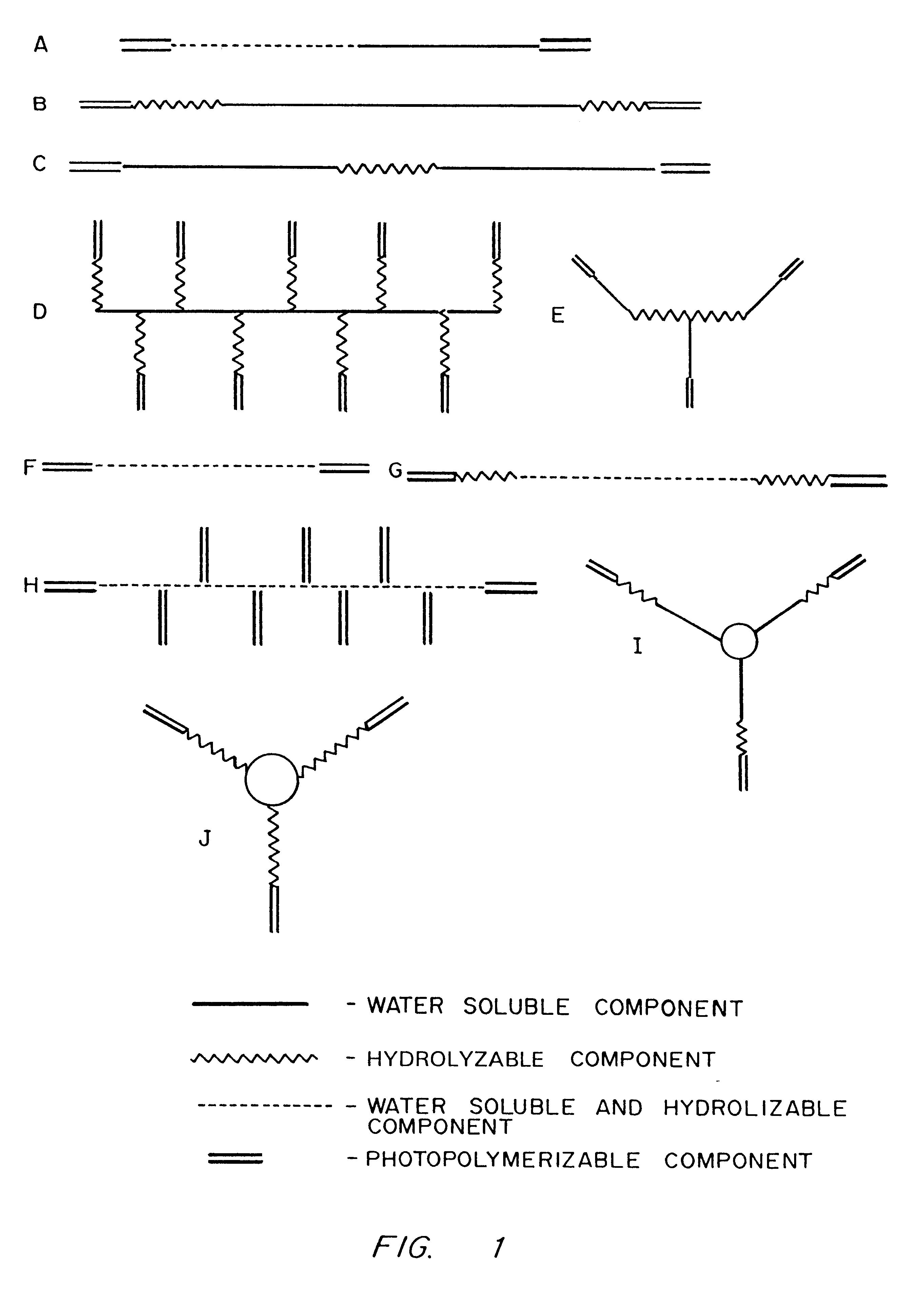
PEG-based biodegradable hydrogels are formed by the rapid laser or UV photopolymerization of water soluble macromers. Macromers, in turn, are synthesized by adding glycolic acid oligomers to the end groups of PEG and then capping with acrylic end groups. The PEG portions of the macromers confer water solubility properties, and subsequent polymerization results in cell-nonadhesive hydrogels. Glycolic acid oligomers serve as the hydrolyzable fraction of the polymer network, while acrylic end groups facilitate rapid polymerization and gelation of the macromers.

In preparation for synthesis, glycolide (DuPont) or DL-lactide (Aldrich) was freshly recrystallized from ethyl acetate. PEG oligomers of various molecular weight (Fluka or Polysciences) were dried under vacuum at 110.degree. C. prior to use. Acryloyl chloride (Aldrich) was used as received. All other chemicals were of reagent grade and used without further p...

example 2

Use of Multifunctional Macromers

30 g of a tetrafunctional water soluble PEG (MW 18,500) (PEG 18.5 k) was dried by dissolving the polymer in benzene and distilling off the water benzene azeotrope. In a glove bag, 20 g of PEG 18.5 k, 1.881 g of glycolide and 15 mg of stannous octoate were charged into a 100 ml round bottom flask. The flask was capped with a vacuum stopcock, placed into a silicone oil bath and connected to a vacuum line. The temperature of the bath was raised to 200.degree. C. The reaction was carried out for 4 hours at 200.degree. C. and 2 hours at 160.degree. C. The reaction mixture was cooled, dissolved in dichloromethane and the copolymer was precipitated by pouring into an excess of dry ethyl ether. It was redissolved in 200 ml of dichloromethane in a 500 ml round bottom flask cooled to 0.degree. C. To this flask, 0.854 g of triethylamine and 0.514 ml of acryloyl chloride were added under nitrogen atmosphere and the reaction mixture was stirred for 12 h. at 0.degr...

example 3

Synthesis of a Photosensitive Macromer Containing DL-lactide

PEG (MW) 20,000) (PEG 20 k) was dried by dissolving in benzene and distilling off the water benzene azeotrope. In a glove bag, 32.43 g of PEG 20 k, 2.335 g of DL-lactide and 15 mg of stannous octoate were charged into a 100 ml round bottom flask. The flask was capped with a vacuum stopcock, placed into a silicone oil bath and connected to a vacuum line. The temperature of the bath was raised to 200.degree. C. The reaction was carried out for 4 hours at 200.degree. C. The reaction mixture was cooled, dissolved in dichloromethane and the copolymer was precipitated by pouring into an excess of dry ethyl ether. It was redissolved in 200 ml of dichloromethane in a 500 ml round bottom flask cooled to 0.degree. C. To this flask, 0.854 g of triethylamine and 0.514 ml of acryloyl chloride were added under nitrogen atmosphere and the reaction mixture was stirred for 12 hours at 0.degree. C. The triethyl amine hydrochloride was separa...


PUM

PropertyMeasurementUnit
solubilityaaaaaaaaaa
molecular weightaaaaaaaaaa

Abstract

Hydrogels of polymerized and crosslinked macromers comprising hydrophilic oligomers having biodegradable monomeric or oligomeric extensions, which biodegradable extensions are terminated on free ends with end cap monomers or oligomers capable of polymerization and cross linking are described. The hydrophilic core itself may be degradable, thus combining the core and extension functions. Macromers are polymerized using free radical initiators under the influence of long wavelength ultraviolet light, visible light excitation or thermal energy. Biodegradation occurs at the linkages within the extension oligomers and results in fragments which are non-toxic and easily removed from the body. Preferred applications for the hydrogels include prevention of adhesion formation after surgical procedures, controlled release of drugs and other bioactive species, temporary protection or separation of tissue surfaces, adhering of sealing tissues together, and preventing the attachment of cells to tissue surfaces.

Description

FIELD OF THE INVENTIONThe present invention relates to photopolymerizable biodegradable hydrogels for use as tissue adhesives and in controlled drug delivery.BACKGROUND OF THE INVENTIONThis is a continuation-in-part of U.S. patent application Ser. No. 07 / 843,485, filed Feb. 28, 1992, now abandonded entitled "Photopolymerizable Biodegradable Hydrogels as Tissue Contacting Materials and Controlled Release Carriers" by Jeffrey A. Hubbell, Chandrashekhar P. Pathak, and Amarpreet S. Sawhney.Hydrogels as Controlled-release CarriersBiodegradable hydrogels can be carriers for biologically active materials such as hormones, enzymes, antibiotic, antineoplastic agents, and cell suspensions. Temporary preservation of functional properties of a carried species, as well as controlled release of the species into local tissues or systemic circulation, are possible. Proper choice of hydrogel macromers can produce membranes with a range of permeability, pore sizes and degradation rates suitable for a...

Claims


Application Information

Patent Timeline
Patent Type & Authority Patents(United States)
IPC IPC(8): A61K9/16A61L27/52A61L24/04A61L27/38A61L29/08A61L27/00A61L27/34A61L26/00A61L24/00A61L29/00A61L31/14A61L31/10A61L31/08C08F291/00C08F290/06C08F290/00C09J153/00C09D153/00C12N5/00C12N5/06C12N11/04C12N11/00A61K35/12A61K9/50A61K9/00A61K38/48A61K47/30A61K47/32A61L31/16B82B1/00C12N5/071
CPCA61K9/0024A61K9/1647A61K47/32A61L24/0031A61L24/0042A61L24/046A61L26/0019A61L27/34A61L27/36A61L27/38A61L27/3839A61L27/52A61L29/085A61L31/10A61L31/145A61L31/148A61L31/16C08F290/00C08F290/06C08F291/00C09D153/00C09J153/00C12N5/0012C12N5/0677C12N11/04A61K9/1635C12Y304/21068A61K38/47A61K38/49C12Y302/01017C08L71/02Y10T428/29A61K2035/128A61L2300/424A61L2300/602C12N2533/30C12N2533/32C12N2533/40C12N2533/74Y10T428/2913Y10T428/2967Y10T428/31786A61K9/50B82Y5/00
Inventor HUBBELL, JEFFREY A.PATHAK, CHANDRASHEKHAR P.SAWHNEY, AMARPREET S.DESAI, NEIL P.HILL, JENNIFER L.
Owner BOARD OF REGENTS
Why Eureka
Try Eureka