Method of producing fused biomaterials and tissue

a biomaterial and tissue technology, applied in the field of producing fused biomaterials and tissue, to achieve the effect of reducing the likelihood of peritonitis

Inactive Publication Date: 2000-07-11
GREGORY KENTON W +1
View PDF18 Cites 119 Cited by
  • Summary
  • Abstract
  • Description
  • Claims
  • Application Information

AI Technical Summary

Benefits of technology

In addition to the above, the biomaterial of the invention can be used as a patch material for use in intestinal or colon repairs which frequently do not heal well with current techniques, particularly when the patient has nutritional or other problems or when the patient is in shock, such as in the case of multiple gunshot wounds or other abdominal injuries (see FIG. 3). The use of such a patch can, for example, seal off intestinal contents and thereby reduce the likelihood of peritonitis. In addition, a patch can be used on a solid organ, such as the liver, when lacerations have occurred. Similarly, the biomaterial of the invention can be used to repair or replace portions of the urinary system i.e., from the calyces of the kidney on down to the urethra. The patch can also be used to seal a defect in a cardiac chamber, such as an atrial septal defect, as well as bronchial or rectal fistulas. The biomaterial can also be used as a cerebrovascular patch for an aneurysm. The biomaterial can be sealed in place with targeted laser fusio...

Problems solved by technology

However, their use is of...

Method used


Image

  • Method of producing fused biomaterials and tissue
  • Method of producing fused biomaterials and tissue
  • Method of producing fused biomaterials and tissue

Examples


example 2

Tissue Welding of Sheets of Elastin-Based Biomaterial

Pre-welding procedure: A 1 mg / ml ICG solution was applied to fresh swine aorta that had been carefully trimmed of adventitia, washed in a sterile 0.9% NaCl solution, and cut into 1 cm2 squares. The 1 mg / ml ICG solution was applied to the lumenal side of the aorta for .about.3 min and wiped off. (ICG was obtained from Sigma and contained 90% dye and 10% sodium iodide. Absorption coefficient measured at 780 nm with a 7.25.times.10.sup.-6 M solution was found to be 175,000 M.sup.-1 cm.sup.-1. The adsorption maximum shifts to 805 nm when ICG is bound to serum proteins (Landsman et al, J. Appl. Physiol. 40 (1976). A small amount of cryoglobulins, containing approximately 40 mg / ml fibrinogen and 10 mg / ml fibronectin doped with ICG, was also applied and the biomaterial placed on it. The two materials were placed between two glass slides. This was submerged in a 0.9% saline solution.

Welding Procedure: Sheets of biomaterial made as describ...

example 3

Preparation of Elastin-Based Biomaterial from Artery Digest

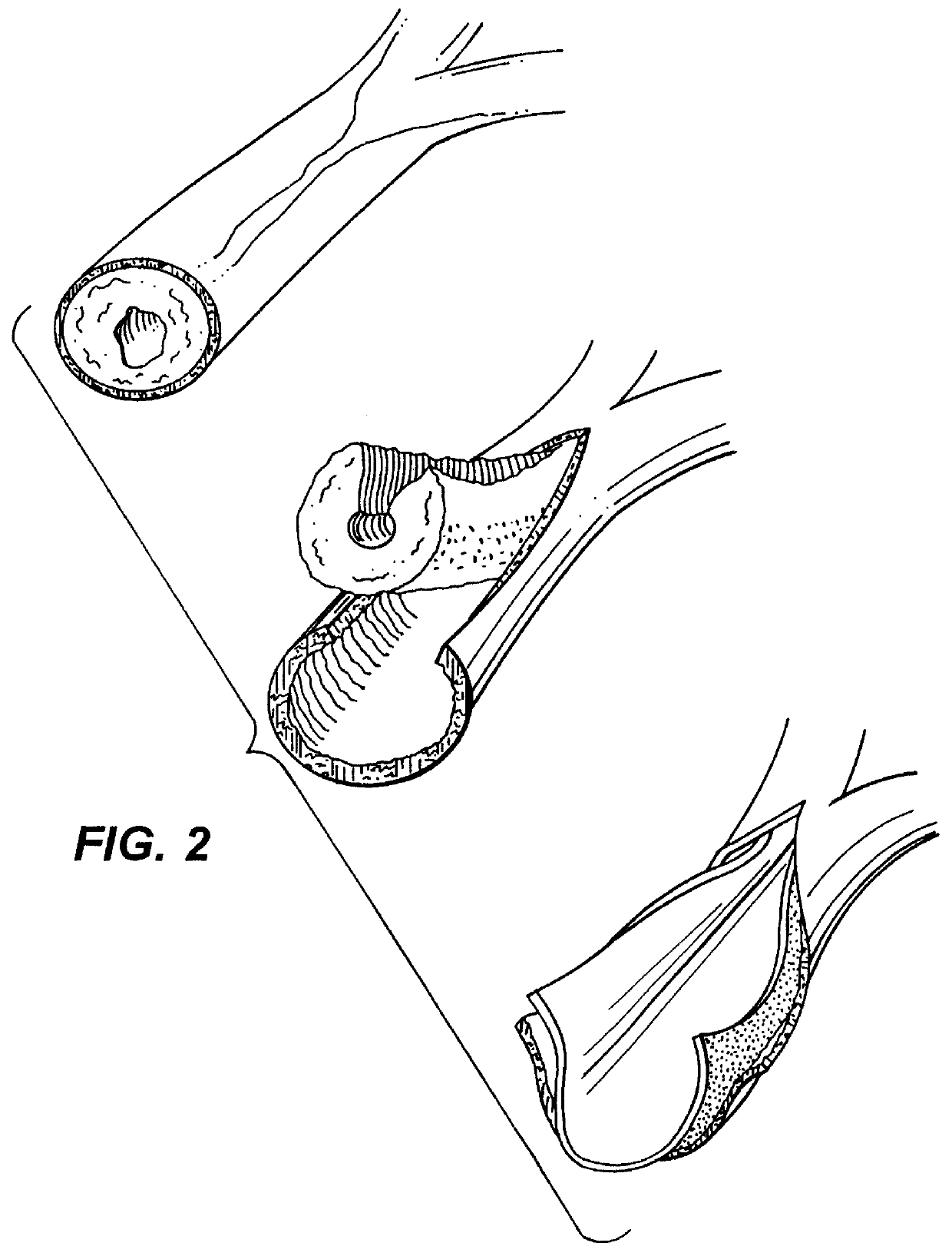
Fresh 4 cm lengths of porcine carotid artery were dissected clean and washed in two changes of 0.9% saline overnight. Vessels were then placed in 0.5M NaOH and sonicated for 120 minutes (a modified method of Crissman, R. 1987) See Crissman, Rogert S. "Comparison of Two Digestive Techniques for Preparation of Vascular Elastic Networks for SEM Observation", Journal of Electron Microscopy Techniques 6:335-348 (1987). Digested vessels were then washed in distilled water and autoclaved at 225.degree. F. for 30 minutes. Digested vessels appear translucent, pearly white in color and collapsed when removed from water indicating the absence of collagen and other structurally supportive proteins.

Welding of the artery digests to porcine aorta was accomplished through the following methods. Fresh porcine aorta was coated with 5 mJ / ml ICG for 5 minutes. Excess ICG solution was blotted off. One.times.one cm sections of NaOH-sonicated dige...

example 4

Preparation of Elastin-Based Biomaterial & Fusion to Porcine Aorta

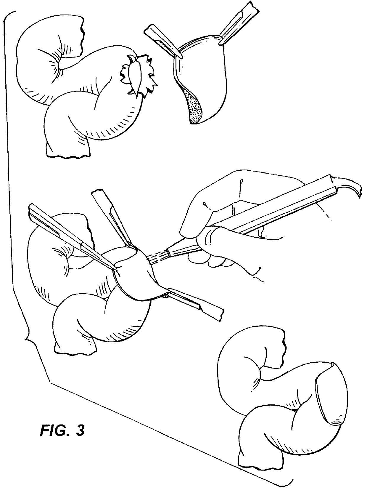
Materials: Bovine nuchal elastin powder (Sigma St. Louis Mo.) Was sifted with a 40 .mu.m screen and swollen with phosphate buffer. Elastin fragments were then reacted with 67 mg of fibrinogen (Sigma):in phosphate buffer, 2 m acid soluble Type 1 collagen (Sigina), 2.8 mg thiourea, 2 mM Ca.sup.2+, 1 mM Mg.sup.2 + and 75 units of thrombin and injected into molds and heated to 77.degree. C. One mm thick sheets and tubes of this biomaterial were removed and stored in 33% ethanol for later use.

Indocyanine green dye was dissolved in de-ionized water to provide a 1% solution and applied to the lumenal surface of fresh porcine aorta. The dye was in place for 5 minutes then the residual dye was blotted off. The elastin biomaterial was placed on the ICG stained aorta and covered with a glass coverslip. Laser energy was applied with a condenser which collected the output of an array of gallium arsenide diode lasers emitting light...


PUM

PropertyMeasurementUnit
Thicknessaaaaaaaaaa
Angleaaaaaaaaaa
Temperatureaaaaaaaaaa

Abstract

It is a general object of the invention to provide a method of effecting tissue repair or replacement using a biomaterial. It is a specific object of the invention to provide a biomaterial suitable for use as a stent, for example, a vascular stent, or as a conduit replacement, as an artery, vein or a ureter replacement. The biomaterial can also be used as a stent or conduit covering or lining. The present invention relates to a method of repairing, replacing or supporting a section of a body tissue. The method comprises positioning a biomaterial at the site of the section and bonding the biomaterial to the site or to the tissue surrounding the site. The bonding is effected by contacting the biomaterial and the site, or tissue surrounding the site, at the point at which said bonding is to be effected, with an energy absorbing agent. The agent is then exposed to an amount of energy absorbable by the agent sufficient to bond the biomaterial to the site or to the tissue surrounding the site.

Description

The present invention relates to the biomaterials in tissue repair and replacement. The invention further relates to methods of securing the biomaterials to existing tissue.Collagen, fibrin, elastin and various other elastin-based biomaterials are known biomaterials. Collagen is an insoluble fibrous protein that occurs in vertebrates as the chief constituent of connective tissue fibrils and in bones. Fibrin is a white insoluble fibrous protein formed from fibrinogen by the action of thrombin especially in the clotting of blood. Elastin is an extracellular matrix protein that is ubiquitous in mammals. Other biomaterials include silicone, poly(etherurethane urea),poly(etherurethane), poly(esterurethane), poly(ethylene), poly(prolene), poly(tetrafluoroethylene), polyvinylidene fluoride, polycarbonate, poly(ethylene terephthlate), poly(methyl methacrylate), polystyrene, poly(vinylchloride), poly(2-hydroxyethylmethacrylate),poly (vinylpyrrolidone), poly(acrylonitrile), polygycolide, poly...

Claims


Application Information

Patent Timeline
IPC IPC(8): A61F2/06A61F2/00A61L27/22A61L27/38A61L27/00A61L27/50A61L27/36A61L31/00A61L31/04A61L31/14B32B9/02B32B5/02A61F2/04
CPCA61F2/0063A61F2/06A61F2/07A61L27/227A61L27/3641A61L27/3691A61L27/3804A61L27/3839A61L27/50A61L31/005A61L31/047A61L31/14B32B5/02B32B9/02A61F2/042A61F2/91A61F2002/072A61F2002/075
Inventor GREGORY, KENTON W.
Owner GREGORY KENTON W
Why Eureka
Try Eureka