[0015] One
advantage of the present invention is that it uses a culture independent approach for the
rapid detection of
Bifidobacterium species DNA in order to determine the source of fecal
pollution in environmental water samples. Methods for detecting Bifidobacterium spp. in
municipal sewage and
animal waste water have been developed, however, a method for detecting Bifidobacterium spp. as markers of fecal contamination in environmental water sources has not previously been shown to be effective. In certain embodiments, for example, the
present method can detect and determine the source of Bifidobacterium spp. in water samples having an enterococcal count of approximately 22 CFU / 100 ml within 24 hours.
[0016] Although several methods of isolating
bacteria may be compatible with the present invention, the isolation of
bacteria from the environmental
water sample is preferably performed by capturing the bacteria on a
solid adsorbent. Isolation of bacteria according to certain embodiments of this invention may be obtained by
filtration of the
water sample through a filter membrane with a pore size of 0.20-0.44 μm. More preferably, a filter membrane with a pore size of 0.22 μm can be used. In such preferred embodiments, the pore size should be sufficiently small so that bacteria are prevented from flowing through the filter and instead become trapped on the filter. The
filter material may be, but is not limited to,
nitrocellulose,
cellulose,
polycarbonate, Teflon, nylon,
polycarbonate,
polyester, polyethersulfone, or
polypropylene. Appropriate housing for the
membrane filter will generally be determined by the
sample volume to be processed; shape of the filter and size or
diameter of the filter and can be determined by one of ordinary skill in the art.
Filtration can be conducted under appropriate
atmospheric pressure or under vacuum. In order to ensure sufficient capturing of the bacteria on the filter, a flow rate in the range of 20-80 cm / Hg, should be considered. More preferably, a flow rate of 40 cm / Hg can be used. Sample volumes in the range of 0.5 ml to more than 1 L are compatible with the current method, but smaller or larger volumes may be suitable. In one embodiment, a
sample volume of 100 mls is used. In other embodiments of the present invention, multiple filter membranes may also be employed in the
filtration step. For example, a larger pore size membrane may be stacked on top of a smaller pore size membrane in order to trap larger particulate matter and other elements that may interfere with the downstream extraction and isolation of
bacterial DNA. The membrane filters may comprise the same or different materials. Isolation of
Bacterial DNA [0017] Isolation of
bacterial DNA can be accomplished using any number of
DNA extraction methods known in the art. For example, isolation of bacteria DNA
cell lysis may be effected by brief
exposure to extremes of pH, organic solvents, chaotropic agents like
urea and
guanidine HCl, detergents like
sodium dodecyl sulfate (SDS) and Triton X-100, osmotic shock,
lysozyme digestion,
protease digestion, or the use of shear forces. The DNA can be separated from lysed
cellular debris and other potential interfering substances through such means as
organic solvent extraction, acid
precipitation,
ultrafiltration,
solid-phase extraction, HPLC, LiCl
precipitation,
protease digestion, RNase digestion, or
polyethylene glycol
precipitation and the like. In one embodiment, the DNA may be extracted using a modification of the “Alternative Protocol” of the UltraClean™
Soil DNA kit (MO BIO Laboratories, Carlsbad, Calif., Product # 12800). For example, the filter containing the trapped bacteria can be placed in a
Petri dish. The bead solution may be initially separated from the beads and placed in the
Petri dish containing the filter. Solutions S1 and IRS may then be added directly to the
Petri dish. In such embodiments, the amounts of solutions S1 (MO BIO's
lysis solution) and IRS (MO BIO's inhibitor removal solution) are preferably added in the proportions indicated by the kit's protocol and sufficient to completely cover the filter membrane. The dish is then vortexed vigorously for 5-30 minutes. The solution can then be removed and returned to the bead tubes. The reminder of the isolation step follows the manufacture's protocol, or protocols known to those of ordinary skill in the art. Nested PCR for Detection of B. adolescentis
[0018] A nested PCR protocol should be used to detect B. adolescentis in environmental water samples. In such a protocol, the first step should comprise amplification of the isolated
bacterial DNA using a universal eubacteria 16S rRNA primer in combination with a Bifidobacterium
genus specific primer. Suitable primer pairs can be determined by one of skill in the art using such tools as BLAST and any number of readily available primer design programs such as Primer3 (Steve Rosen and Helen J. Skaletsky (2000) Primer3 on the WWW for general users and for biologist programmers. In: Krawetz S, Misener S (eds)
Bioinformatics Methods and Protocols: Methods in
Molecular Biology. Humana Press, Totowa, N.J., pp 365-386). An example of a preferred universal eubacteria primer is 785R (SEQ ID NO: 1) and an example of a preferred Bifidobacterium
genus specific primer is IM26F (SEQ ID NO: 2).
[0019] The PCR reaction can be performed using any number of commercially available PCR kits and protocols available and know in the art. In one preferred embodiment, the PCR reaction is performed in 50 μl volume with 0.3 mM dNTP, 4 mMgCl2, 1.5 U
Taq DNA polymerase, 1×PCR reaction buffer, and approximately 30 ng of template DNA. The concentrations of all of the above components can be varied to further optimize the PCR reactions, if needed, and can be determined by one of ordinary skill in the art. In addition, PCR enhancing agents such as DMSO,
betaine,
formamide,
glycerol, nonionic detergents,
bovine serum albumin,
polyethylene glycol,
tetramethylammonium chloride and the like can be added to further increase yield, specificity, and consistency as needed. The PCR reaction can be run on any suitable PCR thermocycler. The choice of denaturation, annealing and extension temperatures, the length of time for each step in a
thermal cycle, and the total number of cycles can be determined by one of ordinary skill in the art and will be specific to the primers used and the target sequence to be amplified. In a PCR reaction utilizing the 785R and IM26F primer pair, the following conditions can be used: initial denaturing at 94° C. for 5 minutes; 30 cycles of 94° C. for 30 seconds, 48° C. for 30 seconds, and 72° C. for 30 seconds; and final elongation at 72° C. for 5 minutes. The amplified product from this PCR reaction is then used as the template for a second PCR mixture For the second PCR reaction, the template from the first reaction is amplified using B. adolescentis
species specific primers. The choice and design of the primers can be determined by one of ordinary skill in the art using the methods referred to above. An example of a preferred B. adolescentis specific primer pair is ADO1 (SEQ ID NO: 3) and ADO2 (SEQ ID NO: 4) (6). A volume of 1 to 5 μl of the first PCR reaction can be used. The PCR reaction can be performed using any number commercially available kits and protocols. In one embodiment, 1 μl of PCR product from the first reaction was added to added to a 50 μl reaction mixture containing the same concentrations of MgCl2, reaction buffer, dNTP, and
Taq polymerase as used in the first reaction. PCR enhancing agents like those listed above may also be added to the reaction as needed. The choice of denaturation, annealing and extension temperatures, the length of time for each step in a
thermal cycle and the total number of cycles can be determined by one of ordinary skill in the art and will be specific to the primers used and the target sequence to be amplified. In a PCR reaction utilizing the ADO1 and ADO2 primer pair, the following conditions can be used: initial denaturing at 94° C. for 5 minutes; 45 cycles of 94° C. for 30 seconds, 48° C. for 30 seconds, and 72° C. for 30 seconds; and final elongation at 72° C. for 5 minutes.
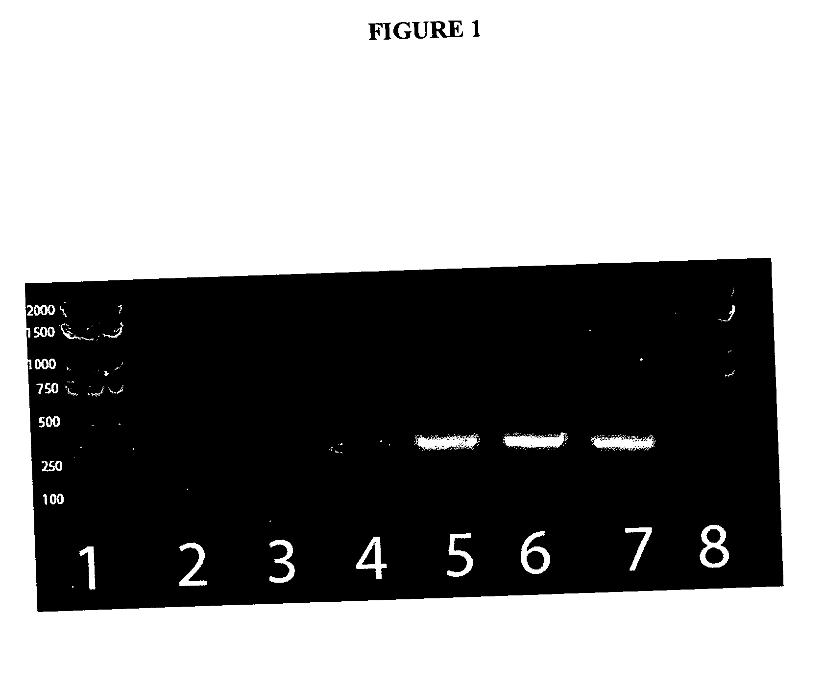
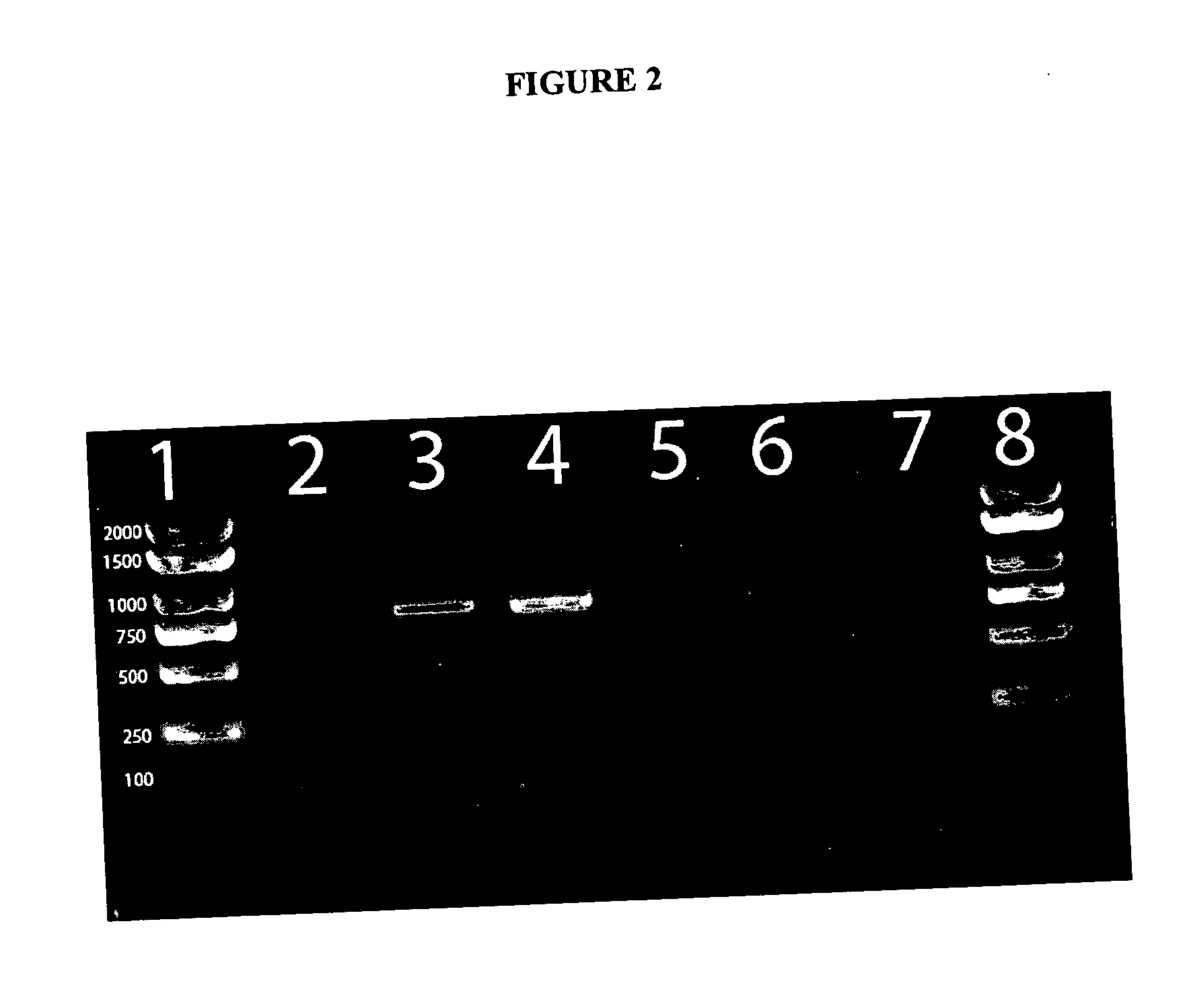
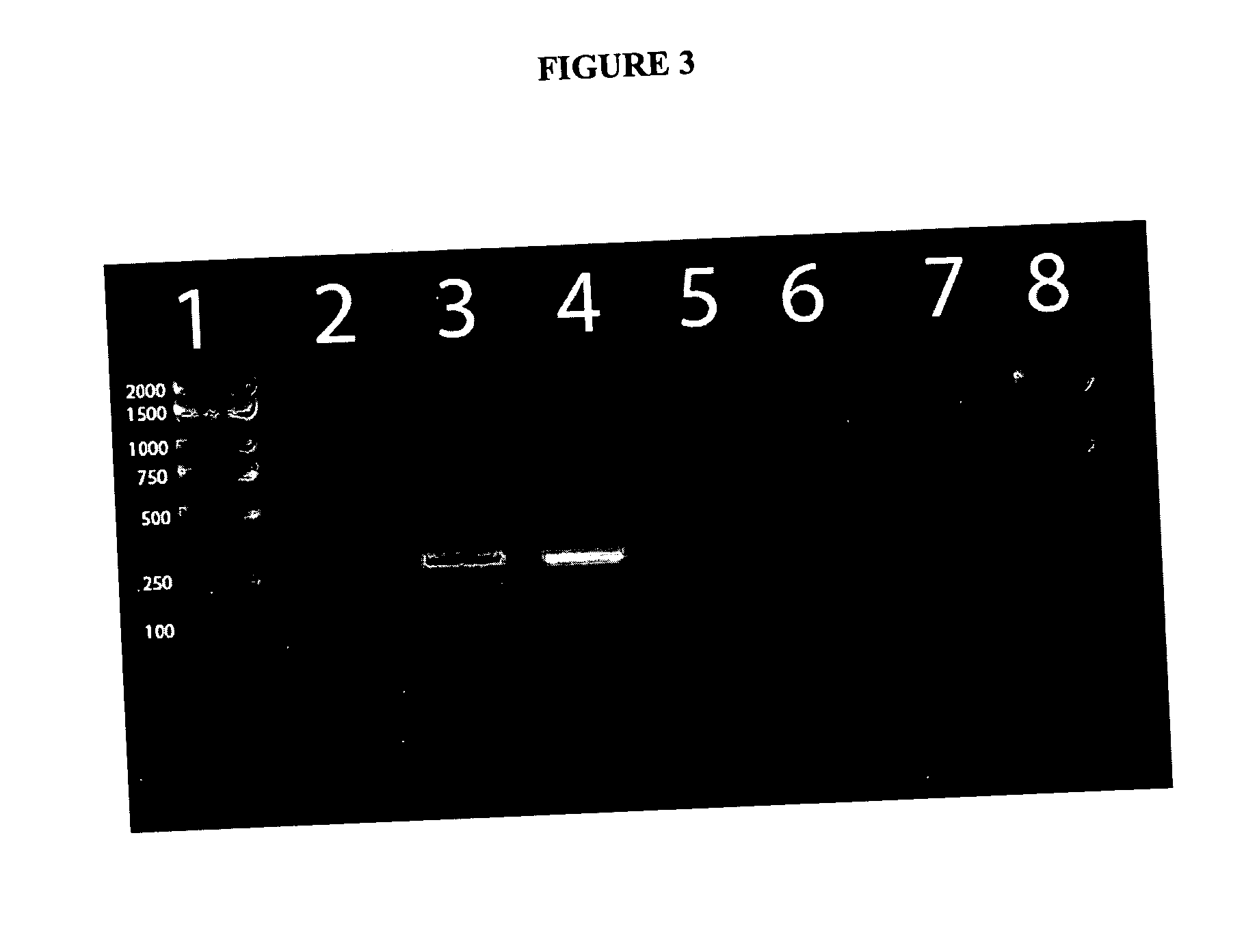
[0020] The products of both PCR reactions can be analyzed by
gel electrophoresis on
polyacrylamide or
agarose gels or other suitable medium and visualized by
staining using appropriate
staining agents such as
ethidium bromide. In certain embodiment of the present invention, the PCR product amplified using primers 785R and IM26F result is 777 bp and the product amplified by the ADO1 and ADO2 primer pair is 279 bp. The above method is also compatible with real time PCR and can be modified for such by one of ordinary skill in the art using any number of commercially available kits and real time PCR thermocylers. The use of real time PCR has the
advantage of not only detecting B. adolescentis, but also providing a rough approximation of the amount of B. adolescentis in the sample (M.W. Pfaffl Nucleic Acids Res. 2001, 29(9):e45). The detection of B. adolescentis indicates 15 the presence of human fecal contamination in the
water sample. The above PCR reactions can be combined into a single reaction, however, previous experiments have shown improved sensitivity and consistency when the nested PCR is conducted as two separate reactions. DNA-DNA Hybridization for
Source Determination of Fecal
Contamination 

