In addition, since chondrocytes are confined in a small area called a lacuna, once
cartilage is damaged, the migration and
recovery of chondrocytes are difficult.
Moreover, since cartilage is avascular tissue, there are no blood vessels for the supply of nutrients, and thus the migration of stem cells is hindered and the regenerative capacity of tissue is reduced.
These characteristics mean that it is almost impossible that once-damaged cartilage is naturally repaired.
However,
drug therapy only has an effect of non-specifically relieving pain or an
inflammatory response itself, and a cartilage protective agent may serve to temporarily protect a joint by simply supplying
nutrition to chondrocytes or
softening impact.
Osteochondral graft is a therapeutic method of
transplanting bone-cartilage
connective tissue that has been previously taken from a patient's own cartilage tissue in a region which is subjected to less load by his / her
body weight into a
cartilage damage site, and this method has a
disadvantage that it cannot be used when a damage site is large.
The first commercialized technique is autologous chondrocytes implantation, which is a technique of detaching a little bit of healthy cartilage from a region of a patient's cartilage tissue, which is subjected to less load by
body weight, isolating chondrocytes therefrom, and then injecting them again into the damage site after
in vitro culture, and has disadvantages that there is a hassle of undergoing two surgeries, cartilage in a healthy region is damaged, due to a lower number of obtained chondrocytes, the cells should be cultured for a certain period, dedifferentiation in which the
phenotype of chondrocytes is not maintained occurs during the culture period,
cell viability after implantation is reduced, and the implanted chondrocytes are not uniform and are localized in a specific region due to gravity so that they are not well distributed.
Particularly, a hydrogel-type
scaffold has poor supply of
oxygen and nutrients so that
cell viability and cartilage differentiation are reduced; a membrane-type
scaffold cannot form three-dimensional cartilage tissue; and when a three-dimensional
sponge or mesh-type
scaffold is used, manufactured artificial cartilage has low
bonding strength to
host tissue so that it is difficult to regenerate cartilage.
In addition, all scaffolds are degradable, and natural biomaterials with a high degradation rate have a high possibility of cells being lost while degradation occurs, in terms of clinical application, xenogeneic and allogeneic natural materials may trigger immune responses, and since
synthetic materials sometimes have harmful degradation products, they are not free from safety issues.
Research on a method of manufacturing artificial cartilage tissue having a three-dimensional structure without a scaffold has been continuously conducted, and since this method forms tissue depending on only cells and their ability to synthesize the
extracellular matrix, it is difficult to form tissue suitable for a damage size requiring implantation, and thus this method is very limited in direct clinical application.
In addition, since the shape and depth of
cartilage damage are not uniform, when artificial cartilage having a three-dimensional structure, which has been manufactured in a laboratory, is larger than the damage site, an
implant needs to be trimmed to the shape of the damage, whereas when a cartilage
implant is smaller than the damage site, it has to be implanted according to a damaged shape by being pieced together like a mosaic.
Currently developed tissue-
engineering cartilage products are implanted by the above-cited method, but cannot match the thickness of the damage.
However, since this culture method cannot regulate the number of cells forming one
cell aggregate and fusion between the formed chondrogenic tissues may occur, there will be variations in the size of chondrogenic tissue and the degree of cartilage differentiation, and thus such a cell aggregate cannot be standardized as a cell therapeutic agent.
This method has an
advantage of regulating the number of cells forming chondrogenic tissue, but due to a difference in ability of naturally forming a cell aggregate according to the state of cells, the method cannot ensure stable acquirement of uniform chondrogenic tissues.
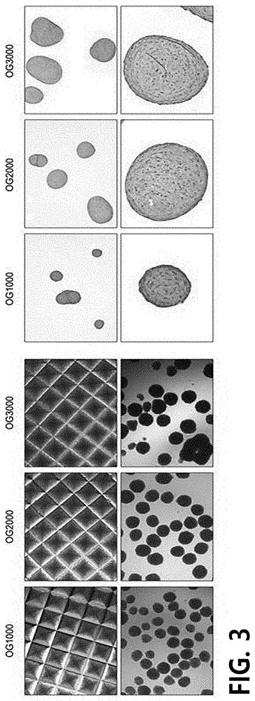
The method of manufacturing micro-tissue using a micromold is evaluated in various cells with a commercialized mold, but since it is also a method of inducing natural
cell aggregation, it cannot ensure stable acquirement of uniform chondrogenic tissue.
In addition, since the formed cell aggregate is very small, it has poor
physical strength and is difficult to
handle, and therefore, it is limited in use as a three-dimensional chondrocyte-based therapeutic agent.
However, the evaluation of the availability of the cartilage structure formed by pellet culture as a cell therapeutic agent was not performed.
Although the pellet
system is a useful method for forming high-quality chondrogenic tissue, it has been considered that it is difficult to be applied in regeneration of a cartilage damage site because of difficulty in forming a pellet with a sufficient size.
In addition, general pellet culture had a
disadvantage that there is difficulty in
mass culture because a method of putting a cell suspension into a capped tube (
conical tube,
storage tube, microcentrifuge tube, etc.), centrifuging, and performing three-dimensional culture to form one pellet per tube was used.
However, because of a low differentiation rate and an unstable
phenotype, the mesenchymal stem cells are most likely to have undesirable differentiation or transformation during differentiation, and therefore, a large amount of mesenchymal stem cells has to be collected from a patient.
In addition, due to the expression of hypertrophy-related genes after
in vivo implantation, the mesenchymal stem cells cause
apoptosis and vascular penetration, resulting in
calcification of chondrocytes.
When cartilage tissue forming a joint is damaged, arthritis with swelling, fever and pain is triggered.
Like this, arthritis is a
disease with very
high incidence in a wide range of
age groups, and due to the fact that once-damaged tissue is not naturally regenerated or repaired, it becomes a cause of limiting the social activity of a patient and reducing his / her
quality of life for a long time.
Currently used agents for arthritis are mostly therapeutic agents requiring surgery, and these agents take a long time for repair or do not have distinct cartilage regeneration
efficacy.
Since human embryonic stem cells are produced from an
embryo that is able to develop into a human, they have many ethical problems, but are known to exhibit excellent cell proliferation and differentiation, compared with adult stem cells.
A disadvantage that a culture technique for maintaining and proliferating undifferentiated hiPSCs is very complicated, and it takes a considerably long time to fully differentiate
human induced pluripotent stem cells into specific cells has become one of the biggest impediments to the development of related techniques including a differentiation-inducing technique.
Various methods of preparing chondrocytes from hiPSCs have been reported, but to differentiate chondrocytes from human induced pluripotent stem cells, a required period increases, unpacking of chondrocyte pellets occurs in the cultured pellets, chondrocytes that are not fully mature are obtained, or hiPSCs differentiate into non-chondrocytes as well as the chondrocytes, and there is a limit that there is diversity in the degree of differentiation for each chondrocyte.


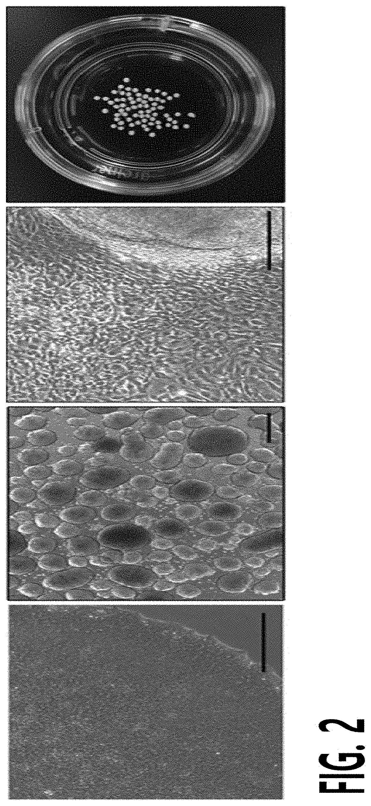