
Implantation of encapsulated biological materials for treating diseases

a technology of encapsulated biological materials and diseases, applied in the field of diseases and their composition, can solve the problems of not being able to be transported into the cells where it is needed and utilized, wasting the appearance of many patients with poorly controlled insulin-dependent diabetes, and unable to meet the needs of the body

Inactive Publication Date: 2008-09-23
NOVOCELL
View PDF119 Cites 84 Cited by
  • Summary
  • Abstract
  • Description
  • Claims
  • Application Information

AI Technical Summary

Benefits of technology

[0048]In one embodiment, the invention is directed to a composition for cellular therapy, which includes a plurality of encapsulating devices including a polyethylene glycol (PEG) coating, said PEG having a molecular weight between about 900 and about 3,000 Daltons; and a plurality of cells encapsulated in the encapsulating devices, wherein said

Problems solved by technology

In diabetes, glucose saturates the blood stream, but it cannot be transported into the cells where it is needed and utilized.
As a result, the cells of the body are starved of needed energy, which leads to the wasted appearance of many patients with poorly controlled insulin-dependent diabetes.
Prior to the discovery of insulin and its use as a treatment for diabetes, the only available treatment was starvation followed predictably by death.
Death still occurs today with insulin treatment from over dosage of insulin, which results in extreme hypoglycemia and coma followed by death unless reversed by someone who can quickly get glucose into the patient.
Also, death still occurs from major under dosage of insulin, which leads to hyperglycemia and ketoacidosis that can result in coma and death if not properly and urgently treated.
While diabetes is not commonly a fatal disease thanks to the treatments available to diabetics today, none of the standard treatments can replace the body's minute-to-minute production of insulin and precise control of glucose metabolism.
Therefore, the average blood glucose levels in diabetics generally remain too high.
Diabetes is the leading cause of new blindness, renal failure, premature development of heart disease or stroke, gangrene and amputation, and impotence.
Since the incidence of diabetes is rising, the costs of diabetes care will occupy an ever-increasing fraction of total healthcare expenditures unless steps are taken promptly to meet the challenge.
The medical, emotional and financial toll of diabetes is enormous, and increase as the numbers of those suffering from diabetes grows.
When the number of beta cells drops to a critical level (10% of normal), blood glucose levels no longer can be controlled and progression to total insulin production failure is almost inevitable.
The multiple daily injections of insulin do not adequately mimic the body's minute-to-minute production of insulin and precise control of glucose metabolism.
Blood sugar levels are usually higher than normal, causing complications that include blindness, heart attack, kidney failure, stroke, nerve damage, and amputations.
Eventually, however, the beta cells are gradually exhausted because they have to produce large amounts of excess insulin due to the elevated blood glucose levels.
Ultimately, the overworked beta cells die and insulin secretion fails, bringing with it a concomitant rise in blood glucose to sufficient levels that it can only be controlled by exogenous insulin injections.
These conditions, together with high blood sugar, increase the risk of heart attack, stroke, and circulatory blockages in the legs leading to amputation.
However, high levels of glucose are toxic to beta cells, causing a progressive decline of function and cell death.
This form of diabetes is due to one of several genetic errors in insulin-producing cells that restrict their ability to process the glucose that enters via special glucose receptors.
Beta cells in patients with MODY cannot produce insulin correctly in response to glucose, which results in hyperglycemia.
The currently available medical treatments for insulin-dependent diabetes are limited to insulin administration and pancreas transplantation with either whole pancreata or pancreatic segments.
However, controlling blood sugar is not simple.
Despite rigorous attention to maintaining a healthy diet, exercise regimen, and always injecting the proper amount of insulin, many other factors can adversely affect a person's blood-sugar including stress, hormonal changes, periods of growth, illness, infection and fatigue.
Insulin-dependent diabetes is a life threatening disease, which requires never-ending vigilance.
Pancreas transplantation is usually only performed when kidney transplantation is required, which makes pancreas-only transplantations relatively infrequent operations.
Although pancreas transplants are very successful in helping people with insulin-dependent diabetes improve their blood sugar control without the need for insulin injections and reduce their long-term complications, there are a number of drawbacks to whole pancreas transplants.
Some risks in taking these immuno-suppressive drugs are the increased incidence of infections and tumors that can be life threatening in their own right.
The risks inherent in the operative procedure, the requirement for life-long immunosuppression of the patient to prevent rejection of the transplant, and the morbidity and mortality rate associated with this invasive procedure, illustrate the serious disadvantages associated with whole pancreas transplantation for the treatment of diabetes.
Due to its relatively large size, there are few sites in the body able to accommodate it for the treatment of a disease like diabetes.
Since it grows into the body and the contained cells are not expected to survive for more than a few years, multiple cell removals and reloading of new cells is required for the long-term application of this device.
It has proven quite difficult to flush and reload this type of device while at the same time maintaining the critical cell compartment distance for oxygen diffusion.
For the diabetes product, it has been quite difficult to place this device into the intraperitoneal cavity of large animals, while maintaining its integrity.
This has been due to the difficulty in securing it in the abdomen so that the intestines cannot cause it to move or wrinkle, which may damage or break the device.
This type has shown efficacy in large animal diabetic trials, but has been plagued by problems in the access to the vascular site.
Both thrombosis and hemorrhage have complicated the development of this approach with it currently being abandoned as a clinically relevant product.
Due to low packing densities, the required cell mass for encapsulation causes the length of this type of hollow to approach many meters.
Therefore, this approach was abandoned for treating diabetes since it was not clinically relevant.
In addition, sealing the open ends of the fiber is not trivial and strength has been a problem depending upon the extravascular site.
However, nearly 25 years have passed since these first reports without the ability to demonstrate clinical efficacy.
One of the problems associated with microcapsules is their relatively large size in combination with low packing densities of cells, especially for the treatment of diabetes.
The permselectivity of pure alginate capsules has been difficult to control with the vast majority being wide open in terms of molecular weight cutoff.
However, polylysine and most other similar molecules invite an inflammatory reaction requiring an additional third coating of alginate to reduce the host's response to the capsule.
In addition, it has been difficult to produce very pure alginates that are not reactive within the host after implantation.
Trying to reduce the size of the alginate microcapsules causes two major problems.
First, the production of very large quantities of empty capsules without any cells.
Second, the formation of smaller capsules results in poorly coated cells.
There is no force to keep the contained cells within the center of the microcapsule, which causes the risk of incomplete coatings to go up exponentially with the decrease in the size of the capsules.
The stringent requirements of encapsulating polymers for biocompatibility, chemical stability, immunoprotection and resistance to cellular overgrowth restrict the applicability of prior art methods of encapsulating cells and other biological materials.
These biomaterials are considered biocompatible if they produce a minimal or no adverse response in the body.
Most synthetic materials, however, do not elicit such a reaction.
Thus, they can interact with implanted material through any of these various regions, resulting in cellular proliferation at the implant surface.
Due to the inability of those of skill in the art to provide one or more important properties of successful cell encapsulation, none of the encapsulation technologies developed in the past have resulted in a clinical product.
Biocompatibility—The materials used to make an encapsulating device must not elicit a host response, which may cause a non-specific activation of the immune system by these materials alone.
This process produces many cytokines that will certainly diffuse through the capsule and most likely destroy the encapsulated cells.
Most devices tested to date have failed in part by their lack of biocompatibility in the host.
Small pores capable of keeping out immune cytokines also cause the death of the encapsulated cells from a lack of diffusion of nutritional elements and waste products.
Relevant Size—Many devices are of such a large size that the number of practical implantation sites in the host is limited.
There is little tolerance for a reduction in diffusive distances, due to the initially low oxygen partial pressure.
This would further lower the oxygen concentration to a point where the cells cannot adequately function or survive.
Many device designs have not considered the fact that encapsulated cells have a limited lifetime in the host and require regular replacement.
A major risk of this procedure is the fact that injection of islets into the portal vein leads to increased portal venous pressures depending on the rate of infusion and the amount infused.
Another risk has been elevated portal venous pressures from large volumes of injected islet tissues that are not sufficiently purified.
This also leads to portal venous thrombosis as a complication of this procedure.
Unfortunately, several patients have had bleeding episodes following this procedure.
This reaction can lead to additional problems long term, such as, bowel obstruction.
Attempts at subcutaneous implantation of encapsulated islets have been unable to produce sustainable results in the treatment of diabetes, probably due to some or all of the scientific challenges described above.
However, this study only examined subcutaneous implantation of the encapsulated islet cells over a one-week period.
One of skill in the art would be unable to determine the desired volume of encapsulated cells needed to administer to a subject.

Method used


Image

  • Implantation of encapsulated biological materials for treating diseases
  • Implantation of encapsulated biological materials for treating diseases
  • Implantation of encapsulated biological materials for treating diseases

Examples


example 1

Isolating Islet Cells in Mice

[0253]Donor mice [C57BL / 6] with an age range of 18 weeks old and average size of 33 grams were obtained from supplier. Pancreas was exposed with euthanasia laparotomy. The pancreata were distended with Sigma collagenase, Type V. The pancreata were removed and kept in cold collagenase during transport to the isolation laboratory. The isolation process combined 30 pancreata for the digestion process. The digestate was washed with 10% fetal bovine serum in RPMI and centrifuged. The COBE was prepared for purification and a continuous gradient marker was used to make the gradient densities. The gradient was loaded into the COBE and the pancreata digestate was loaded on top to perform the purification process. The purified islets were collected and washed in RPMI media. The islets were cultured in T75 flasks in modified ICM media supplemented with 10% fetal bovine serum until ready for encapsulation.

Isolating Islet Cells in Primates

[0254]Juvenile Cynomolgus pr...

example 2

Preparation of Conformal Coating Materials

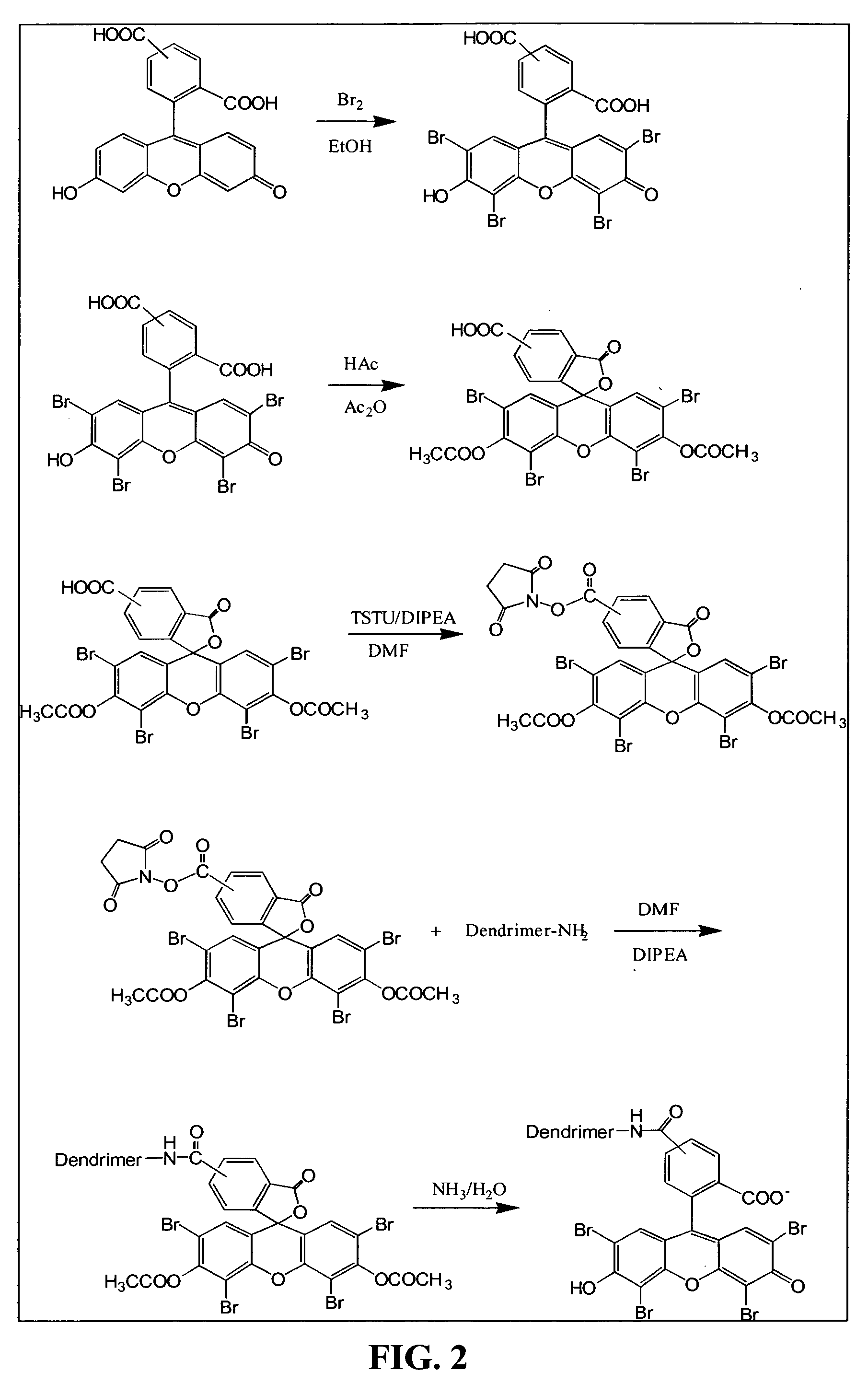
[0256]Depending on the type of cell being encapsulated, the cells were coated directly by a conformal coating or enclosed in a matrix, such as alginate, and then coated with a permselective PEG capsule. FIG. 2 illustrates the synthesis of dendrimer eosin Y conjugate, Dendrimer-EY, a preferred embodiment of this coating, and described as follows.

[0257]The dendrimer used for encapsulation was PAMAM Dendrimer generation 4, which was purchased from Dendritech (FIG. 3). 5(6)-Carboxyeosin was made by bromination of 5(6)-Carboxyfluorescein. The hydroxyl group and 1-carboxyl group were then protected by forming an acetate. The protected 5(6)-Carboxyeosin was activated by N,N,N′N′-Tetramethyl-O-(N-Succinnimidyl)uranium tetrafluoroborate (TSTU). Without further purification, the activated 5(6)-Carboxyeosin diacetate was mixed with PAMAM Dendrimer to form Dendrimer-EY conjugate. The protection group was then removed by reacting with aqueous ammonia. Th...

example 3

Encapsulation of Islets

Encapsulating Mouse Islets

[0262]A preferred method of coating mouse islets is described as follows. Fifteen milliliters of 20 mM low ionic HEPES buffer (containing 1.8 mM CaCl2 and 260 mM Manitol, pH=7.0) was added to a 15 ml conical tube, containing 10 μ1 of islets. The supernatant was removed after centrifugation. 15 ml of Den-EY solution (0.1 mg / ml to 0.4 mg / ml in low ionic HEPES buffer) was added into the pellet and the tube was kept horizontal for 10-30 minutes at room temperature. The stained islets were washed twice with low ionic 20 mM HEPES buffer, which was sparged with Argon for at least 30 minutes. The stained islet pellet was mixed with 10 ml of photoactive polymer solution, which was also sparged with Argon and pre-equilibrated in a 8° C. waterbath for at least 30 minutes. The photoactive polymer solution was made in 20 mM HEPES buffer, pH=8.0, which contained up to 20% PEG, 100 mM TEoA, 32 mg / ml AMPS and 2 μl / ml NVP, and 13% Nycodenz. The suspen...


PUM

PropertyMeasurementUnit
Massaaaaaaaaaa
Massaaaaaaaaaa
Diameteraaaaaaaaaa

Abstract

The present invention relates to compositions and methods of treating a disease, such as diabetes, by implanting encapsulated biological material into a patient in need of treatment. This invention provides for the placement of biocompatible coating materials around biological materials using photopolymerization while maintaining the pre-encapsulation status of the biological materials. Several methods are presented to accomplish coating several different types of biological materials. The coatings can be placed directly onto the surface of the biological materials or onto the surface of other coating materials that hold the biological materials. The components of the polymerization reactions that produce the coatings can include natural and synthetic polymers, macromers, accelerants, cocatalysts, photoinitiators, and radiation. This invention also provides methods of utilizing these encapsulated biological materials to treat different human and animal diseases or disorders by implanting them into several areas in the body including the subcutaneous site. The coating materials can be manipulated to provide different degrees of biocompatibility, protein diffusivity characteristics, strength, and biodegradability to optimize the delivery of biological materials from the encapsulated implant to the host recipient while protecting the encapsulated biological materials from destruction by the host inflammatory and immune protective mechanisms without requiring long-term anti-inflammatory or anti-immune treatment of the host.

Description

RELATED APPLICATIONS[0001]This application claims priority to U.S. provisional application No. 60 / 419,015, filed Oct. 11, 2002 which is incorporated herein by reference in its entirety.FIELD OF THE INVENTION[0002]The present invention relates to compositions and methods of treating a disease, such as diabetes, by implanting encapsulated biological material into a patient in need of treatment.BACKGROUND OF THE INVENTION[0003]Diabetes mellitus is a disease caused by the loss of the ability to transport glucose into the cells of the body, because of either a lack insulin production or diminished insulin response. In a healthy person, minute elevations in blood glucose stimulate the production and secretion of insulin, the role of which is to increase glucose uptake into cells, returning the blood glucose to the optimal level. Insulin stimulates liver and skeletal muscle cells to take up glucose from the blood and convert it into glycogen, an energy storage molecule. It also stimulates ...

Claims


Application Information

Patent Timeline
IPC IPC(8): A61K9/16A61F2/02A61K9/00A61K9/50A61K35/12A61K45/00C12N5/00C12N5/071C12N11/08
CPCA61K9/5031A61K9/5036C12N5/0677C12N11/04C12N11/08C12N5/0012A61K9/0024A61K2035/126C12N2533/74C12N2533/30A61P1/16A61P17/00A61P25/00A61P3/00A61P37/00A61P5/00A61P7/00A61P9/00A61P3/10C12N11/089A61K9/50A61K35/12
Inventor SCHARP, DAVIDLATTA, PAULYUE, CHENGYUNYU, XIAOJIEHUBBELL, JEFFREY ALAN
Owner NOVOCELL
Learn More