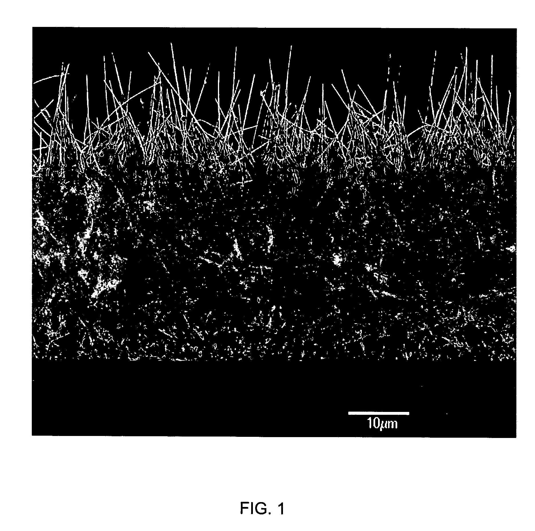
[0008] The plurality of nanostructured components enhance one or more of adhesion, non-adhesion, friction, patency or biointegration of the device with one or more tissue surfaces of a body of a patient depending on the particular application of the device. The nanofibers (or other nanostructured components) on the surfaces of the medical device can optionally be wholly or partially coated with any number of materials including
biocompatible polymers, which may be flowable (e.g., for injecting into the body). The
polymer can protect the wires during
insertion into the body of a patient, and then, in certain embodiments, can be soluble to
expose the nanowires in situ for their intended application (e.g., adhesion, cellular integration, and the like). In one embodiment, the nanowires can be embedded (e.g., potted) in a plastic or
polymer matrix material such as PTFE, and then the material can be partially etched or otherwise partially removed (either in situ or ex situ) such that the plastic or
polymer matrix can protect most of the length of each
nanofiber, leaving only portions of the nanowires such as their ends exposed for their desired intended application (e.g., adhesion, cellular integration, anti-bifouling etc.). Thus, for example, nanostructures such as nanotubes and nanowires can be easily applied to low
melting temperature plastics and polymers for various medical device applications as described more fully herein.
Polymer chains can be formed in situ in a dilute
aqueous solution primarily consisting of a
monomer and an
oxidizing agent. In one embodiment the polymer is created in the solution and subsequently spontaneously adsorbed onto the
nanofiber surfaces as a uniform, ultra-thin film of between approximately 10 to greater than 250 angstroms in thickness. UV initiated
polymerization can also be used to perform
polymerization or any other suitable method can be used as would be known in the art. In one preferred embodiment of the present invention nanofibers are coated with
fibrinogen and / or
fibrin, and there is a second
coating comprising a biocompatible polymer thereon, e.g. for wound dressings.
[0010] The nanofibers or nanowires may be attached to the one or more surfaces of the body structure of the medical device by growing the nanofibers or nanowires directly on the one or more surfaces, or the nanofibers or wires may be attached to the one or more surfaces of the body structure by attaching (e.g., via a covalent linkage) the nanofibers or nanowires to the one or more surfaces using one or more functional moieties. The body structure of the medical device may comprise a variety of materials, and the plurality of nanostructured components may optionally be incorporated into the material(s) of the body structure. The nanofibers (or other
nanostructure) may be stiffened by
sintering the fibers together. Additionally the nanostructures may be coated with a
monomer that is subsequently polymerized (either in situ or ex situ) resulting in a structure having various porosities depending on the
polymerization process. Additionaly the monomers / and or polymers may be crosslinked The step of adding or
coating the
nanostructure with
biocompatible polymers may be done prior to incorporating the nanofibers into the material of the body structure to provide enhanced rigidity and strength.
[0011] The medical device may further comprise one or more biologically compatible or bioactive coatings applied to the one or more nanostructured surfaces, and / or the nanofibers or nanowires may be incorporated into a matrix material (e.g., a polymer material) to provide greater durability for the fibers or wires.
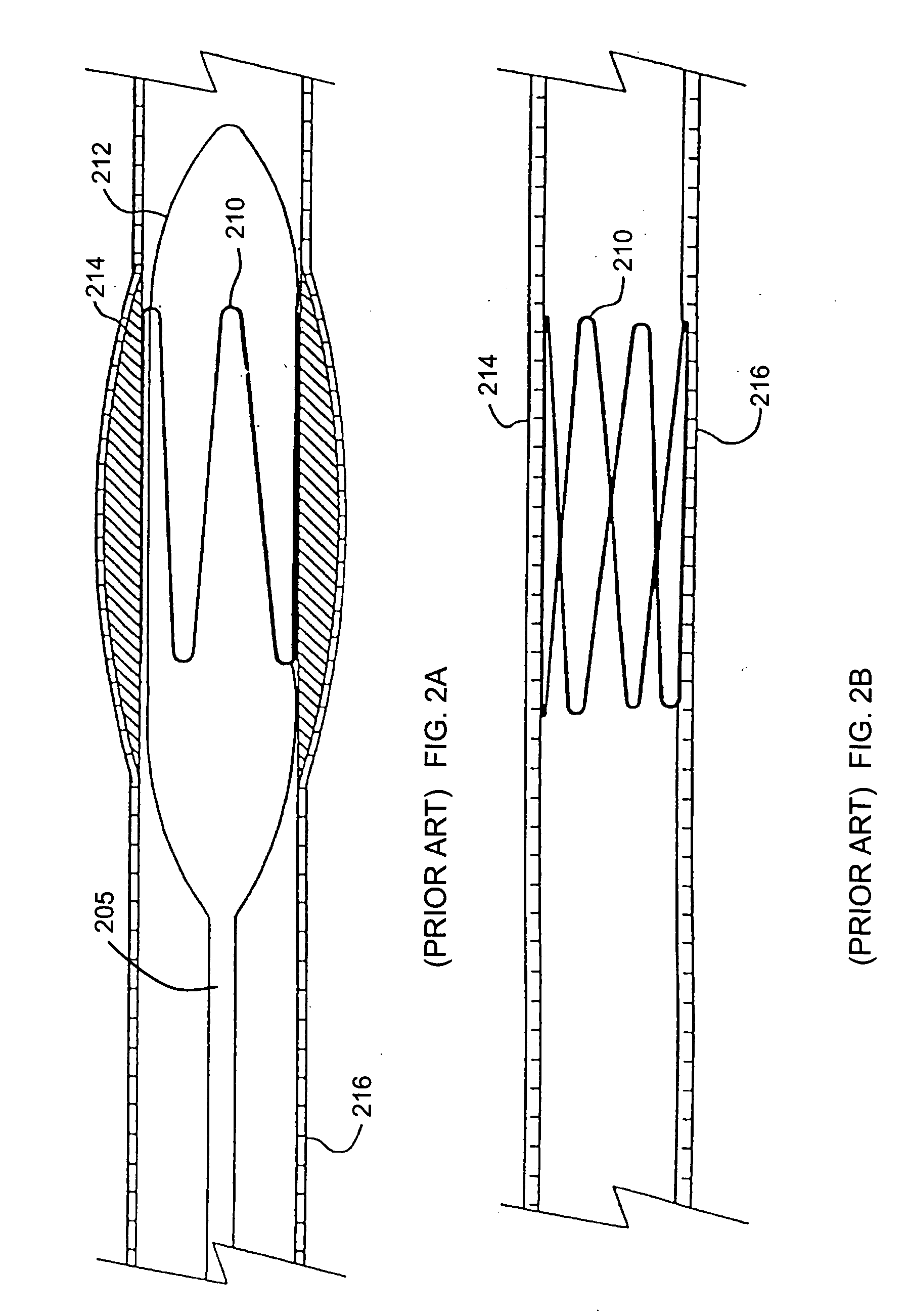
[0018] In another embodiment the
aneurysm repair device has a
nanostructure composite coating and / or nanostructured surface associated therewith. The nanostructures may comprise nanofibers, nanotubes or nanoparticles and / or combinations thereof, and including woven and nonwoven fibrous mats or mesh made of nanofibers and nanotubes and / or having nanostructures thereon. The nanostructures associated therewith, including the fibrous mats may be coated or uncoated, or have multiple coatings thereon. The specific coatings are described herein and vary depending on the desired purpose of the device or method. In one particular embodiment, an
aneurysm coil is disclosed having nanostructures associated therewith which is designed to be placed at the site of an
aneurysm (e.g., in the brain) with the goal of inducing thromobogenesis. The resulting clot formed by the presence of the coil in the vessel would plug the vessel, eliminating the possibility that it could rupture. In contact with blood, the nanostructures (e.g., nanowires grown on the surface of the coil) would aid in
clot formation by helping to induce a thrombogenic response in the vessel.
Fibrin could also be coupled to the surface of the nanostructures to aid in
clot formation. To overcome any potential physical or mechanical damage to the wires during
insertion of the coil into the vessel at the site of the aneurysm, the nanosturctures can be encapsulated (potted) in a
biodegradable polymer such as
polylactic acid or polyglycolic acid or a mixture thereof. This would allow, for example, the nanostructures, grown on the coil, to be placed in the body without any appreciable damage.
[0023] In another embodiment of the invention, a bioengineered
scaffold device for providing a
scaffold for nerve regeneration is disclosed which comprises a base membrane or matrix having a plurality of nanostructured components associated therewith. The membrane or matrix may comprise one or more of the following materials: natural or synthetic polymers, electrically conducting polymers, conjugated polymers capable of
electron transfer, electroluminescent polymersmetals,
metal alloys, ceramics, glass or
silicone. The plurality of nanostructured components may comprise nanowires, nanofibers, nanotubes and nanoparticles. The nanostructured surface of the membrane or matrix may be impregnated or bound with one or more drugs, cells, fibroblasts, nerve growth factors (NGF),
cell seeding compounds, neurotrophic growth factors or
genetically engineered cells producing such factors, VEGF,
laminin or other drugs or substances to encourage axonal elongation and functional nerve performance.
[0028] Methods of use are also disclosed for treating patients with any one or more of the medical devices disclosed herein, which include, for example, a method of therapeutically treating a patient comprising contacting the patient with a medical device comprising a surface and plurality of nanofibers associated with the surface. Methods are disclosed for administering a
drug compound to a body of a patient which comprises, for example, providing a
drug-eluting device comprising at least one surface, a plurality of nanofibers and / or nanotubes associated with the surface, and a
drug compound associated with the plurality of nanofibers and / or nanotubes; introducing the drug-eluting device into a body of a patient; and delivering the
drug compound into the body of the patient. The drug-eluting device in one embodiment comprises a
coronary stent, although any device which would benefit from local
drug delivery at the site of
disease (e.g.,
lesion) could be used in the methods of the invention. Where a
coronary stent is used as the drug-eluting device, the
drug compound may comprise
paclitaxel or
sirolimus, for example, or a variety of other medications including without limitation one or more of the following: anti-inflammatory immunomodulators such as
Dexamethasone, M-
prednisolone,
Interferon,
Leflunomide,
Tacrolimus, Mizoribine, statins, Cyclosporine,
Tranilast, and Biorest; antiproliferative compounds such as Taxol,
Methotrexate, Actinomycin, Angiopeptin,
Vincristine, Mitomycin, RestenASE, and PCNA
ribozyme; migration inhibitors such as Batimastat,
Prolyl hydroxylase inhibitors,
Halofuginone, C-proteinase inhibitors, and
Probucol; and compounds which promote healing and re-endothelialization such as VEGF,
Estradiols, antibodies,
NO donors, and BCP671. The
drug compound may be adsorbed directly to the nanofiber and / or nanotubes surface, or the drug may be disposed inside the
nanotube of the drug-eluting device or otherwise associated with it via the use of one or more
silane groups,
linker molecules or other covalent, ionic, van der Waals etc. attachment means. The nanofiber and / or surface may be configured such that the drug compound elutes slowly over time. This may be accomplished using time released coatings, for example. The plurality of nanofibers optionally are embedded in a biocompatible, non-thrombogenic
polymer coating to provide enhanced durability to the nanofibers.

