
Sustained release implants and methods for subretinal delivery of bioactive agents to treat or prevent retinal disease

a technology of bioactive agents and implants, applied in the field of sustained release implants and to methods for treating eyes, can solve the problems of ineffective treatment, inability to achieve specific therapy, and inability to achieve simple or inexpensive treatment, so as to prevent the progression of the condition

Inactive Publication Date: 2006-11-16
SURMODICS INC
View PDF99 Cites 75 Cited by
  • Summary
  • Abstract
  • Description
  • Claims
  • Application Information

AI Technical Summary

Benefits of technology

[0024] Non-polymer biocompatible materials may also form the core of an implant of the invention. Examples include titanium-nickel alloy wire, titanium alloys, nickel-cobalt base alloys, stainless steel, cobalt-chromium alloys, and biodegradable magnesium alloys. In an exemplary embodiment, the core is titanium nickel wire having the smallest commercially available diameter (e.g., about 10 μm to about 200 μm), thereby thereby maximizing the amount of bioactive agent that the implant may contain. In some embodiments, the core material and core thickness are selected to provide the implant with the desired rigidity and flexibility.
[0026] In some embodiments, the outer surface of the core is coated along a portion of its length with a coating layer comprising a biocompatible polymer matrix and one or more bioactive agents and the implant additionally includes an uncoated length of core that provides a handling portion (e.g., uncoated regions of about 10 mm in length, or less) by which the implant may be grasped, docked with a surgical instrument, or used for easy device retrieval after a period of implantation in the eye.
[0033] In some embodiments, the implants of the invention provide a therapeutic effect by eluting one or more bioactive agents at no more than about 90% excess bioactive agent than that required to provide a therapeutic affect, in other embodiments at no more than about 80% excess, in other embodiments at no more than about 70% excess, in other embodiments at no more than about 60% excess, in other embodiments at no more than about 50% excess, in other embodiments at no more than about 40% excess, in other embodiments at no more than about 30% excess, in other embodiments at no more than about 20% excess, in other embodiments at no more than about 10% excess, in other embodiments at no more than about 5% excess, in other embodiments at no more than about 1% excess bioactive agent than that required to provide a therapeutic affect.
[0043] The present invention provides a direct approach for delivering one or more bioactive agents to a treatment site, thereby reducing the required dosage for effective treatment. Further, such methods may reduce the side effects associated with intravitreal delivery of bioactive agents. The present methods further provide an improved route for the delivery of large molecular weight drugs and proteins to a treatment site.
[0054]“Therapeutically effective amount” refers to that amount of a bioactive agent alone, or together with other substances, that produces the desired effect (such as treatment of a medical condition such as a disease or the like, or alleviation of pain) in a patient. During treatment, such amounts will depend upon such factors as the particular condition being treated, the severity of the condition, the individual patient parameters including age, physical condition, size and weight, the duration of the treatment, the nature of the particular bioactive agent thereof employed and the concurrent therapy (if any), and like factors within the knowledge and expertise of the health practitioner. A physician or veterinarian of ordinary skill can readily determine and prescribe the effective amount of the bioactive agent required to treat and / or prevent the progress of the condition.

Problems solved by technology

Roughly 80 percent of the AMD cases involve non-neovascular conditions, for which there are no effective treatments.
Perhaps the best-known therapy is photodynamic therapy (PDT), however, while this therapy has received significant intention in both the ophthalmic and financial investment communities, it is useful in only about 20 percent of neovascular AMD cases.
In addition, this particular therapy is not a simple or inexpensive treatment.
Its systemic side effects, however, include peripheral neuropathy, central nervous system depression, and embryotoxicity.
In addition, these systemic side effects have limited the dosage administered to patients for the treatment of subretinal neovascularization.
Systemic inhibition of angiogenesis in older patients can also interfere with the development of collateral circulation, which has a role in the prevention of central nervous system as well as cardiac ischemic events.
However, delivery of drugs, proteins and the like to the eye(s) of mammals so as to achieve the desired therapeutic or medical effect, especially to the retina and / or the choroid, has proven to be challenging, most of which is owed to the geometry, delicacy and / or behavior of the eye and its components.
Oral ingestion of a drug or injection of a drug at a site other than the eye can provide a drug systemically; however, such a systemic administration does not provide effective levels of the drug specifically to the eye.
In many ophthalmic disorders involving the retina, posterior tract, and optic nerve, adequate levels of the drug cannot be achieved or maintained by oral or parenteral routes of administration.
Such further and repeated administrations of such drugs, however, may produce undesired systemic toxicity.
In addition, topical eye drops may drain from the eye through the nasolacrimal duct and into the systemic circulation, further diluting the medication and risking unwanted systemic side effects.
Furthermore, delivery of drugs in the form of topical eye drops is also of limited utility because the drug cannot easily cross the cornea or sclera and be made available to the vitreous, retina, or other subretinal structures such as the retinal pigment epithelium (“RPE”) or choroidal vasculature and / or is highly unstable and therefore not easily formulated for topical delivery.
Direct delivery of drugs to the eye by a topical insert has also been attempted; however, this method is not desirable.
Consequently, this technique demands a certain degree of manual dexterity that can be problematic for geriatric patients who are particularly susceptible to certain eye disorders that appear age related (e.g., age related macular degeneration).
Also, in many instances such topical inserts may cause eye irritation and such inserts are prone to inadvertent loss due to eyelid laxity.
In addition, these devices provide a source of drug only to the cornea and anterior chamber, and thus do not provide any significant pharmacologic advantage over topical eye drops or ointments.
Thus, such devices have limited utility for providing an effective source of drugs to the vitreous or tissues located in the posterior segment of the eye.
In addition, it also is well known that many therapeutic drugs cannot easily diffuse across the retina.
In addition to the relative effectiveness of drug delivery across the barrier, complications or side effects have been observed when using the direct injection into vitreous technique with some therapeutics.
When these compounds were used to treat neovascularization of the posterior segment by direct injection, these compounds were observed to cause undesirable side effects in many patients.
Moreover, a risk exists that the use of corticosteroids in patients with normal intraocular pressure will cause elevations in pressure that result in damage to ocular tissue.
Since therapy with corticosteroids is frequently long term, a potential exists for significant damage to ocular tissue as a result of prolonged elevations in intraocular pressure attributable to that therapy.
Such a course of treatment also increases the duration and cost as well as the realistic risks of corneal ulceration, cataract formation, intraocular infection, and / or vitreous loss that accompany these procedures.
As noted in U.S. Pat. No. 5,273,530, the penetration of the anterior part or segment of the eye, using the transcorneal or the transscleral route creates the risks of corneal ulceration, cataract formation and other anterior penetration problems.
Thus, there are a number of drawbacks with currently available methods for treating eye disorders and diseases.
For example, in the case of these posterior segment eye diseases, traditional routes of drug administration such as topical or oral dosing often fall short of reaching the disease site.
Subsequently, this approach often requires frequent, large dose injections which have been associated with complications such as glaucoma and cataract formation.
Furthermore, large molecular weight molecules (>70 kD) are virtually incapable of traversing the tight junction complexes of the retinal pigment epithelium and retinal capillaries.
Microparticle injections have improved the sustained release capabilities of conventional injections, but this still does not resolve the widespread distribution of the medication via intraocular convection.
In the case of steroids, this distribution is known to lead to adverse effects such as glaucoma and cataract.

Method used


Image

  • Sustained release implants and methods for subretinal delivery of bioactive agents to treat or prevent retinal disease
  • Sustained release implants and methods for subretinal delivery of bioactive agents to treat or prevent retinal disease
  • Sustained release implants and methods for subretinal delivery of bioactive agents to treat or prevent retinal disease

Examples


example 1

Materials Used:

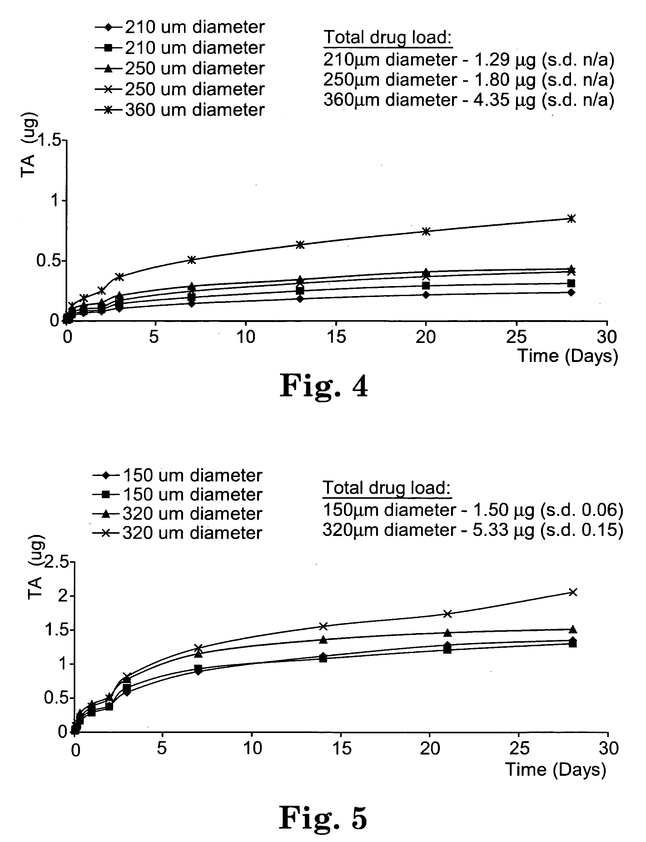
[0168] Poly(caprolactone) (Average Mw 80,000, [—O(CH2)5CO—]n, Melt index 125° C. / 0.3 MPa, Sigma Aldrich Biochemicals, St. Louis, Mo.) [0169] Triamcinolone acetonide (TA) (Purity 99%, Mn 434.5, C24H31FO6, Sigma Aldrich Biochemicals, St. Louis, Mo.) [0170] Prednisolone Purity 99%, C21H28O5, Mn 360.5, Sigma Aldrich Biochemicals, St. Louis, Mo.) [0171] Chloroform (purity 99.8%, CHCl3, A.C.S. spectroscopic grade, Sigma Aldrich Chemicals) [0172] Ether (purity 99%, Mn 74.12, (C5H5)2O A.C.S. reagent, Sigma Aldrich Chemicals) [0173] Balanced salt solution (Sterile, preservative free, Akorn, Inc., Somerset, N.J.) [0174] Bovine serum albumin (Molecular biology grade, Sigma Aldrich Biochemicals, St. Louis, Mo.)

Abbreviations: [0175] PCL: poly(caprolactone) biodegradable implant [0176] TA: triamcinolone [0177] PCL / TA: biodegradable triamcinolone loaded poly(caprolactone) implants

Implant Preparation:

[0178] The implants used in the example were prepared as follows. PCL was sol...

example 2

[0211] All procedures abided by the Guide for the Care and Use of Laboratory Animals, the USDA Animal Welfare Regulations (CFR 1985) and Public Health Service Policy on Humane Care and Use of Laboratory Animals (1996) and the Institution's policies governing the use of vertebrate animals for research, testing, teaching or demonstration purposes.

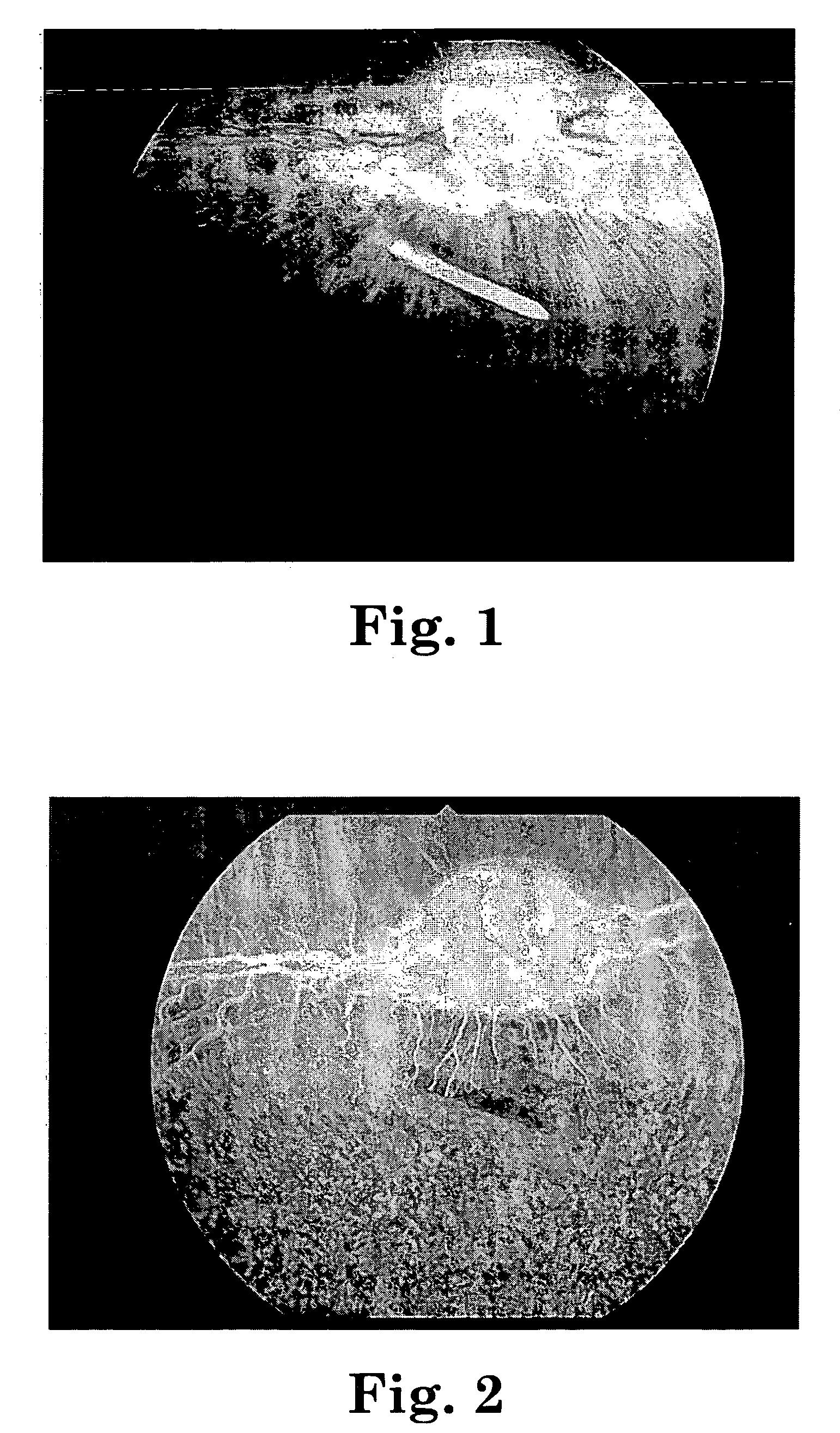
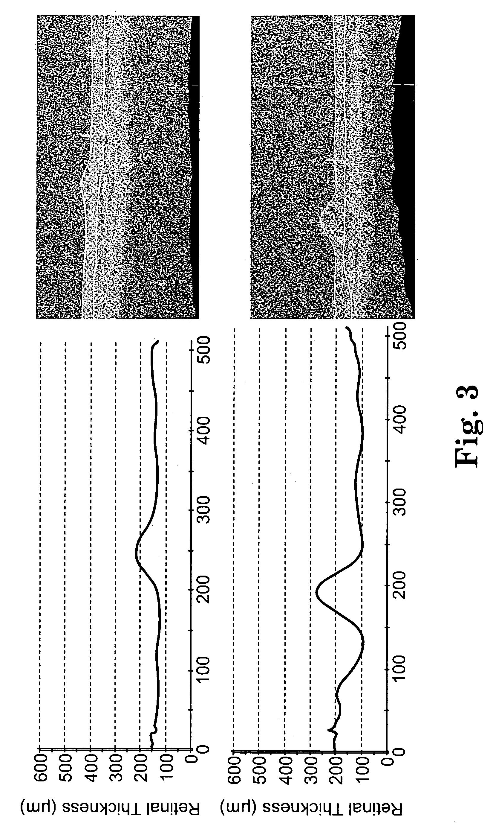
[0212] Seven pigmented rabbits (J1-J7) underwent standard pars plana vitrectomy, and insertion of a drug delivery device into the subretinal space. The animals were anesthetized with an intramuscular injection of 0.3 mL of ketamine hydrochloride (100 mg / mL; Fort Dodge Lab., Fort Dodge, Iowa) and 0.1 mL of xylazine hydrochloride (100 mg / mL; Miles Inc, Shawnee Mission, Kans.) per kilogram of body weight. Pupils were dilated with 1 drop each of 2.5% phenylephrine and 1% tropicamide. A 3-mm peritomy was made at the superotemporal and superonasal quadrant of the right eye. Sclerotomies were created with a 20-gauge microvitreoretinal blade 1 to 2 ...

example 3

Materials Used and Abbreviations:

Core:

[0219] NIT: 80 μm etched Nitinol wire, commercially available from Nitinol Devices and Components (Freemont Calif.).

Bioactive Agent: [0220] RAP: Rapamycin, commercially available from LC Laboratories, Woburn Mass.

Polymers: [0221] pEVA: polyethylene vinyl acetate copolymer (33% wt. vinyl acetate and 67% wt. polyethylene), commercially available from Aldrich Chemical Co. [0222] pBMA: poly(n-butyl methacrylate), commercially available from Aldrich Chemical Co.

Solvent: [0223] CHCl3: chloroform solvent, commercially available from Burdick & Jackson.

Implant Preparation:

Coating Solution Preparation:

[0224] A coating solution was prepared by first adding 25.0 parts pEVA and 25.0 parts pBMA to an aliquot of CHCl3 solvent. In order to dissolve the pEVA, the components were heated to 30° to 40° C. for approximately 1 hour. After the pEVA and pBMA had dissolved in the CHCl3, the resulting polymer / solvent solution was allowed to cool to room te...


PUM

PropertyMeasurementUnit
total diameteraaaaaaaaaa
lengthaaaaaaaaaa
diameteraaaaaaaaaa

Abstract

The invention relates to sustained release implants and to methods for treating eyes, particularly the eyes of mammals having eye disorders or diseases. By using the implants and methods described herein, the delivery of the one or more bioactive agents can be localized at a desired treatment site, particularly the choroid and the retina.

Description

[0001] This application claims the benefit of U.S. Provisional Application Ser. No. 60 / 669,701, filed Apr. 8, 2005, entitled “SUSTAINED DELIVERY DEVICES FOR THE CHOROID AND RETINA AND METHODS FOR SUBRETINAL ADMINISTRATION OF BIOACTIVE AGENTS TO TREAT AND / OR PREVENT RETINAL DISEASES,” which is incorporated herein by reference in its entirety.FIELD OF THE INVENTION [0002] The present invention relates to sustained release implants and to methods for treating eyes, particularly the eyes of mammals having eye disorders or diseases. More particularly the present invention relates to implants and methods for administering one or more bioactive agents subretinally by the use of one or more implants. By using the implants and methods described herein, the delivery of the one or more bioactive agents can be localized at a desired treatment site, particularly the choroid and the retina. BACKGROUND OF THE INVENTION [0003] There are a number of vision-threatening disorders or diseases of the ey...

Claims


Application Information

Patent Timeline
Patent Type & Authority Applications(United States)
IPC IPC(8): A61F2/00C08F20/00
CPCA61F2/14A61F9/0017A61K9/0051A61L27/44A61L27/54A61L2430/16A61L2300/406A61L2300/41A61L2300/416A61L2300/604A61L2300/606A61L2300/252A61P27/02
Inventor VARNER, SIGNE E.FOX BEELEY, NATHAN ROBERTDEJUAN, EUGENE
Owner SURMODICS INC
Learn More