
Compositions and means for diagnosing microbial infections

Active Publication Date: 2009-03-12
SSI DIAGNOSTICA AS
View PDF11 Cites 18 Cited by
  • Summary
  • Abstract
  • Description
  • Claims
  • Application Information

AI Technical Summary

Benefits of technology

[0069]Another advantage of the present invention is that when the platform is used to test urine for infections the number of colonies formed on the semi-solid microbial growth medium directly correlates with the number of bacteria per volume of urine added to the platform. Thereby making it easy to determine the concentration of bacteria in the urine.
[0070]In a preferred embodiment of the present invention, the semi-solid medium comprises tryptophan. The advantage of including tryptophan is that it enables easy detection of Enterobacteria of the Proteus-Morganella-Providencia and at the same time tryptophan does not interfere with determination of antibiotic susceptibility.
[0071]In another preferred embodiment, the semi-solid medium in the composition according to the invention comprises a galactose polymer (Agar-agar) or Gelatin.
[0072]Preferably the composition according to the invention comprises a microbial growth medium selected from the group consisting of: Iso-sensitest agar, Danish Blood Agar, Discovery medium and Mueller-Hinton medium. For those media which do not inherently comprise tryptophan it may be a further advantage to include tryptophan in these media. The concentration of tryptophan in the medium may be between 0.25-3.0 g / liter.
[0073]The Iso-sensitest agar is currently available from Oxoid and its composition is specified in The Oxoid Manual 1998. The Iso-sensitest agar is composed by 11 g / l Hydrolysed casein, 3 g / l Peptones, 2 g / l Glucose, 3 g / l Sodium Chloride, 1 g / l soluble starch, 2 g / l Disodium hydrogen Phosphate, 1 g / l Sodium Acetate, 0.2 g / l Magnesium glycerophosphate, 0.1 g / l Calcium gluconate, 0.001 g / l cobaltous sulphate, 0.001 g / l Cupric sulphate, 0.001 g / l Zinc sulphate, 0.001 g / l Ferrous Sulphate, 0.002 g / l Magnesium Chloride, 0.001 g / l Menadione, 0.001 g / l Cyanobalamin, 0.02 g / l L-Cysteine hydrochloride, 0.02 g / l Tryptophan, 0.003 g / l pyridoxine, 0.003 g / l pantothenate, 0.003 g / l nicotinamide, 0.0003 g / l Biotin, 0.00004 g / l Thiamine, 0.01 g / l Adenine, 0.01 g / l Guanine, 0.01 g / l Xanthine, 0.01 g / l Uracil, 8 g / l agar, in distilled water.
[0074]As for the Danish Blood Agar this is known by the skilled person to have the following composition: 2 g Na2HPO4.12H2O, 625 g tryptone, 250 g starch, 833.6 g Potassium Chloride, 2.5 g detergent, 74.8 g meat broth (Oxoid CM975K), 800 g D(+)Glucose-monohydrate, 1.75 g Xanthin, 1.75 g Guanin, 17.5 g Magnesium Sulphate 7H2O, 19.2 g CaCl2.2H2O, 2,720 g Agar, 5 N HCl to pH 7.4, solution of vitamins, and 12.5 l horse blood per 250 liter distilled water.

Problems solved by technology

Although most infections are uncomplicated lower UTI, i.e. bladder infections, which often cure themselves without antibiotic treatment, UTI can also be complicated with ascending renal infections which can spread to the blood leading to septicemia with septic shock.
The bladder infection leads to local pain, which can be felt behind the pubic bones or perhaps in the loins—but this is often a sign of renal involvement, as well as pain during voiding.
Also, the irritation in the bladder will lead to frequent voiding.
Due to the common colonisation of the external part of the urethra, it is difficult to obtain a sample during voiding without contamination of the sample.
But since these latter methods are rather cumbersome and often painful for the patient in the large majority of cases and also demands hospital admission, in most cases the urine is collected as a Mid Stream Urine (MSU), i.e. the meatus is cleaned with a cotton swab wetted with sterile saline, the patient then voids a small first part of the urine to cleanse the urethra and then voids—mid-stream—a sample collected in a sterile container—to end by voiding the rest of the urine volume in the toilet.
a) Direct methods1. Microscopy: Bacteria can be visualized in the urine sample by either phase contrast microscopy of a wet smear, or by simple light microscopy of a Gram-coloured preparation. In the wet smear, the form and possible movement of the bacteria can be seen but naturally not the Gram-type of the bacteria. This can be discerned by the light microscopy of the Gram-stain, and in both cases leucocytes can be seen and roughly quantified. The problem with microscopy is the lack of specificity, i.e. only a presumptive bacterial diagnosis can be given, and the sensitivity i.e. bacteria can only be visualized when present in numbers of at least 105 bacteria / ml of urine. Furthermore, microscopy will not reveal the antibiotic susceptibility of the bacteria. Phase contrast microscopy has a positive predictive value of 58% as related to quantitative culture with ≧105 bacteria / ml as criteria for UTI4.2. Culture: The gold standard of diagnosis of UTI. In the laboratory this is performed by a quantitative culture, i.e. a standardized loop applying 1 or 10 μl urine on an agar plate. This allows quantification of bacteria by counting the number of colonies (colony forming units, CFU) on the incubated plate. If low numbers of bacteria are anticipated (e.g. suprapubic puncture or catheter sample) up to one ml of urine may be cultured allowing the counts of down to 1-2 CFU / ml urine. The culture can lead to further diagnosis of the bacteria by biochemical and other types of laboratory workup. Furthermore, a susceptibility test can be performed. Sensitivity of the method is by definition 100%, but in some cases bacteria may not grow if special media, atmosphere or temperature conditions are not used (some Aerococcus strains may only grow on blood agar and in CO2), or if the patient has started antibiotic treatment bacteria may not grow due to antibiotic suppression. The specificity is not 100%, since the positive culture may still contain contaminants—the result should be combined with the symptoms and signs as well as the presence of increased numbers of leucocytes in the urine. The quantitative loops have a variation of + / −½ log CFU / ml, which means that only a rough estimate of the counts can be made5. The disadvantage of quantitative cultures in the laboratory is that the urine must be kept in a condition, where the bacteria do not multiply prior to culture; this can be obtained by cooling the urine during transportation (i.e. Other culture methods: Dipslides with a plastic plate skeleton carrying agar media on one or both sides that are dipped into the urine have been used for 20-30 years (e.g. Uricult (Orion)). The advantage of these is the ease of inoculation and that the direct inoculation can be quantified by comparing the bacterial growth with pictures of dipslides used for cultures with known quantities of bacteria. The dipslide can also be used as a transport medium. A susceptibility test has been developed, where antibiotic containing discs are placed on the agar surfaces after inoculation (e.g. Sensicult (Orion)). The positive predictive value (PPV) of the susceptibility test has in some cases been as low as 0.6, which can be explained by the lack of standardized inoculum and difficulty in interpreting a zone around the disc on a rounded surface, as the agar plate is not completely flat. Also, the small surface of the dipslide (i.e. approx. 2×5 cm) makes it difficult to evaluate the growth characteristics of bacteria, and especially whether there are more than one species. Furthermore, evaluation of susceptibility is difficult without the knowledge of the bacterial species. A dipslide with chromogenic agar on one of the sides (DipStreak) has recently been marketed by Novamed in Israel.3. Other methods: During the later years several methods have been tried for quantifying bacteria in urine such as turbidity measurements by spectrophotometry, cyto-centrifugation, ATP-measurement and others. So far, to our knowledge, none of these have been applied in routine laboratories and especially not so in primary care due to their cost and demand for technical skill.
b) Indirect methods:1. Dipstick for nitrite and leucocyte-esterase: Determination of nitrite in the urine is used for diagnosis of UTI, since this substance can only be present if nitrate-reductase producing bacteria are present in the urine. Nitrate stems from protein-metabolism and is found in urine in varying concentrations depending on the intake of protein the day up to the sampling. Nitrite can be removed, however, if there are bacteria present, which produce nitrite-reductase (e.g. E. coli can contain both types of enzymes). The test is not very sensitive, since the reaction needs about 3 hours of incubation, some bacteria produce nitrite-reductase, and some urinary pathogens do not produce nitrate-reductase at all, e.g. staphylococci. The leucocyte-esterase test is more relevant, since it is the easiest way to prove the presence of leucocytes; the enzyme can only stem from leucocytes, and the amount of enzyme is correlated to the numbers of leucocytes. Whole leucocytes lyse easily and rapidly, which means that the urine microscopy for leucocytes must be performed within an hour after sampling in order to achieve a relevant quantitative microscopy, while the enzyme test can be performed several hours later due to the stability of the enzyme. The presence of leucocytes, however, is only predictive of infection in 50% of patients, since leucocyturia can be found in many patients without infection. Together, the test for the two enzymes combined (i.e. either one or both positive) has a rather low PPV (60-80%) due to the above mentioned factors.2. Symptoms alone: In many cases in general practice, the general practitioner (GP) will initiate treatment based on symptoms alone.
This can be due to reservations against diagnostic workup due to cost, geography or tradition, knowledge of susceptibility of the pathogens and / or use of broad spectrum antibiotics suspected to cover all possible pathogens.

Method used


Image

  • Compositions and means for diagnosing microbial infections
  • Compositions and means for diagnosing microbial infections
  • Compositions and means for diagnosing microbial infections

Examples


example 1

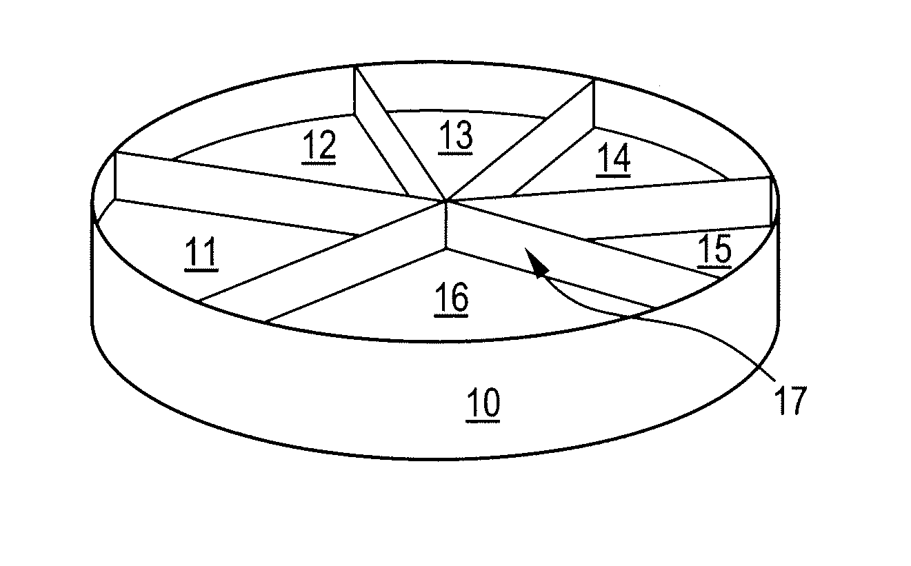
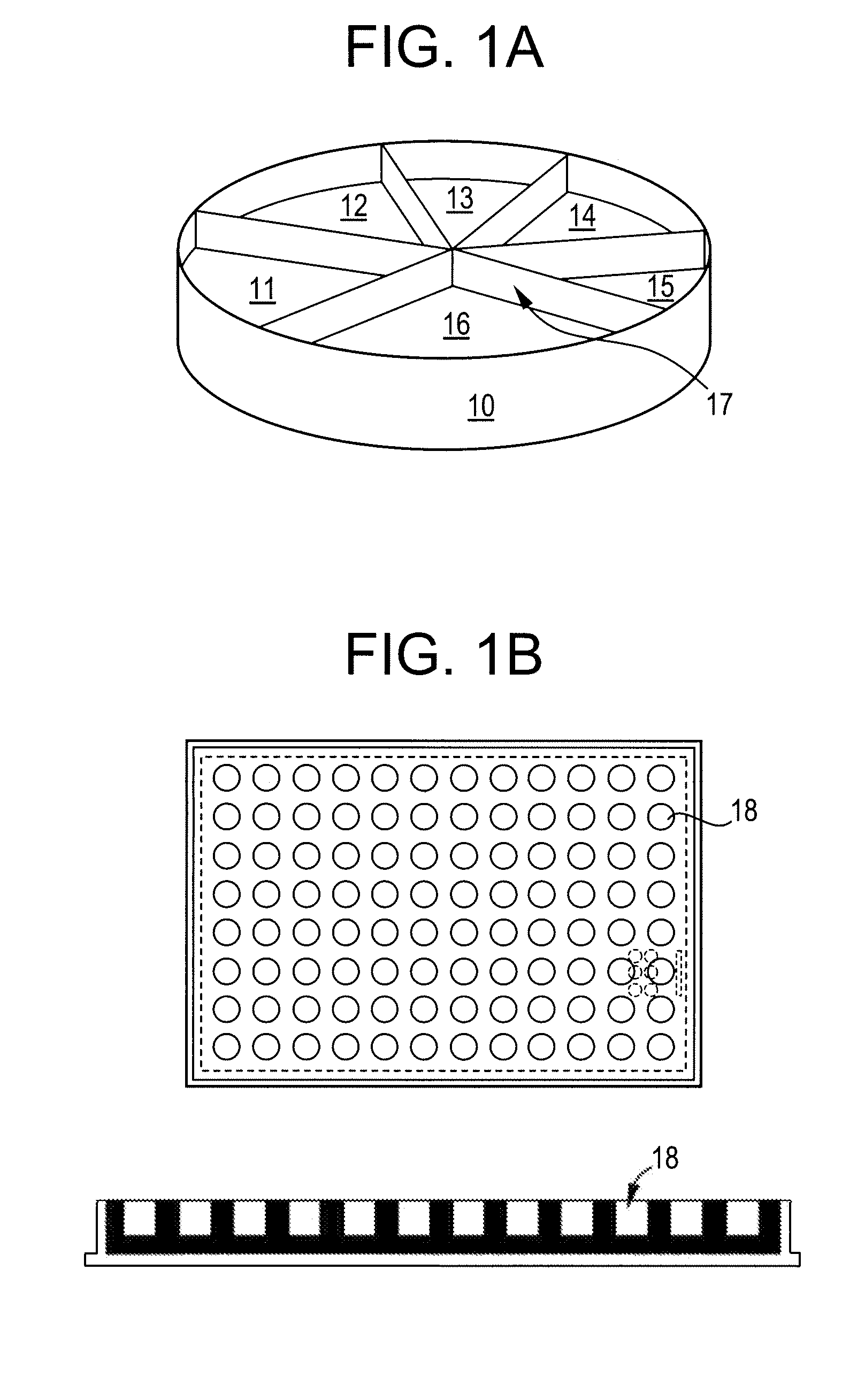
Preparation of a Platform Based on a 6-Compartment Petri Dish

Flexicult

[0221]Iso-sensitest agar was produced and combined with 1.5 g / l of a mixture of chromogenic substrate comprising 5-Bromo-4-chloro-3-indolyl-β-D-galactopyranoside, 6-Chloro-3-indolyl-β-D-galactopyranoside, 5-Bromo-4-chloro-3-indolyl-β-D-glucopyranoside, 5-Bromo-4-chloro-3-indolyl-β-D-glucuronide, 5-Bromo-6-chloro-3-indolyl-phosphate.

[0222]Alternatively Discovery agar (Oxoid, CM 1087) was used.

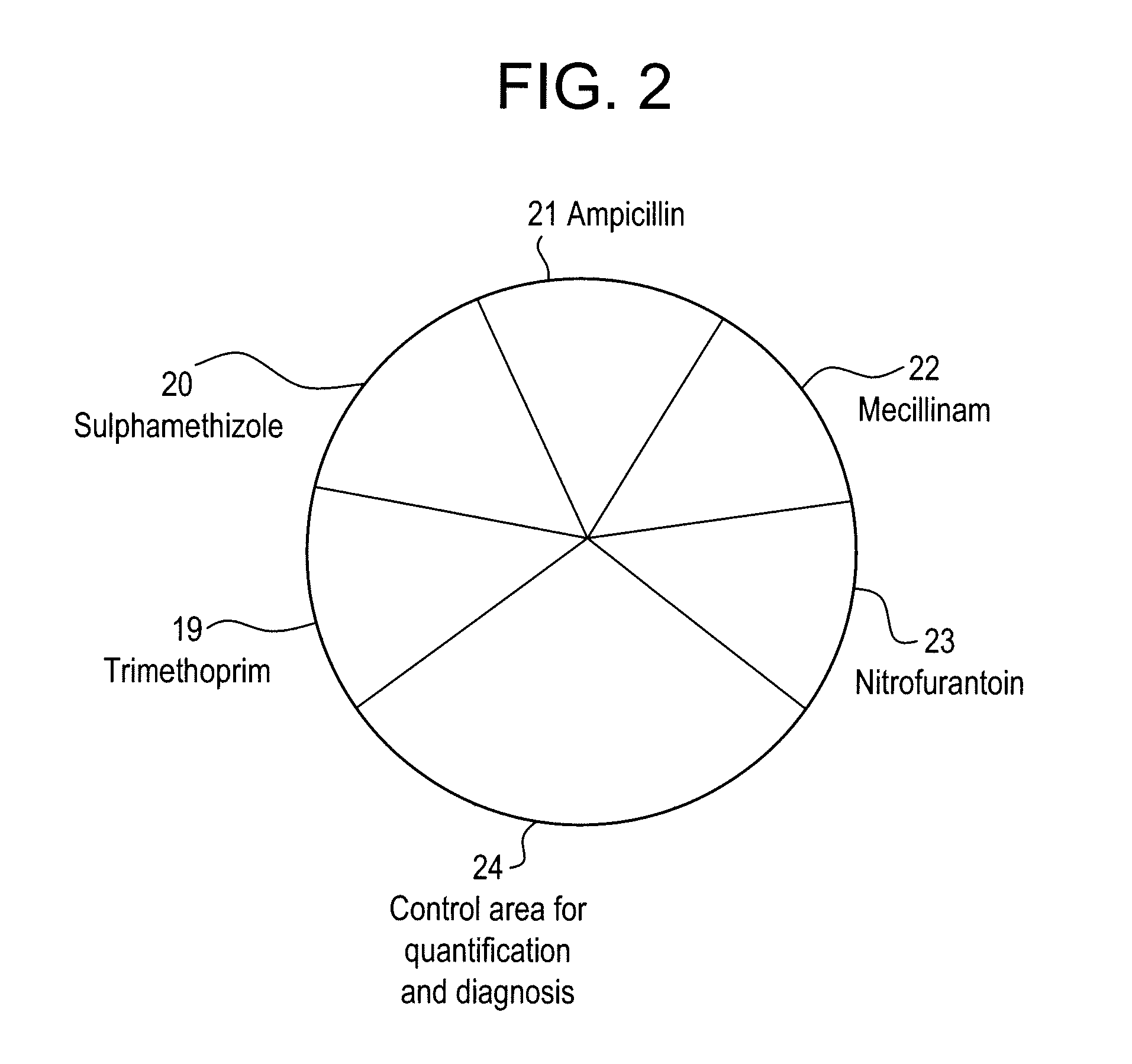
[0223]Antibiotic solutions were produced: trimethoprime, sulphamethizole, ampillicine, nitrofurantoine, and mecilliname.

[0224]Preparation of hot plates and temperature regulation via PT-100 sensor:

1. The bottles with Iso-sensitest / Discovery agar were melted at 105° C. for 60 min. After melting, the bottles were placed in a water bath at 56° C. for approx. 60 min.

2. The equipment for the filling of Flexicult was prepared: Variomag (Multitherm Stirring Block Thermostat and Telemodul 40 CT were interconnected and then connected t...

example 2

Stability Test

[0240]The stability of the chromogenic agar in Flexicult has been determined by testing Flexicult agars kept up to 4 months in refrigerator ([0241]Agar stability (water content / dryness, form)[0242]Colourisation of bacterial colonies[0243]Susceptibility / resistance towards the antibiotics (sulfamethizole, trimethoprim, ampicillin, nitrofurantoin and mecillinam.

example 3

Clinical Validation

[0244]The Flexicult agar kit has been tested in two GP clinics. Both clinics have 9-10 GP's working in the clinic and perform routine urine culture (either by dipslide or agar plate with subsequent antibiotic disc diffusion susceptibility in certain cases), performed by clinical biochemistry technician staff.

[0245]For the study, the Flexicult plate was inoculated with urine from patients with suspected UTI as based on history and symptoms in parallel with the routine culture systems for each clinic. The plates were incubated at 35-37° C. in ambient atmosphere for 18-24 h (tests performed Friday, which could not be read Saturday, were not included). Plates were read by the technicians and the results recorded (by the use of material provided together with the plates from the Statens Serum Institut: A picture scheme showing the quantities of bacteria on the Flexicult plate, see FIG. 3, and a picture scheme showing the different colony types of urinary pathogens, see...


PUM


Abstract

The present invention pertains to the need for novel, reliable, fast and inexpensive approaches to diagnosing, including detecting and characterising microbial infections in humans and animals or methods for detecting and characterising microbial infections in various environments, such as in a food or feed sample.The present invention provides compositions, platforms, kits and methods for diagnosing, detecting and / or characterising a microbial infection or contamination. In particular the present invention relates to such compositions, platforms, kits and methods for diagnosing, detecting and / or characterising a urinary tract infection.

Description

CROSS-REFERENCE TO RELATED PATENT APPLICATIONS[0001]European Patent Office Priority Application 07115480.1, filed Aug. 31, 2007 including the specification, drawings, claims and abstract, is incorporated herein by reference in its entirety. This application claims priority from Provisional Application U.S. Application 60 / 969,260, filed Aug. 31, 2007, incorporated herein by reference in its entirety.TECHNICAL FIELD OF THE INVENTION[0002]The present invention relates to compositions, platforms, kits and methods for diagnosing, detecting and / or characterising a microbial infection or contamination. In particular the present invention relates to such compositions, platforms, kits and methods for diagnosing, detecting and / or characterising a urinary tract infection.BACKGROUND OF THE INVENTIONUrinary Tract Infections (UTI)[0003]Urinary tract infection is one of the most common infections in general practice as well as the most common nosocomial infection in the western world. The overall ...

Claims


Application Information

Patent Timeline
IPC IPC(8): C12Q1/18C12Q1/44C12Q1/04
CPCC12Q1/045C12Q1/08C12Q1/14G01N2800/202C12M41/46C12M23/10C12M23/12C12M23/34C12M41/36G01N2800/348
Inventor FRIMODT-MOLLER, NIELS
Owner SSI DIAGNOSTICA AS
Learn More