Compositions and their use to treat complications of aneurysmal subarachnoid hemorrhage

a technology of aneurysmal subarachnoid hemorrhage and composition, which is applied in the direction of drug composition, biocide, cardiovascular disorder, etc., can solve the problems of increased blood pressure, ischemic brain damage, and increased blood pressure, so as to achieve mild or severe effects

Inactive Publication Date: 2018-10-09
EDGE THERAPEUTICS
View PDF96 Cites 2 Cited by
  • Summary
  • Abstract
  • Description
  • Claims
  • Application Information

AI Technical Summary

Benefits of technology

The patent describes a method for treating cerebral arteries at risk of interruption due to subarachnoid hemorrhage (SAH) in humans. The method involves administering a flowable sustained release particulate composition to the subarachnoid space near the affected artery. The composition contains microparticles with a therapeutic agent dispersed throughout, and a pharmaceutical carrier. The composition is injected locally, either intracisternally or intraventricularly, to contact the at-risk artery without causing unwanted side effects. The therapeutic agent is released from the microparticles over a period of 6 days to 14 days, with a concentration in plasma and cerebrospinal fluid that is low to prevent systemic side effects. The composition can be injected through a needle, cannula, or catheter. The viscosity of the composition can vary depending on the injection method. The invention provides a safe and effective way to treat cerebral artery interruption due to SAH.

Problems solved by technology

The mass effect of an intracerebral hematoma may compromise the blood supply of adjacent brain tissue; or SAH may cause reactive vasospasm of cerebral surface vessels, leading to further ischemic brain damage.
Aneurysms occasionally can rupture into the brain, causing an intracerebral hematoma, and into the cerebral ventricles, causing intraventricular hemorrhage.
Vasoconstriction usually results in an increase of blood pressure and may be slight or severe.
Bleeding due to SAH may result in brain damage, brain shift, decreased cerebral perfusion and hydrocephalus.
SAH is a medical emergency and may lead to death or severe disability even if recognized and treated at an early stage.
The group of people at risk for SAH is younger than the population usually affected by stroke, but the risk still increases with age.
The body releases large amounts of adrenaline and similar hormones as a result of the bleeding, which leads to a sudden increase in the blood pressure.
CT angiography (“CTA”) (visualizing blood vessels with radiocontrast on a CT scan) to identify aneurysms is generally the first step, although the more invasive catheter angiography (injecting radiocontrast through a catheter advanced to the brain arteries) is the gold standard test but has a higher risk of complications.
Delay in diagnosis of minor SAH without coma (or mistaking the sudden headache for migraine or some other less serious illness) contributes to poor outcome.
During the hospital stay, occurrence of delayed ischemia resulting from vasospasm, development of intracerebral hematoma or intraventricular hemorrhage (bleeding into the ventricles of the brain), and presence of fever on the eighth day of admission also worsen the prognosis.
However, outcome overall is still poor, and current rescue therapies, such as hemodynamic therapy, endovascular balloon or pharmacological angioplasty, are associated with substantial morbidity, and are expensive and labor intensive.
SAH that does not show an aneurysm by complete catheter angiography may be referred to as “angiogram-negative SAH.” This carries a better prognosis than SAH from an aneurysm; however, it still is associated with a risk of ischemia, rebleeding and hydrocephalus.
Aneurysmal SAH may lead to damage of the hypothalamus and the pituitary gland, two areas of the brain that play a central role in hormonal regulation and production.
Patients who survive SAH also are at risk of secondary complications.
It is the most common cause of focal ischemia after SAH; it adversely affects outcome in patients with SAH as it accounts for up to 23% of SAH-related disability and death.
Cerebral vasospasm ultimately can lead to brain cell damage, in the form of cerebral ischemia and infarction, due to interrupted blood supply.
Conversely, the incidence of vasospasm and DCI is increased by the utilization of antifibrinolytic drugs which prolong the exposure of arteries to clot and possibly cause ischemia by other mechanisms.
When operations were preferentially performed during the peak period for vasospasm, outcomes were generally worse.
In addition, hypovolemia and an impaired cerebral autoregulatory function may concurrently interfere with cerebral perfusion and contribute to DCI due to angiographic vasospasm.
The cumulative effects of these processes can lead to reduction in cerebral blood flow so severe as to cause cerebral ischemia leading to infarction.
Additionally, a period of severe constriction could lead to morphologic changes in the walls of the cerebral arteries, which may cause them to remain narrowed without the continued presence of vasoactive substances.
Hydrocephalus (a condition marked by an excessive accumulation of CSF resulting in dilation of the cerebral ventricles and raised intracranial pressure) may complicate SAH in both the short- and long-term, and may be detected on CT scanning.
Fluctuations in blood pressure and electrolyte disturbances, as well as pneumonia and cardiac decompensation, occur in about 50% of hospitalized patients with SAH, and may worsen prognosis.
In the heart, a decrease in calcium available for each beat results in a decrease in cardiac contractility.
However, because calcium channel antagonists result in a decrease in blood pressure, the baroreceptor reflex often initiates a reflexive increase in sympathetic activity leading to increased heart rate and contractility.
Most calcium channel antagonists are not the preferred choice of treatment in individuals with cardiomyopathy due to their negative inotropic effects.
Some calcium channel antagonists can also cause a lowering of the heart rate and may cause heart block (which is known as the “negative chronotropic effect” of calcium channel antagonists).
Dihydropyridine calcium channel antagonists often are used to reduce systemic vascular resistance and arterial pressure, but are not used to treat angina (with the exception of amlodipine, which carries an indication to treat chronic stable angina as well as vasospastic angina) since the vasodilation and hypotension can lead to reflex tachycardia.
The binding of endothelin to ETA increases vasoconstriction and the retention of sodium, leading to increased blood pressure.
However, suppression of TRPC6 channels in cerebral vascular smooth muscle does not attenuate the UTP-induced membrane depolarization and vasoconstriction.
An activity inhibitor may interfere with the ability of the TRP channel to bind an agonist such as UTP.
Alternatively, an activity inhibitor may interfere with a component upstream or downstream of the TRP channel but which interferes with the activity of the TRP channel.
DCI and cerebral infarction are associated with poor outcome.
According to this data, both clinical grade and clot thickness are independently related to risk of infarction, and infarction is associated with poor outcome.
Since DCI is a well-documented risk factor for poor outcome, it follows that clinical grade at presentation alone cannot adequately predict patients at risk for DCI and poor outcome, and that the volume of the initial hemorrhage must be taken into account when making a judgment about which patients to treat.
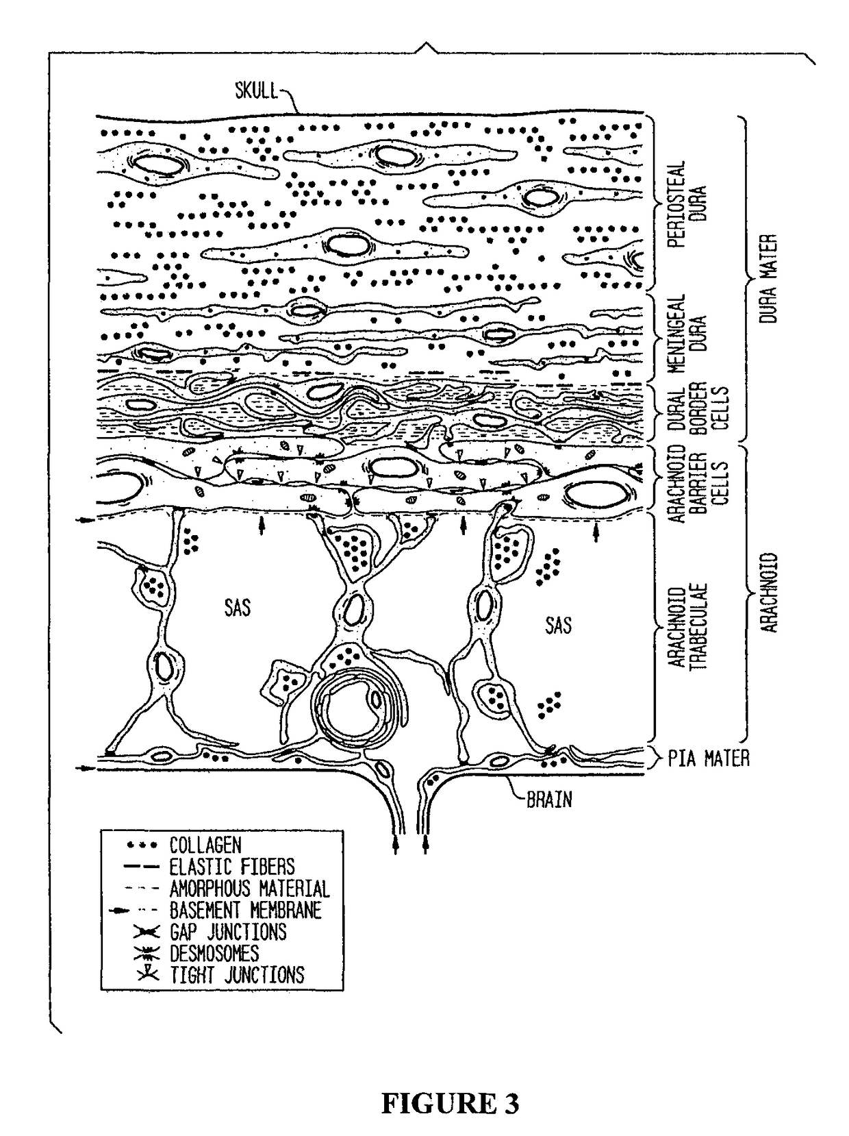
The limited permeability of the brain capillary endothelial wall, constituting the blood brain barrier (BBB), poses challenges to the development of methods of drug delivery to target sites in the brain.
However, such localized intracranial or spinal administrations are invasive and are associated with a risk of CNS infections, which increases if more injections have to be given or if a catheter has to be left in place to repeat the injection.
Furthermore, most drugs delivered directly into the cerebrospinal fluid (CSF) are rapidly cleared, exhibiting very short half-lives, thus requiring frequent invasive administrations to maintain therapeutic levels at target sites of the action.
This limits the practical applicability of localized drug delivery to the central nervous system (CNS).
Since each drug has a therapeutic range above which it is toxic and below which it is ineffective, oscillating drug levels may cause alternating periods of ineffectiveness and toxicity.
Polyesters such as lactic acid-glycolic acid copolymers display bulk (homogeneous) erosion, resulting in significant degradation in the matrix interior.
Analgesia (pain control) is important in order to permit good blood pressure control but must be balanced against oversedating patient, which impacts mental status and thus interfere with the ability to monitor the level of consciousness.
Rebleeding is hard to predict but may happen at any time and carries a dismal prognosis.
When the aneurysm has been located, metallic coils are deployed that lead to formation of a blood clot in the aneurysm and obliteration.
Aneurysms of the middle cerebral artery and its related vessels are hard to reach and of less optimal configuration for endovascular coiling and tend to be amenable to clipping, while those of the basilar artery and posterior arteries are hard to reach surgically and tend to be more accessible for endovascular management.
The main drawback of coiling is the possibility that the aneurysm may recur; this risk is lower in the surgical approach.
However, the aforementioned treatments are expensive, time consuming and only partially effective.
For over 35 years, physicians have been trying to prevent or reduce the incidence of adverse consequences of SAH, including angiographic vasospasm and DCI, and have had limited effect due to side effects of current agents or lack of efficacy.
There currently are no FDA approved agents for the prevention of vasospasm or the reduction of delayed ischemic neurologic deficits also known as delayed cerebral ischemia (DCI).
Current methods to prevent vasospasm have failed due to lack of efficacy or to safety issues, primarily hypotension and cerebral edema.
Voltage-dependent calcium channel antagonists may be effective in preventing and reversing vasospasm to a certain extent, however, prior art treatments administer doses too low to exert a maximal pharmacologic effect.
Endothelin-receptor antagonists also may be effective at preventing and reversing angiographic vasospasm to a certain extent, but this reversal or prevention of angiographic vasospasm does not translate into as marked an improvement in outcome as would be anticipated by the reduction in angiographic vasospasm.
Without being limited by theory, it is postulated that the systemic delivery of the voltage-dependent calcium channel antagonists may cause side effects that mitigate the beneficial effects on angiographic vasospasm, such as, for example, systemic hypotension and pulmonary vasodilation with pulmonary edema, which prevent the administration of higher systemic doses.
Dilation of blood vessels in the lungs also may cause lung edema and lung injury.
Nimodipine, an oral calcium channel antagonist, has been shown in clinical trials to reduce the chance of a poor outcome, however it may not significantly reduce the amount of angiographic vasospasm detected on angiography.
Other calcium channel antagonists and magnesium sulfate have been studied, but are not presently recommended.
When administered in the doses used clinically for oral or intravenous administration, nimodipine is associated with dose-limiting hypotension in up to 50% of patients.
Hypotension is deleterious to patients with aneurysmal SAH because it may lower cerebral perfusion pressure and worsen DCI.
While there is some evidence suggesting that nimodipine can have neuroprotective effects, it is not conclusive.
However, the study was limited to patients who had severe head trauma with a Glasgow Coma Scale≤8 and patients with traumatic or chronic lung pathology or brain lesion who required surgical intervention were excluded from this study.
Dreier et al. reported that intravenous administration of nimodipine to rats can reverse cortical spreading ischemia after SAH triggered by hemoglobin in rats to cortical spreading hyperemia, but conceded that no conclusion could be drawn from their study regarding territorial infarctions after SAH, which likely include other pathogenic cascades.
Induced hypertension is believed to be the most important component of this treatment although evidence for the use of this approach is inconclusive, and no sufficiently large randomized controlled trials ever have been undertaken to demonstrate its benefits.
Removal of subarachnoid blood clots with recombinant tissue plasminogen activator (r-t-PA) in patents with aneurysmal SAH has been reported to reduce angiographic vasospasm and DCI but with inconclusive results due to the small number of patients treated and lack of randomized, blinded trials.
Hypomagnesemia is common following aneurysmal SAH and is associated with poor outcome and development of vasospasm.
Current therapies to prevent or reduce the incidence of secondary complications after aSAH, such as DCI and angiographic vasosparm, are risky, only marginally efficacious, expensive and time-consuming.

Method used


Image

  • Compositions and their use to treat complications of aneurysmal subarachnoid hemorrhage
  • Compositions and their use to treat complications of aneurysmal subarachnoid hemorrhage
  • Compositions and their use to treat complications of aneurysmal subarachnoid hemorrhage

Examples


example 1

Exemplary Nimodipine Microparticle Formulation

[0516]A microparticle nimodipine formulation containing a uniform size distribution of microparticles was prepared by combining a polymer solution (e.g., a 50-50 glycolide-lactide blend) with a solvent in the presence of nimodipine and suspended in a buffer. The mixture was added to a surfactant containing aqueous solution to form an emulsion and the solvent extracted to produce the flowable microparticle nimodipine formulation.

[0517]Nimodipine dispersed throughout each microparticles comprising a 50-50 glycolide-lactide (PLGA) blend had an initial drug load of 65%, i.e., 65% nimodipine and 35% polymer. An exemplary nimodipine microparticle formulation comprises 63.2 wt. % of nimodipine and 36.8 wt. % of PLGA.

[0518]The mean particle diameter was about 70 μm, ranging between 40 μm-100 μm. FIG. 13 shows a scanning electron micrograph (SEM) image of a microparticle nimodipine formulation according to the present invention.

example 2

In Vitro Release Kinetic Analysis

[0519]For measuring in vitro release profiles, samples of microparticle nimodipine formulations were analyzed for nimodipine content by high performance liquid chromatography (HPLC) at specific time points (1 hr, 2 hrs, 6 hrs, 24 hrs and then each day for 14 days). The release characteristics in vitro were assessed by mixing nimodipine PLGA microparticles, 10 mg, with 20 ml PBS / sodium dodecyl sulfate. The supernatant fluid (10 ml) was removed and assayed at the specified times. At each time the supernatant fluid removed was replaced with an equal volume of the buffer solution. The assay was optimized to meet regulatory requirements for sink conditions. Exemplary microparticle formulations are capable of releasing in vitro about 50% to 100% of nimodipine within a time frame of 6 to 14 days in vitro. FIG. 14 shows the in vitro cumulative release of exemplary microparticle nimodipine formulations expressed as weight % of the over time.

example 3

Local Delivery of the Microparticle Nimodipine Formulation

[0520]The microparticle nimodipine formulation was combined with a pharmaceutical carrier or an injection vehicle to form the flowable pharmaceutical composition of the described invention. The placebo formulation contained the microparticles created without nimodipine plus vehicle. For example, a vehicle (e.g. water, saline (hydroxyl propyl methyl cellulose (HPMC) in PBS, sodium hyaluronate, etc.) can be mixed with the microparticle nimodipine formulation to form a flowable suspension.

[0521]The microparticle nimodipine formulation was filled into a 10 mL syringe (sterile, latex-free) and closed with a polyprolyene cap. The syringe / cap was then packaged within a laminated foil pouch to protect from light.


PUM


Abstract

The described invention provides a method for treating an interruption of a cerebral artery in a subarachnoid space at risk of interruption caused by brain injury in a mammal, which reduces signs or symptoms of at least one delayed complication associated with brain injury using a flowable sustained release particulate composition.

Description

CROSS REFERENCE TO RELATED APPLICATIONS[0001]This application is a continuation in part of U.S. application Ser. No. 13 / 440,276, entitled “Intraventricular drug delivery system for improving outcome after brain injury affecting cerebral blood flow,” filed Apr. 5, 2012, which claims the benefit of priority of U.S. Provisional Application No. 61 / 471,779 filed Apr. 5, 2011, and is a continuation in part of U.S. application Ser. No. 12 / 137,320, entitled “A Drug Delivery System for the Prevention of Cerebral Vasospasm,” filed Jun. 11, 2008, which claims the benefit of priority of U.S. Provisional Application No. 60 / 976,902 (filed Oct. 29, 2007) and No. 60 / 943,124 (filed Jun. 11, 2007). The content of each of these applications is incorporated by reference herein in its entirety.FIELD OF INVENTION[0002]The present invention relates to a system for therapeutic agents delivered locally to the cerebral arteries to prevent or reduce the incidence or severity of adverse consequences of subarac...

Claims


Application Information

Patent Timeline
Patent Type & Authority Patents(United States)
IPC IPC(8): A61K9/00A61K31/4418A61K9/70A61K9/14A61K9/50A61K31/4422
CPCA61K9/70A61K9/0002A61K9/0019A61K31/4422A61K9/146A61K9/5031A61K31/4418A61K9/0085A61P25/00A61P9/00
Inventor MACDONALD, R. LOCH
Owner EDGE THERAPEUTICS
Why Eureka
Try Eureka