
Methods for Sterilizing Tissue

Inactive Publication Date: 2011-04-21
BURGESS WILSON +3
View PDF99 Cites 8 Cited by
  • Summary
  • Abstract
  • Description
  • Claims
  • Application Information

AI Technical Summary

Benefits of technology

[0028]In accordance with these and other objects, a first embodiment of the present invention is directed to a method for sterilizing one or more tissues that are sensitive to radiation, the method comprising irradiating the one or more tissues with radiation for a time effective to sterilize the one or more tissues at a rate effective to sterilize the one or more tissues and to protect the one or more tissues from the radiation.
[0029]Another embodiment of the present invention is directed to a method for sterilizing one or more tissues that are sensitive to radiation, comprising: (i) applying to the one or more tissues at least one stabilizing process selected from the group consisting of: (a) adding to the one or more tissues at least one stabilizer in an amount effective to protect the one or more tissues from the radiation; (b) reducing the residual solvent content of the one or more tissues to a level effective to protect the one or more tissues from the radiation; (c) reducing the temperature of the one or more tissues to a level effective to protect the one or more tissues from the radiation; (d) reducing the oxygen content of the one or more tissues to a level effective to protect the one or more tissues from the radiation; (e) adjusting or maintaining the pH of the one or more tissues to a level effective to protect the one or more tissues from the radiation; and (f) adding to the one or more tissues at least one non-aqueous solvent in an amount effective to protect the one or more tissues from the radiation; and (ii) irradiating the one or more tissues with a suitable radiation at an effective rate for a time effective to sterilize the one or more tissues.
[0030]Another embodiment of the present invention is directed to a method for sterilizing one or more tissues that are sensitive to radiation, comprising: (i) applying to the one or more tissues at least one stabilizing process selected from the group consisting of: (a) adding to the one or more tissues at least one stabilizer; (b) reducing the residual solvent content of the one or more tissues; (c) reducing the temperature of the one or more tissues; (d) reducing the oxygen content of the one or more tissues; (e) adjusting or maintaining the pH of the one or more tissues; and (f) adding to the one or more tissues at least one non-aqueous solvent; and (ii) irradiating the one or more tissues with a suitable radiation at an effective rate for a time effective to sterilize the one or more tissues, wherein the at least one stabilizing process and the rate of irradiation are together effective to protect the one or more tissues from the radiation.
[0031]Another embodiment of the present invention is directed to a method for sterilizing one or more tissues that are sensitive to radiation, comprising: (i) applying to the one or more tissues at least two stabilizing processes selected from the group consisting of: (a) adding to the one or more tissues at least one stabilizer; (b) reducing the residual solvent content of the one or more tissues; (c) reducing the temperature of the one or more tissues; (d) reducing the oxygen content of the one or more tissues; (e) adjusting or maintaining the pH of the one or more tissues; and (f) adding to the one or more tissues at least one non-aqueous solvent; and (ii) irradiating the one or more tissues with a suitable radiation at an effective rate for a time effective to sterilize the one or more tissues, wherein the at least two stabilizing processes are together effective to protect the one or more tissues from the radiation and further wherein the at least two stabilizing processes may be performed in any order.
[0032]Another embodiment of the present invention is directed to methods for sterilizing one or more tissues that are sensitive to radiation while producing substantially no neo-antigens in the tissue and / or reducing the number of reactive allo-antigens and / or xeno-antigens. Such methods reduce post-implantation complications including, but not limited to, inflammation, immune rejection reactions, calcification, and similar conditions that reduce the implant's ability to function and / or survive in the recipient.

Problems solved by technology

Many biological materials that are prepared for human, veterinary, diagnostic and / or experimental use may contain unwanted and potentially dangerous biological contaminants or pathogens, such as viruses, bacteria, in both vegetative and spore states, (including inter- and intracellular bacteria, such as mycoplasmas, ureaplasmas, nanobacteria, chlamydia, rickettsias), yeasts, molds, fungi, prions or similar agents responsible, alone or in combination, for TSEs and / or single-cell or multicellular parasites.
The tissue in these knee surgery cases was cartilage, which is not sterilized as it is believed such sterilization would damage the implant.
Such procedures, however, are not always reliable, as evidenced by the death of at least one Minnesota man who received a cartilage implant, and are not able to detect the presence of prions or certain viruses, particularly those present in very low numbers.
This reduces the value, certainty, and safety of such tests in view of the consequences associated with a false negative result, which can be life threatening in certain cases, for example in the case of Acquired Immune Deficiency Syndrome (AIDS).
Furthermore, in some instances it can take weeks, if not months, to determine whether or not the material is contaminated.
Moreover, to date, there is no commercially available, reliable test or assay for identifying prions, ureaplasmas, mycoplasmas, and chlamydia within a biological material that is fully suitable for screening out potential donors or infected material (Advances in Contraception 10(4):309-315 (1994)).
Thus, the products of unicellular natural or recombinant organisms or tissues virtually always carry a risk of pathogen contamination.
In addition to the risk that the producing cells or cell cultures may be infected, the processing of these and other biological materials also creates opportunities for environmental contamination.
Interestingly, even products from species as different from humans as transgenic plants carry risks, both due to processing contamination as described above, and from environmental contamination in the growing facilities, which may be contaminated by pathogens from the environment or infected organisms that co-inhabit the facility along with the desired plants.
Mice can harbor serious human pathogens such as the frequently fatal Hanta virus.
Indeed, such rodents are notoriously difficult to control, and may gain access to a crop during sowing, growth, harvest or storage.
Likewise, contamination from overflying or perching birds has the potential to transmit such serious pathogens as the causative agent for psittacosis.
Thus, any biological material, regardless of its source, may harbor serious pathogens that must be removed or inactivated prior to administration of the material to a recipient human or other animal.
This is a result of safety concerns for the workers conducting the tests, and the difficulty and expense associated with facilities for containment and waste disposal.
According to current standards of the U.S. Food and Drug Administration, heat treatment of biological materials may require heating to approximately 60° C. for a minimum of 10 hours, which can be damaging to sensitive biological materials.
Indeed, heat inactivation can destroy 50% or more of the biological activity of certain biological materials.
Unfortunately, this method may also remove products that have a high molecular weight.
Further, in certain cases, small viruses may not be removed by the filter.
This procedure requires that unbound sensitizer be washed from products since the sensitizers are toxic, if not mutagenic or carcinogenic, and cannot be administered to a patient.
The published literature in this area, however, teaches that gamma radiation can be damaging to radiation sensitive products, such as blood, blood products, protein and protein-containing products.
In particular, it has been shown that high radiation doses are injurious to red cells, platelets and granulocytes (Leitman).
This patent concludes that “[i]f the gamma irradiation were applied while the protein material was at, for example, ambient temperature, the material would be also completely destroyed, that is the activity of the material would be rendered so low as to be virtually ineffective.” Unfortunately, many sensitive biological materials, such as monoclonal antibodies (Mab), may lose viability and activity if subjected to freezing for irradiation purposes and then thawing prior to administration to a patient.
Another factor that may feed future transplantation demand is certain poor lifestyle choices in the population, including such factors as poor nutrition (including such trends as the increasing reliance on so-called fast foods and fried foods; insufficient intake of fruits, vegetables and true whole grains; and increased intake of high glycemic, low nutritional value foods, including pastas, breads, white rice, crackers, potato chips and other snack foods, etc.), predilections toward a sedentary lifestyle, and over-exposure to ultraviolet light in tanning booths and to sunlight.
The increasing occurrence of such factors as these have resulted, for example, in increased incidences of obesity (which also exacerbates such conditions as arthritis and conditions with cartilage damage, as well as impairs wound healing, immune function, cancer risk, etc.), type II diabetes and polycystic ovary syndrome (high post prandial glucose values causing damage to such tissues as nerve, muscle, kidney, heart, liver, etc., causing tissue and organ damage even in persons who are not diabetic), many cancers, and hypertension and other cardiovascular conditions, such as strokes and Alzheimer's disease (recent data suggesting that Alzheimer's may be the result of a series of mini-strokes).
Thus, poor lifestyle choices ultimately will increase demand for bone, cartilage, skin, blood vessels, nerves, and the specific tissues and organs so destroyed or damaged.
In addition, there is an epidemic of infection by intracellular microbes for which reliable commercial tests are not available (for example, mycoplasma, ureaplasma, and chlamydia), for example, as a result of sexual contact, coughing, etc.
Such is the case with some Streptococci infections (antibodies produced against M proteins of Streptococci that cross-react with cardiac, joint and other tissues), for example, in which tissue and other cardiac tissue may be attacked to cause reduced cardiac function, and which can result in death if the infection is not properly treated before extensive damage occurs.
Tissue vegetations and mitral regurgitation are common in intravascular infections, although tissue destruction so extensive as to require valve replacement is rare.
Yet another factor in transplantation demand is drug use, particularly the use of illicit drugs, but also including inappropriate and sometimes illegal use of otherwise licit drugs (such as overuse of alcohol / alcoholism causing cirrhosis of the liver, and therefore requiring liver transplantation).
Such drug use often strongly damages or even destroys sensitive tissues and organs such as kidney, liver, lung, heart, brain / nerves, and / or portions thereof.
In addition, intravenous drug use greatly increases the odds of contracting intravascular infections by any one or more of the above-cited infectious agents (among many others), which infections can attack virtually any organ or portion thereof, including the tricuspid valve (located between the right atrium and the right ventricle), the mitral valve (located between the left atrium and the left ventricle), the pulmonary or pulmonic valve (located between the right ventricle and the pulmonary artery), and the aortic valve (located between the left ventricle and the aorta) with any infectious agent that may enter through implanted tissue.

Method used


Image

  • Methods for Sterilizing Tissue
  • Methods for Sterilizing Tissue
  • Methods for Sterilizing Tissue

Examples


example 1

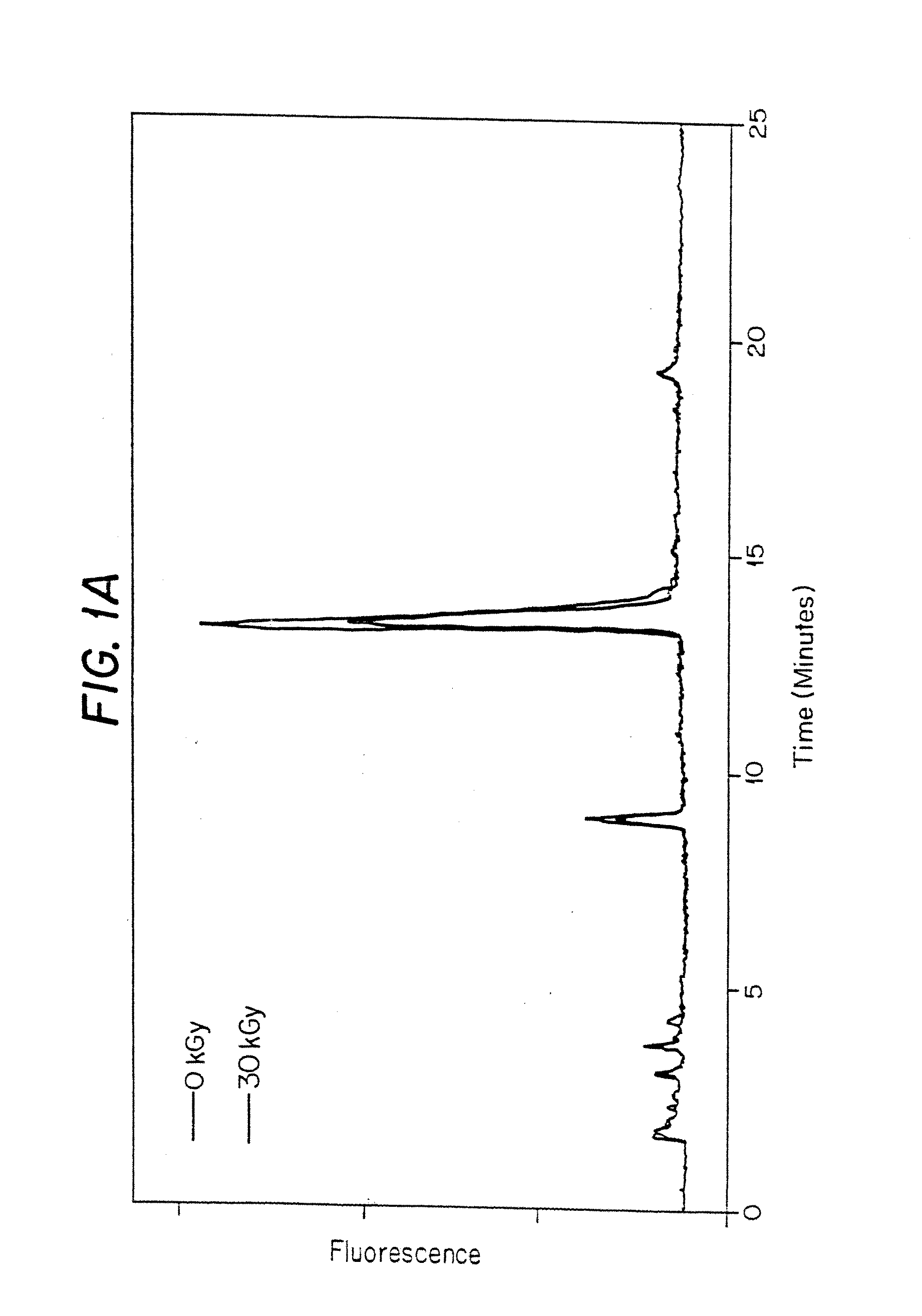
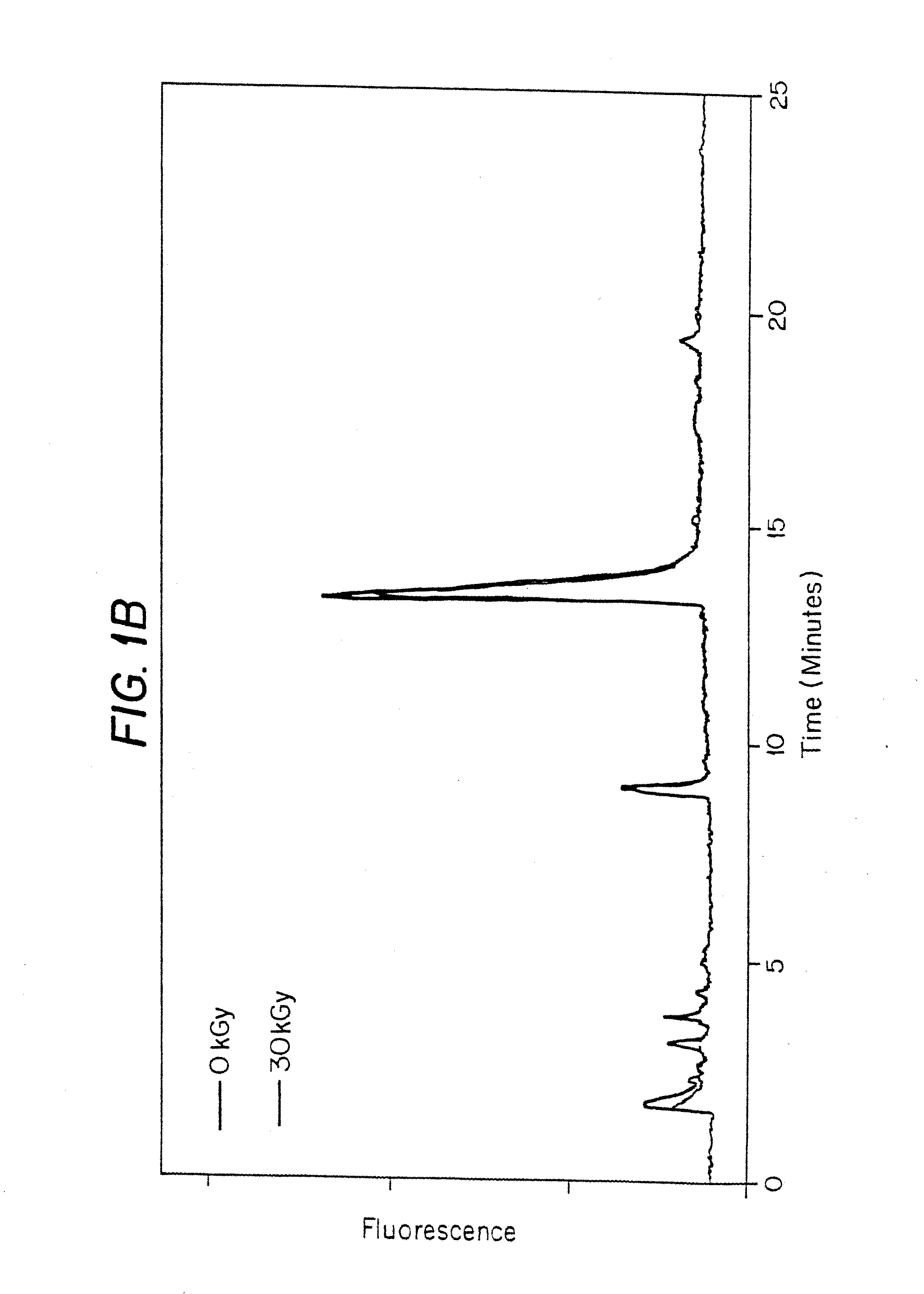
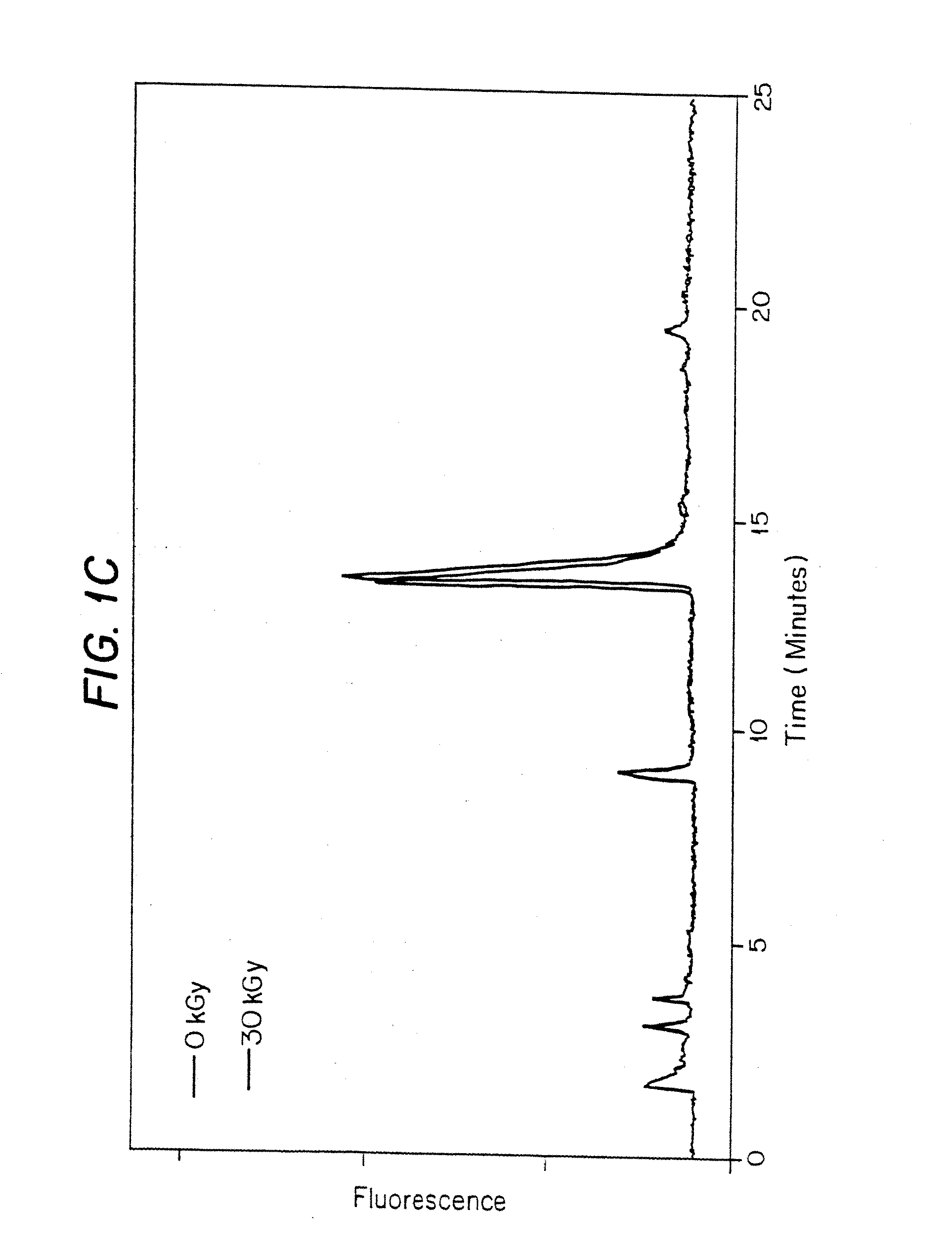
[0118]In this experiment, porcine heart valves were gamma irradiated in the presence of polypropylene glycol 400 (PPG400) and, optionally, a scavenger, to a total dose of 30 kGy (1.584 kGy / hr at −20° C.).

Materials:

[0119]Tissue—Porcine Pulmonary Valve (PV) Heart valves were harvested prior to use and stored.[0120]Tissue Preparation Reagents—[0121]Polypropylene Glycol 400. Fluka: cat# 81350, lot# 386716 / 1[0122]Trolox C. Aldrich: cat# 23, 881-3, lot# 02507TS[0123]Coumaric Acid. Sigma: cat# C-9008, lot# 49H3600[0124]n-Propyl Gallate. Sigma: cat# P-3130, lot# 117H0526[0125]α-Lipoic Acid. CalBiochem: cat# 437692, lot# B34484[0126]Dulbecco's PBS. Gibco BRL: cat# 14190-144, lot# 1095027[0127]2.0 ml Screw Cap tubes. VWR Scientific Products: cat# 20170-221, lot# 0359[0128]Tissue Hydrolysis Reagents—[0129]Nerl H2O. NERL Diagnostics: cat# 9800-5, lot# 03055151[0130]Acetone. EM Science: cat# AX0125-5, lot# 37059711[0131]6 N constant boiling HCl. Pierce: cat# 24309, lot# BA42184[0132]Int-Pyd (Ace...

example 2

[0189]In this experiment, the effects of gamma irradiation were determined on porcine heart valve cusps in the presence of 50% DMSO and, optionally, a stabilizer, and in the presence of polypropylene glycol 400 (PPG400).

Preparation of Tissue for Irradiation:

[0190]1. 5 vials of PV and 3 vials of atrial valves (AV) were thawed on ice.[0191]2. Thaw media was removed and valves rinsed in beaker filled with PBS.[0192]3. Transferred each valve to 50 ml conical containing PBS. Washed by inversion and removed.[0193]4. Repeated wash 3 times.[0194]5. Dissected out the 3 cusps (valves).[0195]6. Stored in PBS in 2 ml screw top Eppendorf Vials (Eppendorfs) and kept on ice.

Preparation of Stabilizers:

[0196]All stabilizers were prepared so that the final concentration of DMSO was 50%.

1 M Ascorbate in 50% DMSO:

[0197]Aldrich: cat# 26, 855-0, lot# 10801HU

200 mg dissolved in 300 μl H2O. Add 500 μl DMSO. The volume was adjusted to 1 ml with H2O. Final pH was≈8.0.

1 M Coumaric Acid:

[0198]Sigma: cat# C-900...

example 3

[0248]In this experiment, frozen porcine AV heart valves soaked in various solvents were gamma irradiated to a total dose of 30 kGy at 1.584 kGy / hr at −20° C.

Materials:

[0249]1. Porcine heart valve cusps were obtained and stored at −80° C. in a cryopreservative solution (Containing Fetal calf serum, Penicillin-Streptomycin, M199 media, and approximately 20% DMSO).[0250]2. Dulbecco's Phosphate Buffered Saline. Gibco BRL: cat#14190-144, lot#1095027[0251]3. 2 ml screw cap vials. VWR: cat# 20170-221, lot #0359[0252]4. 2 ml glass vials. Wheaton: cat# 223583, lot#370000-01[0253]5. 13 mm stoppers. Stelmi: 6720GC, lot#G006 / 5511[0254]6. DMSO. JT Baker: cat# 9224-01, lot# H40630[0255]7. Sodium ascorbate. Aldrich: cat# 26, 855-0, lot 10801HU; prepared as a 2 M stock in Nerl water.[0256]8. Fetal calf serum[0257]9. Penicillin-Streptomycin[0258]10. M199 media[0259]11. DMSO

Methods:

Cryopreservative Procedure:

[0260]Preparation of Solutions

[0261]Freeze Medium:[0262]Fetal calf serum (FCS) (10%)=50 ml[0...


PUM


Abstract

Methods are disclosed for sterilizing tissue to reduce the level of one or more active biological contaminants or pathogens therein, such as viruses, bacteria, (including inter- and intracellular bacteria, such as mycoplasmas, ureaplasmas, nanobacteria, chlamydia, rickettsias), yeasts, molds, fungi, prions or similar agents responsible, alone or in combination, for TSEs and / or single or multicellular parasites. The methods involve sterilizing one or more tissues with irradiation.

Description

BACKGROUND OF THE INVENTION[0001]1. Field of the Invention[0002]The present invention relates to methods for sterilizing tissue to reduce the level of one or more active biological contaminants or pathogens therein, such as viruses, bacteria (including inter- and intracellular bacteria, such as mycoplasmas, ureaplasmas, nanobacteria, chlamydia, rickettsias), yeasts, molds, fungi, prions or similar agents responsible, alone or in combination, for transmissible spongiform encephalopathies (TSEs) and / or single or multicellular parasites. The present invention particularly relates to methods of sterilizing tissue with irradiation, wherein the tissue may subsequently be used in transplantation to replace diseased and / or otherwise defective tissue in an animal.[0003]2. Background of the Related Art[0004]Many biological materials that are prepared for human, veterinary, diagnostic and / or experimental use may contain unwanted and potentially dangerous biological contaminants or pathogens, s...

Claims


Application Information

Patent Timeline
IPC IPC(8): A61L2/00
CPCA61L2/0088A61L2/0035
Inventor BURGESS, WILSONDROHAN, WILLIAM N.MACPHEE, MARTIN J.MANN, DAVID M.
Owner BURGESS WILSON
Learn More