While the growth of cells in two-dimensions is frequently used for the preparation and examination of cultured cells
in vitro, it may lack the characteristics of intact, tissue
in vivo tissue which, for example, includes
cell-cell and cell-matrix interactions.
However, long-term culture and proliferation of cells in such systems has not yet been achieved.
However, numerous complications and the generally unsatisfactory nature of long-term aesthetic results caused the procedure to be rapidly abandoned.
More recently, the use of injectable
silicone became prevalent in the 1960's for the correction of minor defects, although various inherent complications also limited the use of this substance.
Complications associated with the utilization of injectable liquid
silicone include local and systemic inflammatory reactions, formation of
scar tissue around the
silicone droplets, rampant and frequently distant, unpredictable migration throughout the body, and localized tissue breakdown.
Due to these potential complications, silicone is not currently approved for general clinical use.
Although the original proponents of
silicone injection have continued experimental programs utilizing specially manufactured “Medical Grade” silicone (e.g., Dow Corning's MDX 4.4011®) with a limited number of subjects, it appears highly unlikely that its use will be generally adopted by the surgical
community.
However, success has been heretofore limited.
Subsequent undesirable micro-
particulate media migration and serious granulomatous reactions frequently occur with the injection of this material.
However, while these aforementioned materials create immediate augmentation and / or repair of defects, they also have a tendency to migrate and be reabsorbed from the original
injection site.
The poor results initially obtained with the use of non-biological injectable materials prompted the use of various non-immunogenic, proteinaceous materials (e.g.,
bovine collagen and
fibrin matrices).
Clinical protocols calling for repeated injections of atelocollagen are, in practice, primarily limited by the development of immunogenic reactions to the
bovine collagen.
The increased
viscosity, and in particular irregular increased
viscosity resulting in “lumpiness,” not only rendered the material more difficult to utilize, but also made it unsuitable for use in certain circumstances.
However, like
glutaraldehyde, GAG may be released into the tissue causing unforeseen long-term effects on human subjects.
Additionally, a reduction of collagen
blood clotting capacity may also be deleterious in the application in
bleeding wounds, as
fibrin clot contributes to an adhesion of the graft to the surrounding tissue.
It should be noted, however, that there is no quantitative evidence which demonstrates that human collagen injection results in lower levels of
implant degradation than that which is found with
bovine collagen preparations.
Furthermore, the utilization of autologous collagen preparation and injection is limited to those individuals who have previously undergone
surgery, due to the fact that the collagen is produced is derived from the tissue removed during the surgical procedure.
Therefore, it is evident that, although human collagen circumvents the potential for
immunogenicity exhibited by bovine collagen, it fails to provide long-term therapeutic benefits and is limited to those patients who have undergone prior
surgical procedures.
Clinical utilization of the FIBREL product has been reported to often result in an overall lack of
implant uniformity.
Therefore, in conclusion, none of the currently utilized
protein-based injectable materials appears to be totally satisfactory for the augmentation and / or repair of the subjacent
dermis and
soft tissue.
Thus, for large scale repair procedures the amount of
adipose tissue which can be surgically-excised from the patient may be limiting.
In addition, other frequently encountered difficulties with the aforementioned methodologies include non-uniformity of the injectate, unpredictable
longevity of the aesthetic effects, and a 4-6 week period of post-injection
inflammation and swelling.
However, unfortunately, these forms of treatment have all exhibited numerous disadvantages.
For example, split thickness autographs generally show limited
tissue expansion, require repeated surgical operations, and give rise to unfavorable aesthetic results.
Epidermal autographs require long periods of time to be cultured, have a low success (“take”) rate of approximately 30-48%, frequently form spontaneous
blisters, exhibit contraction to 60-70% of their original size, are vulnerable during the first 15 days of engraftment, and are of no use in situations where there is both epidermal and dermal tissue involvement.
Similarly, epidermal allografts (cultured allogenic keratinocytes) exhibit many of the limitations which are inherent in the use of epidermal autographs, in addition to
graft rejection.
However, this too has met with limited success due to, for example,
graft rejection and unfavorable aesthetic results.
However, subsequent attempts to reproduce the living
skin equivalent using human fibroblasts and keratinocytes has met with only limited success.
Additionally skin
thinning is an age-dependent defect.
The use of these components for the treatment of
cellulite confers
drug-use issues (e.g., long-term use at high concentrations) preventing the components for extended and over-the-counter use and hence, limiting marketing.
Commonly, more aggressive approaches to
cellulite combine several types of the therapies described above, along with exercise and low-fat diets, but in general, only very little and temporary progress has been reported for treating this prevalent condition.
This occurs in cases where redundant skin is not available for healing, as in burns over flexor joints surfaces, such as the neck.
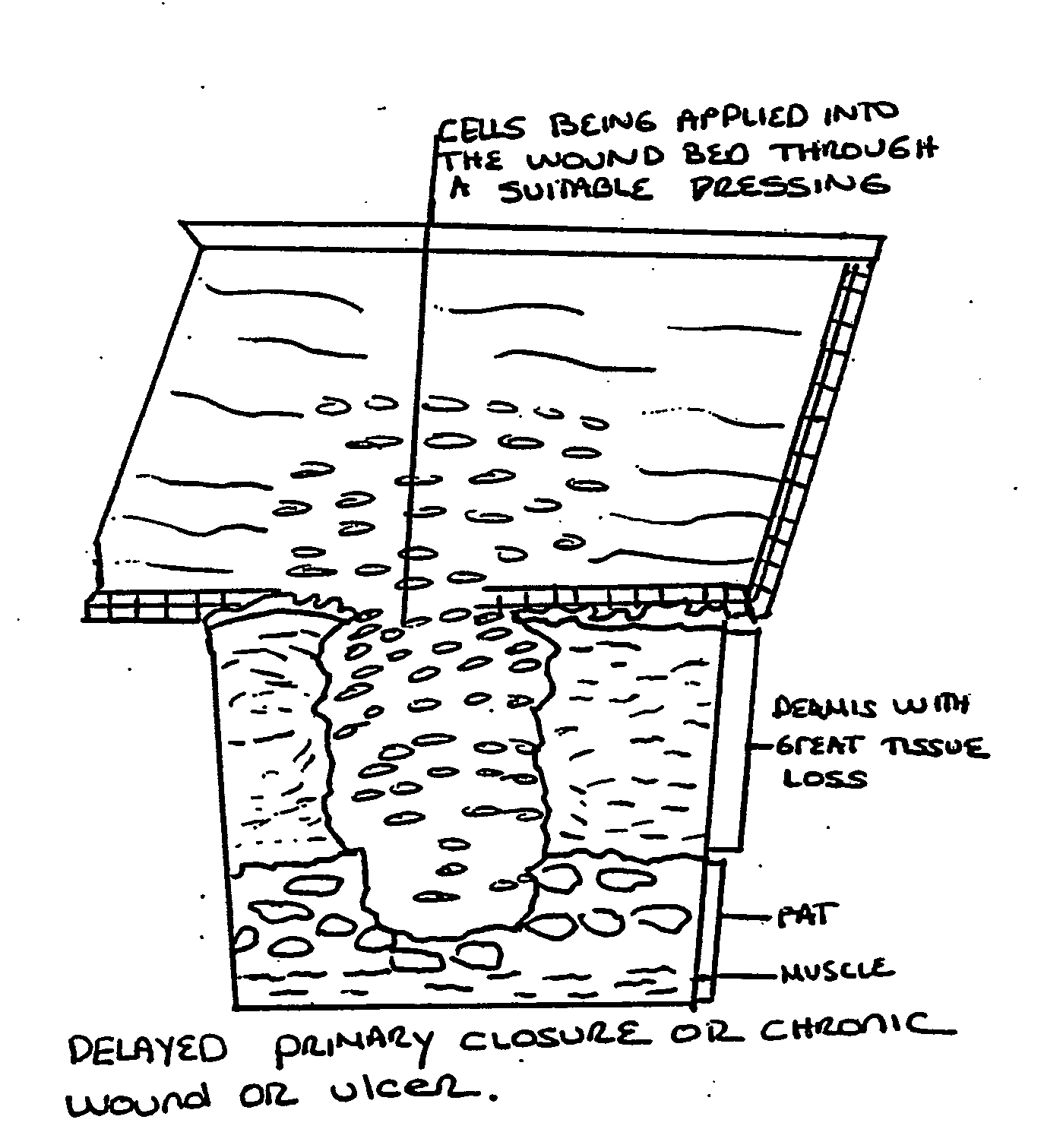
Chronic wounds remain as one of the most expensive and unsolved problems in
medicine.
Usually chronic wounds, such as pressure, diabetic, venous stasis / ischemic ulcers, fail to heal because of a co-existing underlying health problem, such as diabetes or
varicose veins.
In recent years the availability of innumerable types of dressings, that are expensive and only marginally effective, has dramatically increased.
Allografts of
cadaver skin,
foreskin and cross-linked
porcine skin have been used as temporary wound dressings, but cannot provide a permanent dermal replacement, since they are either rejected or do not revascularize, respectively.
Painful hardening of the breasts due to formation of fibrous
scar tissue occurs around the implants.
Poor firmness and less natural looks often result.
They do not prevent
capsular contracture formation, the possibility of rupture, or deflation due to
saline leakage, even in more recent models displaying a leaf valve mechanism that allows custom inflation (Peters W., Can J Plast Surg, 5 (4):241, 1997).
Implants manufactured with a two layered non-porous and porous outer shell made of
spinning polymer fibers are not completely resistant to rupture or impermeable to silicone gel migration (U.S. Pat. No. 5,376,117).
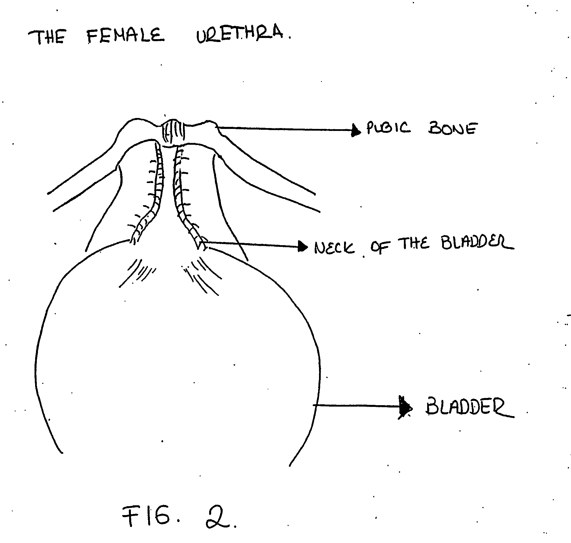
Female
urinary incontinence is a common problem and is particularly prevalent where damage to the bladder or neck of the bladder has occurred during child birth.
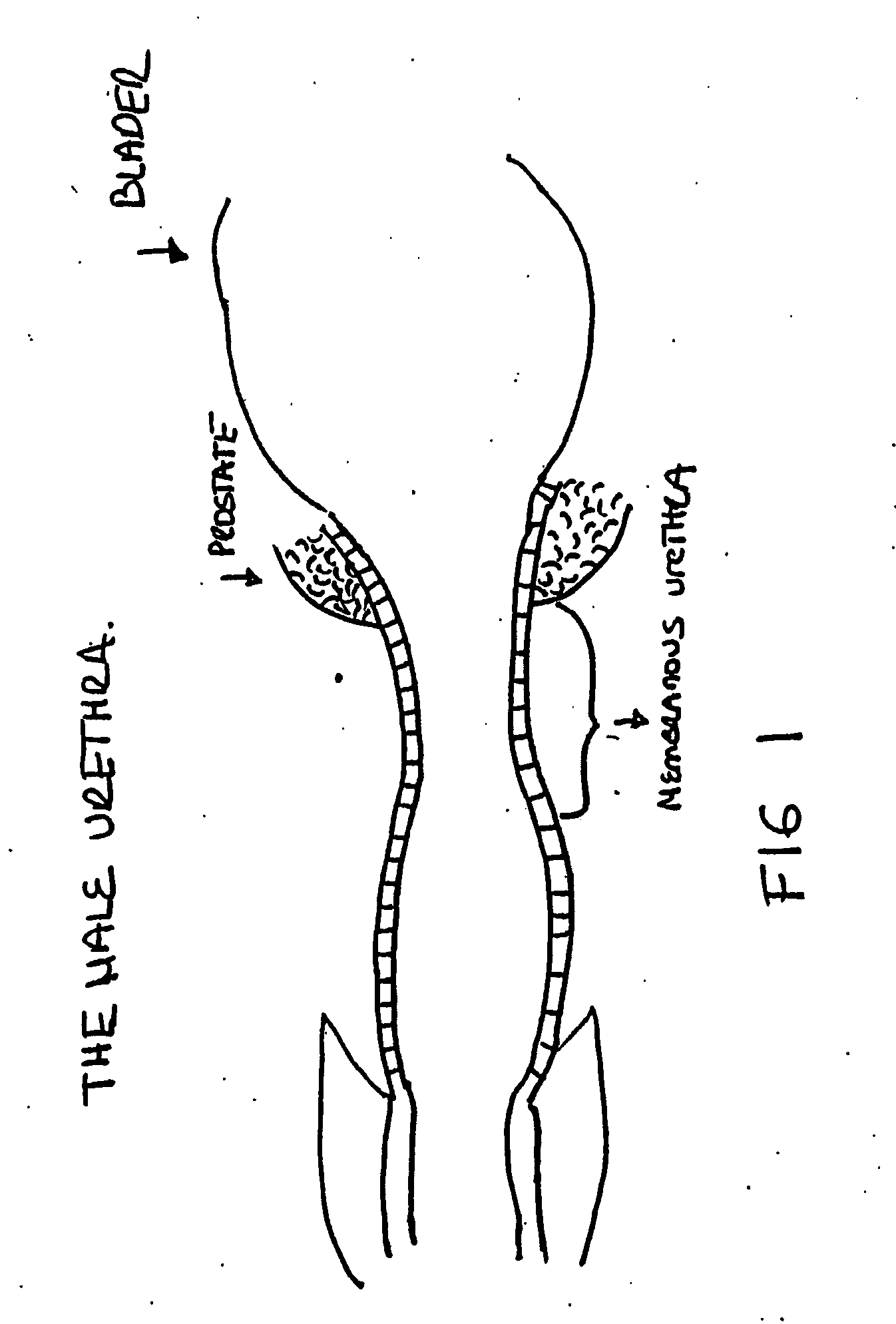
In addition, incontinence in elderly men result is often due to overflow incontinence and detrusor
instability.
The involuntary loss of
urine is unpleasant and embarrassing and can cause other medical problems such as
irritation and burning of the surrounding skin and lower urinary tract resulting in infections of diverse severity.
The most important and common complication of this condition is the frequent episodes of urinary infections commonly requiring chronic antibiotic intake and that can be severe enough to compromise
kidney function.
Beside the pharmacological approach, several
surgical methods are available with poor results, requiring expensive hospitalization and long
recovery times. Frequently the problem is undercorrected and a second or third different surgical technique may be attempted without a permanent outcome.
The surgical implantation of several devices of diverse materials has been attempted with little success.
The implantations are cumbersome, difficult to place and maintain and need frequent adjustments or replacements.
The use of alginate in combination with cultured bovine chondrocytes autografts (Atala et al., J Urol, 150:74, 1993) inherently has two potential problems, adverse immunological reactions and possible
calcification of the chondrocytes.
Side-effects of non-
biological materials have been observed as local and systemic inflammatory reactions, formation of
scar tissue around the site of injection, and rampant and frequent distant, unpredictable migration to the body that may cause life-threatening embolisms.
In any type of graft there are two operative sites, potential infection complications, scar formation from the skin donor site, discomfort, compromised
blood supply for the graft site with subsequent
necrosis,
reabsorption or retention of the graft, potential
hemostasis problems, poor aesthetic results (color and texture differences) and with skin, a particular problem exists which is the presence of hair follicles in the graft.
Many procedures require several weeks to months before the soft tissues are ready for prosthetic or
implant adaptation causing great discomfort to the patient.
Often painless, these diseases develop slowly or progress quite rapidly, causing major damage to the periodontal complex.
As this space increases in depth, the root of the tooth gets exposed, the ligaments and bone get involved and the tooth is no longer stable, becoming loose in its socket.
In the presence of large pockets (>5 mm) the cleaning process may not be enough.
If untreated, several complications may arise.
Another
severe complication is the ripping of the abdominal content, (incarceration) within the
hernia, requiring emergency surgical release.
Complications due to adhesions, intestinal obstruction and fistulization have been reported when not enough care is exercised to prevent the abdominal viscera from contact with the mesh directly.
The most common and severe complications arising from the use of a mesh is infection, since all the
synthetic materials can become sequestered, act like a
foreign body, aggravating and prolonging infections.
Another important problem related to GER in newborns and infants is the failure to thrive caused by the regurgitation or
reflux of considerable amounts of formula after feedings (Avery G et al.
These patients may need aggressive GER treatments to prevent long-term complications from chronic
esophagitis, as stricture formations may cause
stenosis.
Ligaments and tendons are commonly injured during athletic activity and due to the fact that that sports are an increasingly important part of
day to day life in the U.S. the number of
ligament and
tendon injuries have steadily increased over the past few decades.
However, almost any ligament or tendon in the
human body can be injured, torn or ruptured.
ACL injuries are troublesome because they take a long time to heal and often healing very poorly (Lin V. et al., Tiss Engin.
This is a problem common to all ligaments.
This autograft alternative is far from ideal since it can cause
mechanical instability and loss of function of the site from which grafts are taken.
Mosby, St Louis, 1998) of ligaments from cadavers, have the inherent problems described above, in addition to potential
disease transmission of infectious agents from the donor.
Furthermore, in both auto and allografts, the internal part of the grafts show
necrosis shortly after
surgery.
This is a major reason why biological grafts do not provide adequate
mechanical strength until a complete remodeling of the graft is achieved.
In the 1970s, a synthetic
prosthesis made of polymers was introduced as an alternative, but never gained total acceptance because of
mechanical failure, due to fatigue and
abrasive wear.
The lack of long-term studies showing their performance makes it prudent to limit their use to salvage procedures in which autografts and reconstructive procedures have failed (Lin V. et al., Tiss Engl.
In the presence of hormonal abnormalities or changes, inflammation, toxic exposures, and stress, abnormal conditions within the
follicle may occur, inducing either gradual
thinning or rapid loss of the hair.
These follicles may eventually lose their potential for
cycling by the progressive shortening of anagen phase.
However, women abnormally producing higher levels of androgens from the ovaries or the adrenals often show a complete recession of hair on the frontal
scalp.
Many, if not most, do not work or are merely temporary or partial solutions, that are expensive and often are not free of possible dangerous or adverse secondary effects.
As a vasodilator there are safety concerns about possible secondary adverse effects.
Women in reproductive years must be careful not to have any contact with the medication because of known risk of birth defects.
Complications may arise from these procedures and often the cosmetic results do not meet the patient's expectations.
A
hair transplant often may require repetitive procedures that add to the risk of complications and costs.


