Biological component comprising artificial membrane

Inactive Publication Date: 2010-05-11
EMORY UNIVERSITY
View PDF31 Cites 41 Cited by
  • Summary
  • Abstract
  • Description
  • Claims
  • Application Information

AI Technical Summary

Benefits of technology

[0076]The artificial cell membrane of this invention provides an immunoisolation barrier with improved performance characteristics because of its capacity to control interfacial transport by the presence of channels, transporters, and pores, as well as through its ability to act as a template for more complex composite structures with other immunoregulatory macromolecules. In a preferred embodiment of this invention, a membrane-mimetic glycocalyx is provided comprised of a stable, lipid membrane-like bilayer supported on a hydrated islet encapsulating alginate substrate. Transport properties are tailored by the insertion of poly(ethylene oxide) dendritic polymers, to create artificial molecular pores and channels, and maladaptive inflammatory responses are modulated by the incorporation of glycosaminoglycan (GAG) neoglycolipids, as a

Problems solved by technology

An additional approach involves the binding of a heparin-quaternary amine complex, or other antithrombotic agent, to the biomaterial surface However, many of these methods have the disadvantage of being nonpermanent systems in that the surface coating is eventually stripped off or leached away.
For example, heparin, which is complexed to the biomaterial surface, is not only gradually lost from the polymer surface into the surrounding medium but also has limited bioactivity due to catabolism and its inherent instability under physiological conditions.
While these materials have improved stability and promising blood-contacting properties have been reported, a number of limitations exist.
In particular, the ability to engineer surface properties on a molecular level, by taking advantage of the principal of self-organization intrinsic to amphiphilic molecules, is lost.
In addition, the ability to early incorporate diverse biomolecular functional groups into the membrane-mimetic surface is also lost.
A significant limitation in the widespread use of supported biomembranes is their limited stability for most applications outside of a laboratory environment.
Atherosclerosis remains a serious source of morbidity and death despite advances in preventive measures and pharmacological therapeutics.
Although large diameter vascular grafts (6 mm internal diameter) have been successfully developed from polymers such as polytetrafluoroethyl

Method used


Image

  • Biological component comprising artificial membrane
  • Biological component comprising artificial membrane
  • Biological component comprising artificial membrane

Examples


example 1

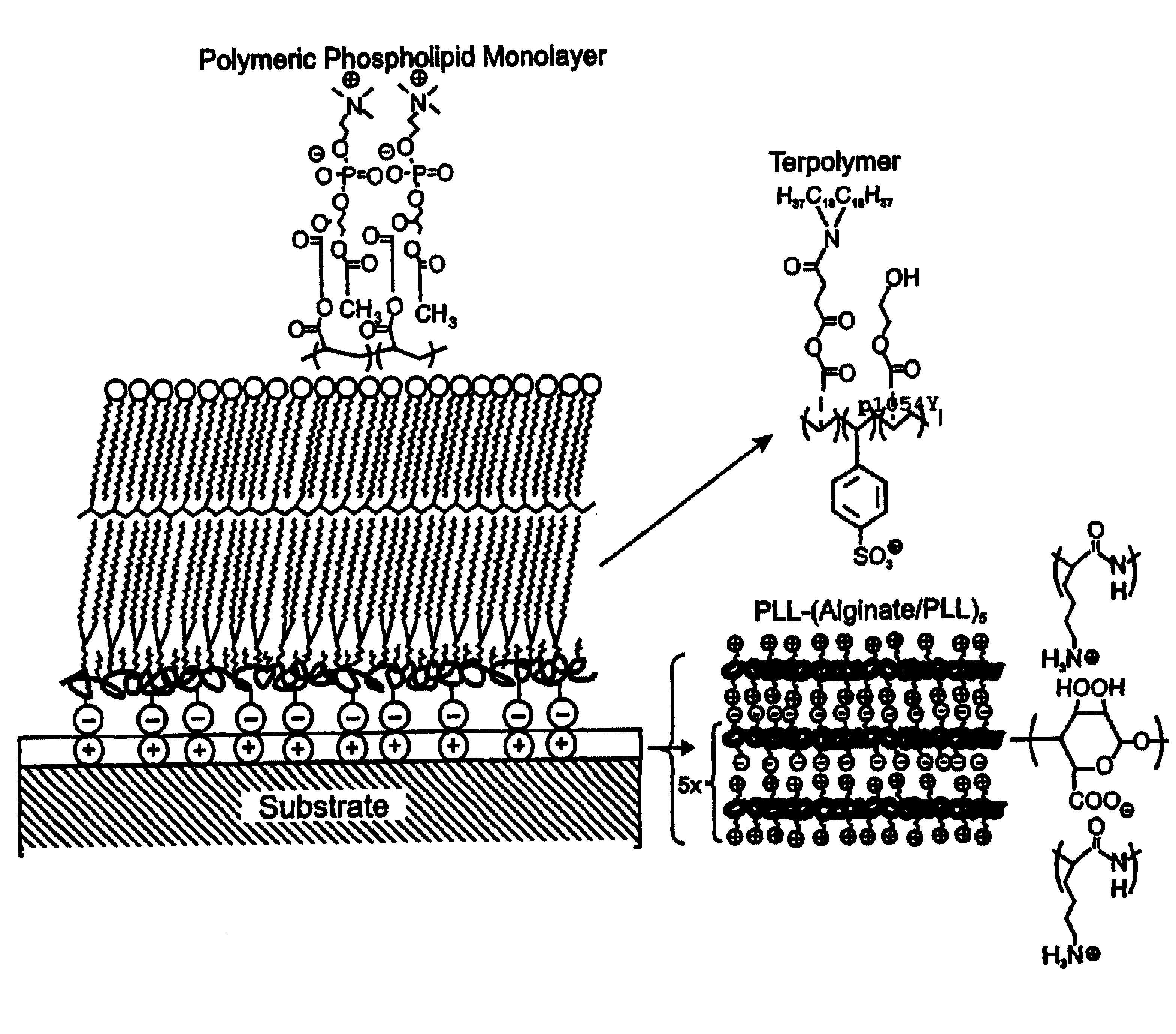
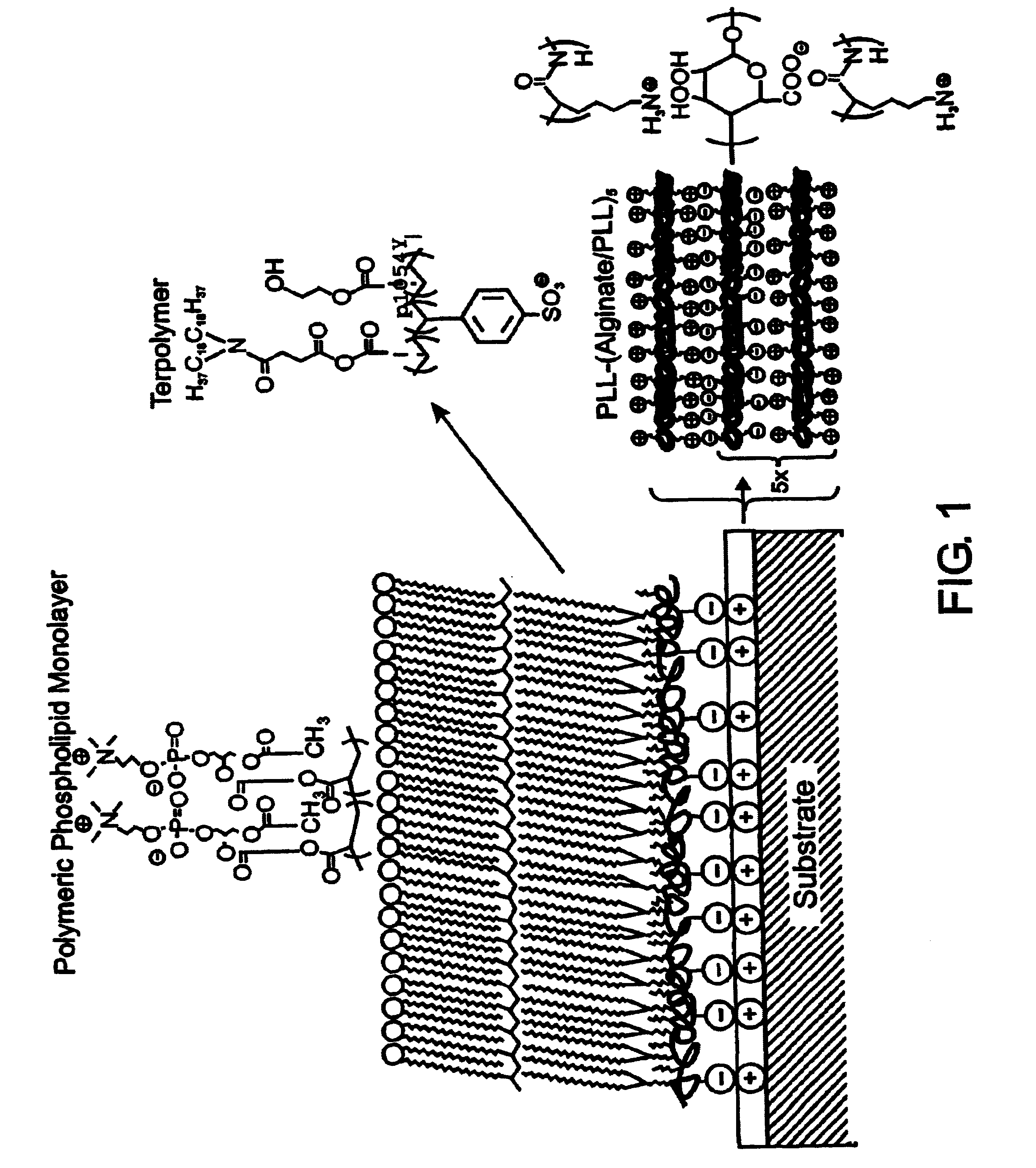
Preparation of Membrane-mimetic Film on Microbeads

[0114]Reagents. All starting materials and synthetic reagents were purchased from commercial suppliers unless otherwise noted. Poly-L-lysine (PLL; 21-479 kD) was purchased from Sigma. Alginate (ca. 41% guluronic acid) was obtained from ISP alginate (San Diego, Calif.) and used as received. N,N-dioctadecylcarbamoyl-propionic acid (DOD), poly(HEA-DOD)6:3, 1-palmitoyl-2-(12-(acryloyloxy)dodecanoyl)-sn-glycero-3-phosphorylcholine (mono-AcrylPC), and 1-palmitoyl-2-(12-(acryloyloxydodecanoyl)-sn-glycero-3-phosphorylethanolamine-fluorescein isothiocyanate (mono-AcrylPE-FITC) were synthesized as previously described. (Marra, K. G., Winger, T. M., Hanson, S. R. & Chaikof, E. L. [1997] Macromolecules 30:6483-6488; Chon, J. H., Marra, K. G. & Chaikof, E. L. [1999] J. Biomat. Sci. Polymer Ed. 10, 95-108; Sells, T. D. & O'Brien, D. F. [1994] Macromolecules 27, 226-233; Sun, X. L., Liu, H., Orban, J. M., Sun, L. & Chaikof, E. L. [2001] Submitted t...

example 2

Cytomimetic Biomaterials: Synthesis and Analysis of Bioactive Membrane-Mimetic Structures Synthesis of Lipopeptide and Glycolipid Conjugates

[0145]Site-specific methods have been developed for the synthesis and purification of lipid-peptide and glycolipid macromolecules in the absence of complex blocking group strategies. (Winger, T. M. et al. [1995] Bioconjug. Chem. 6:323-326; Winger, T. M. et al. [1995] J. Liquid Chromatogr. 18:4117-4125; Winger, T. M. et al. [1995] Biomaterials 16:443-449; Winger, T. M. and Chaikof, E. L. [1997] Langmuir 13:3256-3259; Sun, L. and Chaikof, E. L. [1997] Bioconj. Chem. 8:567-571; Sun, L. and Chaikof, E. L. [1998] Carbohydrate Res. 370: 7-81.) Using these techniques, the orientation of the coupled peptide and carbohydrate has been maintained, which is often required for effective ligand-receptor interactions. The conjugates thus obtained are incorporated into self-assembling systems, such as lipid monolayers for behavioral studies at an air-water inte...

example 3

Stabilization of Membrane-mimetic Biomaterials: Polymerization of Lipid Macro-assemblies and Characterization of Blood Contacting Properties

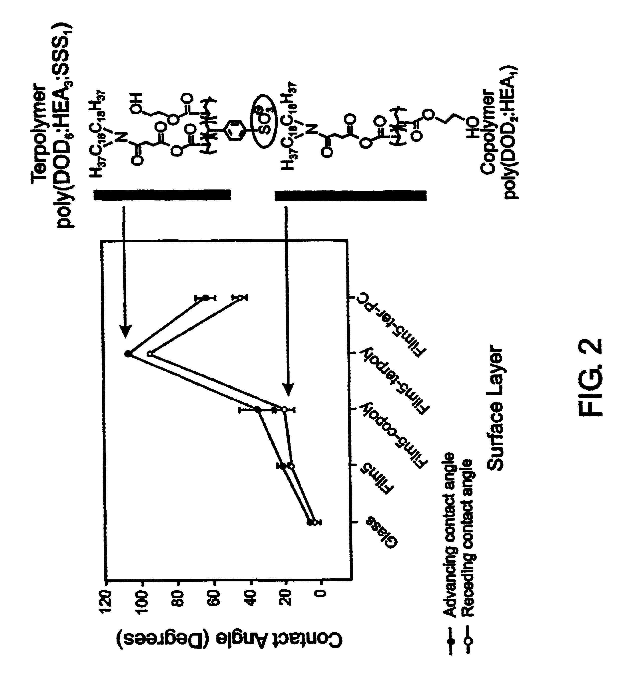
[0151]In situ polymerization of phospholipids on an alkylated surface. An in situ polymerization strategy was developed using monoacrylate phospholipids as a means of stabilizing a self-assembled lipid monolayer at a solid-liquid interface (FIG. 8). (Marra, K. G. et al. [1997] Macromolecules 30:6483-6487.) Phospholipid monomer, 1-palmitoyl-2-(12-(acryloyloxy)dodecanoyl)-sn-glycero-3-phosphorylcholine, was synthesized, prepared as unilamellar vesicles, and fused onto alkylated (OTS) glass. Free-radical polymerization was carried out in aqueous solution at 70° C. using the water-soluble initiator, 2,2′-azobis(2-methylpropionamidine) dihydrochloride (AAPD) (Scheme 2). Under optimized conditions, the supported monolayer displayed advancing and receding water contact angles of 65° and 44°, respectively. Angle-dependent ESCA results confirmed the pres...


PUM

PropertyMeasurementUnit
Fractionaaaaaaaaaa
Percent by moleaaaaaaaaaa
Lengthaaaaaaaaaa

Abstract

A biocompatible biological component is provided comprising a membrane-mimetic surface film covering a substrate. Suitable substrates include hydrated substrates, e.g. hydrogels which may contain drugs for delivery to a patient through the membrane-mimetic film, or may be made up of cells, such as islet cells, for transplantation. The surface may present exposed bioactive molecules or moieties for binding to target molecules in vivo, for modulating host response when implanted into a patient (e.g. the surface may be antithrombogenic or antiinflammatory) and the surface may have pores of selected sizes to facilitate transport of substances therethrough. An optional hydrophilic cushion or spacer between the substrate and the membrane-mimetic surface allows transmembrane proteins to extend from the surface through the hydrophilic cushion, mimicking the structure of naturally-occurring cells. An alkylated layer directly beneath the membrane-mimetic surface facilates bonding of the surface to the remainder of the biological component. Alkyl chains may extend entirely through the hydrophilic cushion when present. To facilitate binding, the substrate may optionally be treated with a polyelectrolyte or alternating layers of oppositely-charged polyelectrolytes to facilitate charged binding of the membrane-mimetic film or alkylated layer beneath the membrane-mimetic film to the substrate. The membrane-mimetic film is preferably made by in situ polymerization of phospholipid vesicles.

Description

CROSS-REFERENCE TO RELATED APPLICATIONS[0001]This application claims priority to PCT Application PCT / US01 / 24020 filed July 30, 2001, which claims benefit of U.S. provisional application Ser. Nos. 60 / 221,828, 60 / 221,618, and 60 / 221,655, all filed on July 28, 2000.STATEMENT REGARDING FEDERALLY SPONSORED RESEARCH OR DEVELOPMENT[0002]This invention was made with government support under NIH Contract No. RR14190 awarded by the National Institutes of Health. The government has certain rights in the invention.BACKGROUND OF THE INVENTION[0003]Several approaches have been proposed for improving the biocompatibility of biomaterials useful in medical applications. For example, modifying the biomaterial surface to provide either low polarity or ionic charge or coating the surface with biological substances such as cells, proteins, enzymes, etc., has been used to prevent undesirable protein adhesion. Another approach involves the incorporation of an anticoagulant into the biomaterial, rendering ...

Claims


Application Information

Patent Timeline
IPC IPC(8): A61L27/00A61F13/00A61K9/127C12N11/02A61F2/02A61F2/06A61F2/08A61F2/16A61F2/24A61F2/28A61K9/50A61L15/32A61L27/22A61L27/24A61L27/50A61L29/00A61L33/00A61M1/10A61M1/16A61M1/18C07F9/10C07F9/655C08F220/28C08F220/36C12N1/00C12N5/07C12N5/071C12N5/10
CPCC12N1/00A61L15/32A61L27/222A61L27/227A61L27/24A61L27/507A61L33/0082B82Y5/00B82Y30/00C07F9/6552C08F220/28C08F220/36A61K9/5031A61K9/1273A61K9/5073C08F220/281
Inventor CHAIKOF, ELLIOT L.FENG, JUNEORBAN, JANINE M.LIU, HONGBOSUN, XUE LONGFAUCHER, KEITH M.
Owner EMORY UNIVERSITY
Why Eureka
Try Eureka